Produktinformationen "Brain stem, deep cerebral and diencephalic structures"
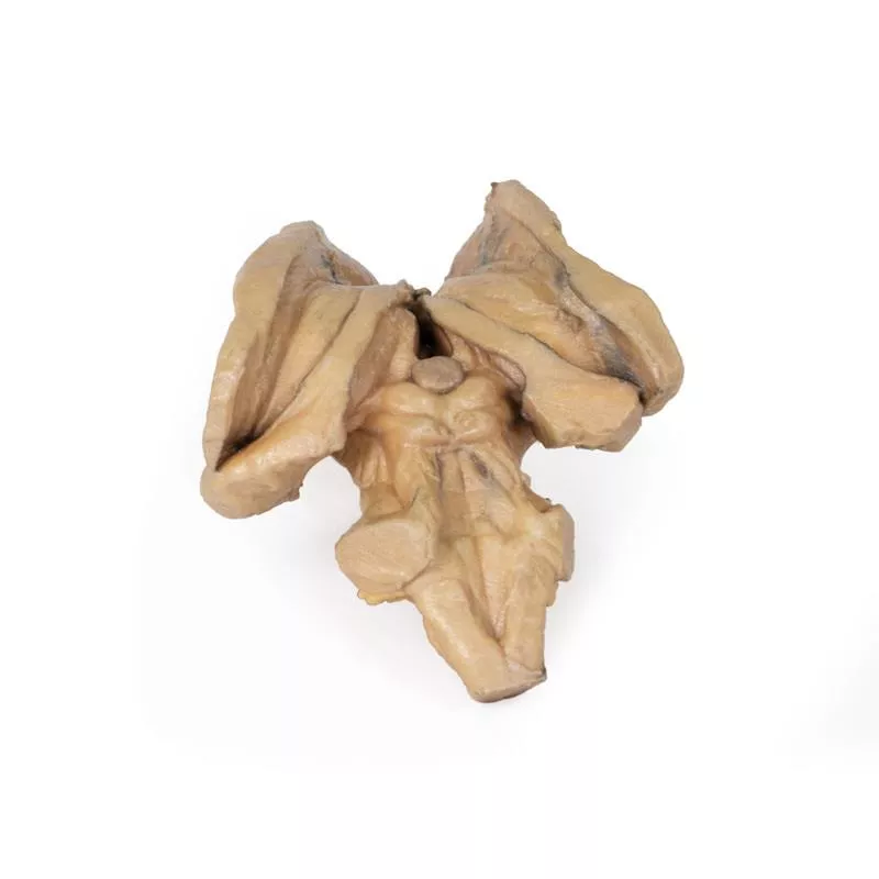
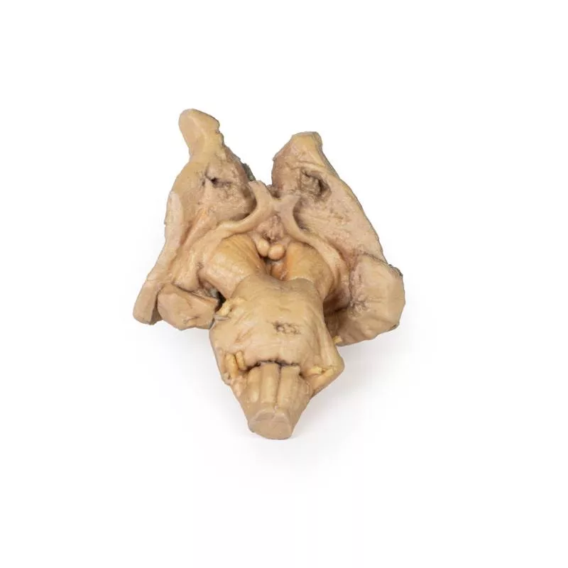
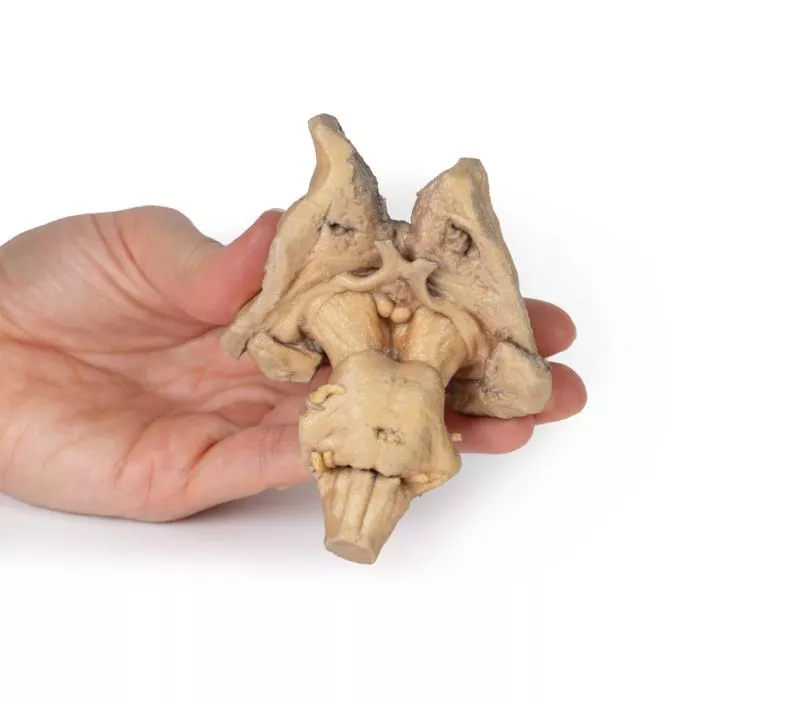
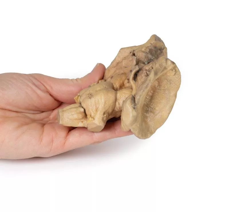
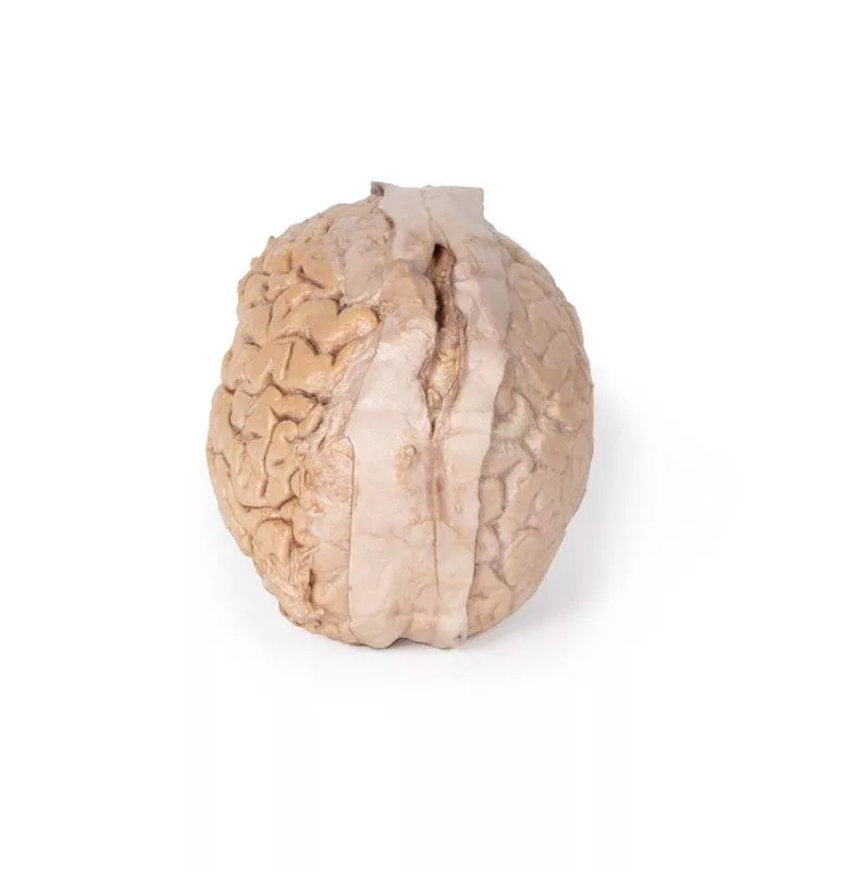
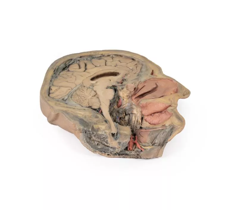
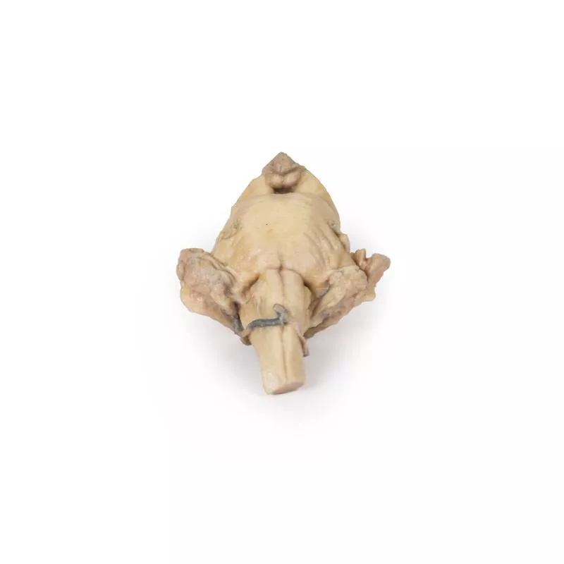
Dieses 3D-Modell bewahrt die verschiedenen tiefen Strukturen des Großhirns und des Zwischenhirns bis hin zur proximalen Medulla oblongata und ergänzt das andere isolierte Hirnstammmodell (MP1101) in unserer Serie.
Oben rechts im 3D-Modell befindet sich der Lentiformkern (Lentikularis) und um ihn herum ist die Corona radiata der inneren Kapsel zu sehen. Auf der linken Seite fehlt der Lentiformkern, aber der Kopf und Körper des Nucleus caudatus sind auf beiden Seiten medial vorhanden, umschließen medial die erhaltenen Ränder der inneren Kapsel und führen zu den Amygdaloidkörpern auf jeder Seite. Die Thalami sind bilateral vorhanden, und der dritte Ventrikel ist in der Mittellinie unterhalb des Epithalamus (Zirbeldrüse) leicht geöffnet.
Anterior sind die Hirnstiele vorhanden, wobei sich die Sehnerven vom erhaltenen Chiasma und den Bahnen aus erstrecken. Die interpedunkuläre Region ist freigelegt, wobei sowohl die Mamillarkörper als auch der durchtrennte Infundibulum sichtbar sind. Kaudal zur interpedunkulären Region befindet sich die Pons, die die Ursprünge der mittleren Kleinhirnstiele sowie die Ursprünge der Hirnnerven V, VII und VIII bewahrt. Der erhaltene Teil der Medulla oblongata weist markante Pyramiden und Oliven auf.
Posterior liegen der Colliculus superior und der Colliculus inferior direkt oberhalb der durchtrennten oberen Kleinhirnstiele, und der vierte Ventrikel ist geöffnet, um die Fossa rhomboidea und die Merkmale des Bodens freizulegen: die Eminentia medialis, den Colliculus facialis, das Dreieck der Hypoglossusnerven, das Dreieck des Vestibularnervs und das Dreieck des Vagusnervs.
Erler-Zimmer
Erler-Zimmer GmbH & Co.KG
Hauptstrasse 27
77886 Lauf
Germany
info@erler-zimmer.de
Achtung! Medizinisches Ausbildungsmaterial, kein Spielzeug. Nicht geeignet für Personen unter 14 Jahren.
Attention! Medical training material, not a toy. Not suitable for persons under 14 years of age.


