Produktinformationen "Superficial Facial nerves & Parotid Gland"
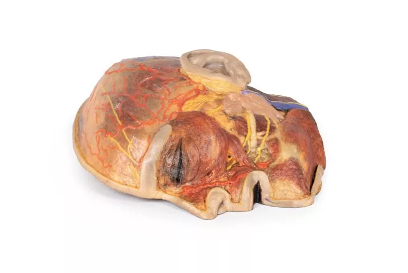
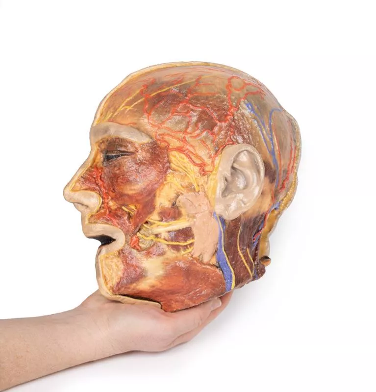
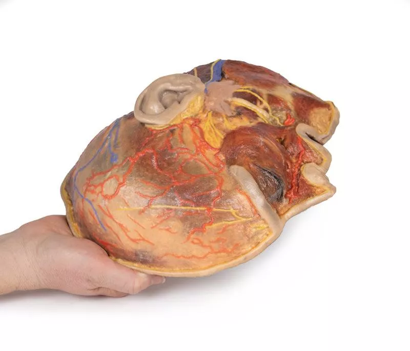
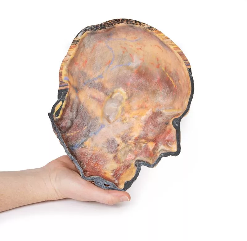
Dieses 3D-Modell bietet eine detaillierte Ansicht der oberflächlichen Anatomie von Gesicht und Kopf und erweitert unser Modell HW 44 um eine umfassendere Präparation der Kopfhaut, der Okzipitalregion und der Bereiche unterhalb des Außenohrs.
Hauptmerkmale:
Erweiterte Gesichtsanatomie
Enthält die Endäste des Gesichtsnervs (CN VII), die von der Ohrspeicheldrüse ausgehen, wobei der Platysma-Muskel erhalten bleibt und sich vom Unterkiefer bis zum Hals erstreckt.
Verbesserte posteriore Präparation
- Breitere Freilegung der hinteren Kopfhaut und der Okzipitalregion
- Enthält die Vena retromandibularis, den Nervus auricularis major und den Nervus occipitalis minor
- Zeigt den Verlauf der Arteria occipitalis und der Vena occipitalis in der Nähe des Trapezmuskels.
Neurovaskuläre Highlights
Verbesserte Darstellung der Arteria supraorbitalis, Arteria supratrochlearis und Arteria temporalis superficialis sowie der entsprechenden Nerven.
Muskulatur
Erhält die Fasern der Muskeln Auricularis und Occipitalis, die in den Epicranius (Occipitofrontalis) integriert sind.
Erler-Zimmer
Erler-Zimmer GmbH & Co.KG
Hauptstrasse 27
77886 Lauf
Germany
info@erler-zimmer.de
Achtung! Medizinisches Ausbildungsmaterial, kein Spielzeug. Nicht geeignet für Personen unter 14 Jahren.
Attention! Medical training material, not a toy. Not suitable for persons under 14 years of age.


