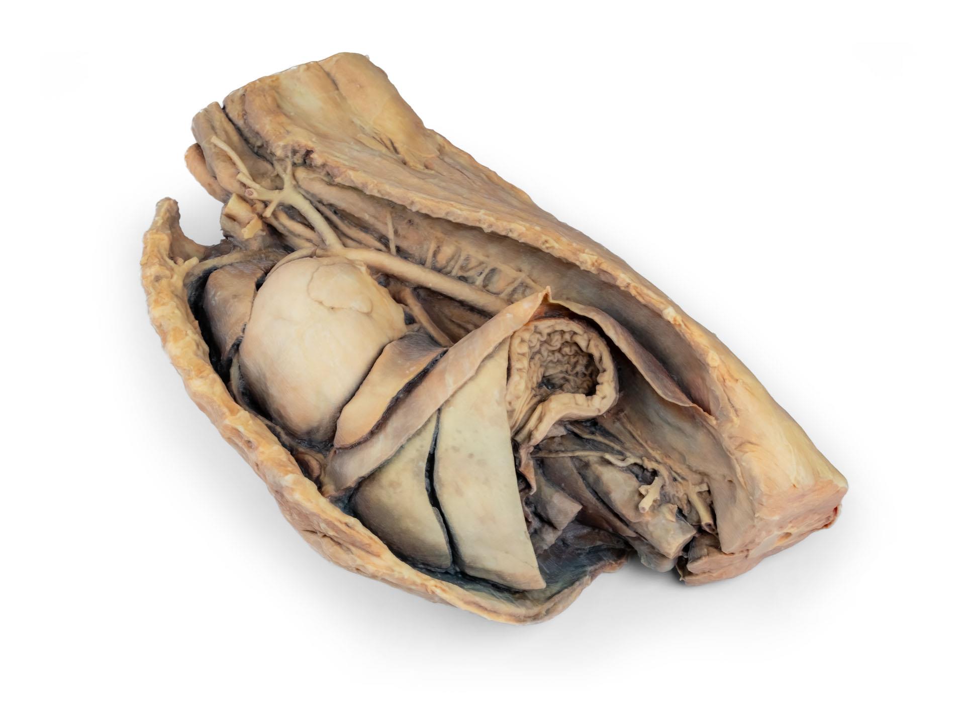
Product information "Canine heart with dissection"
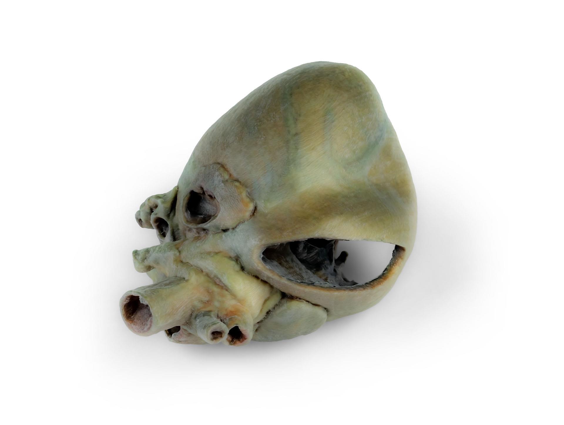
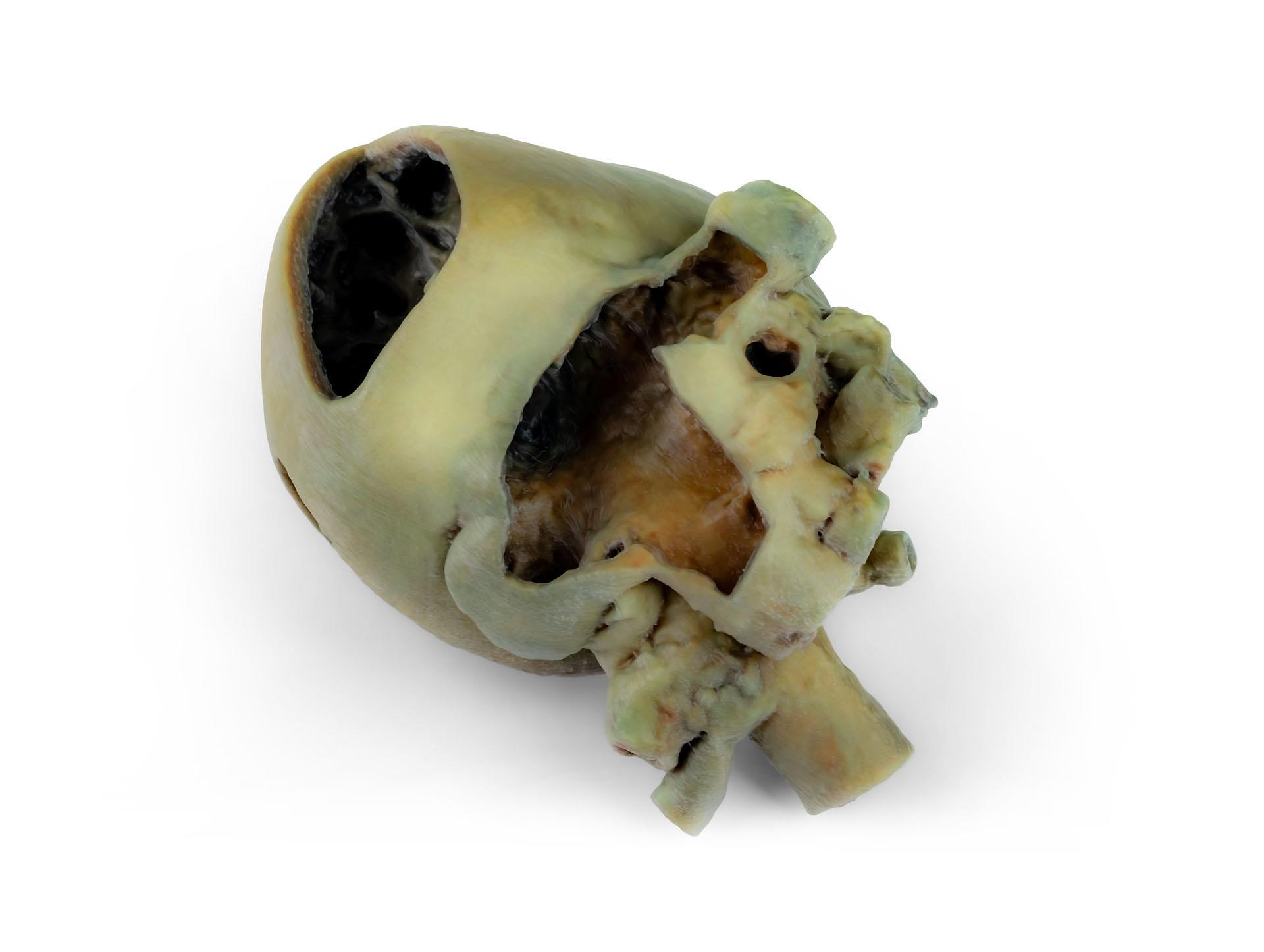
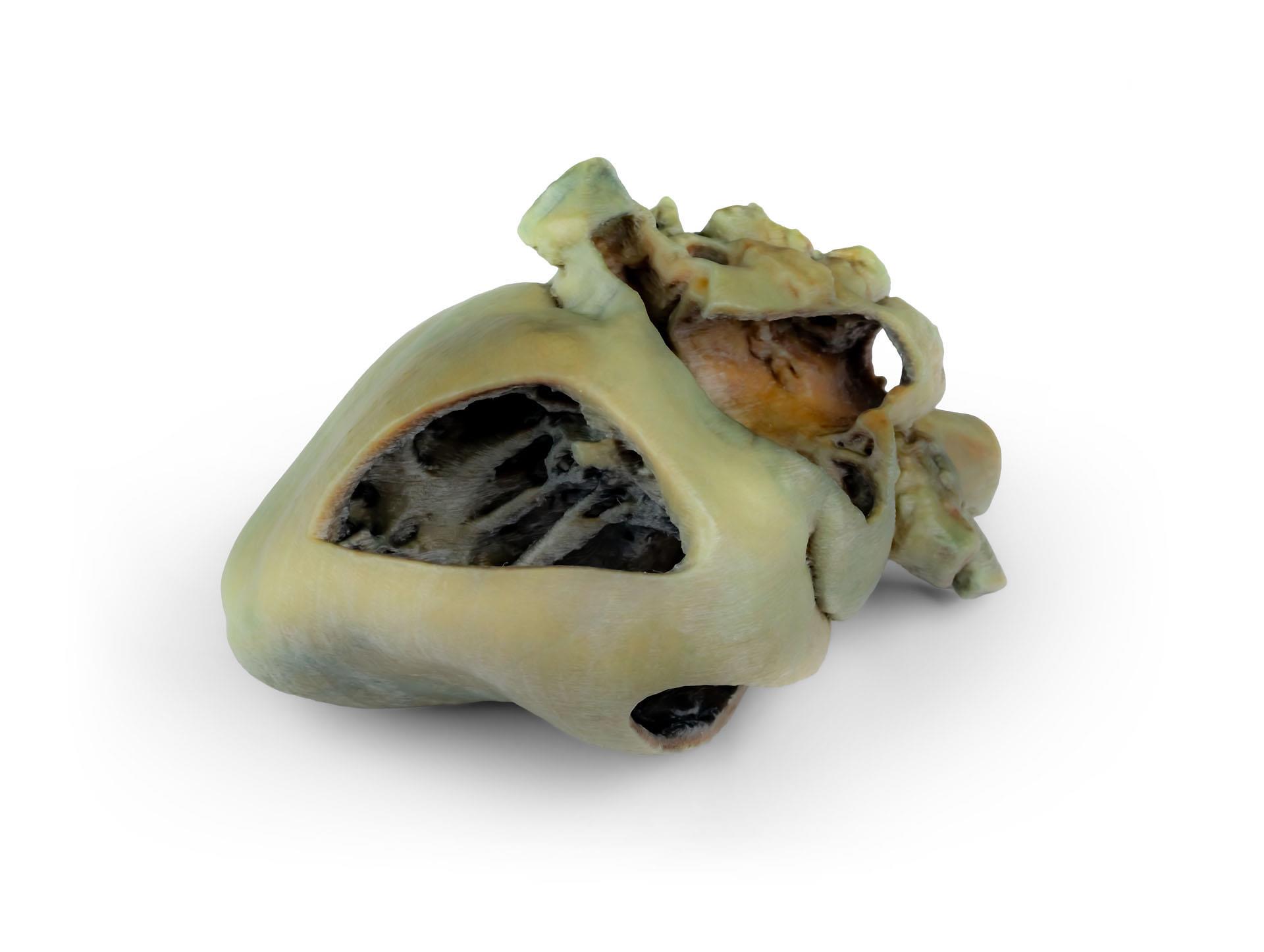
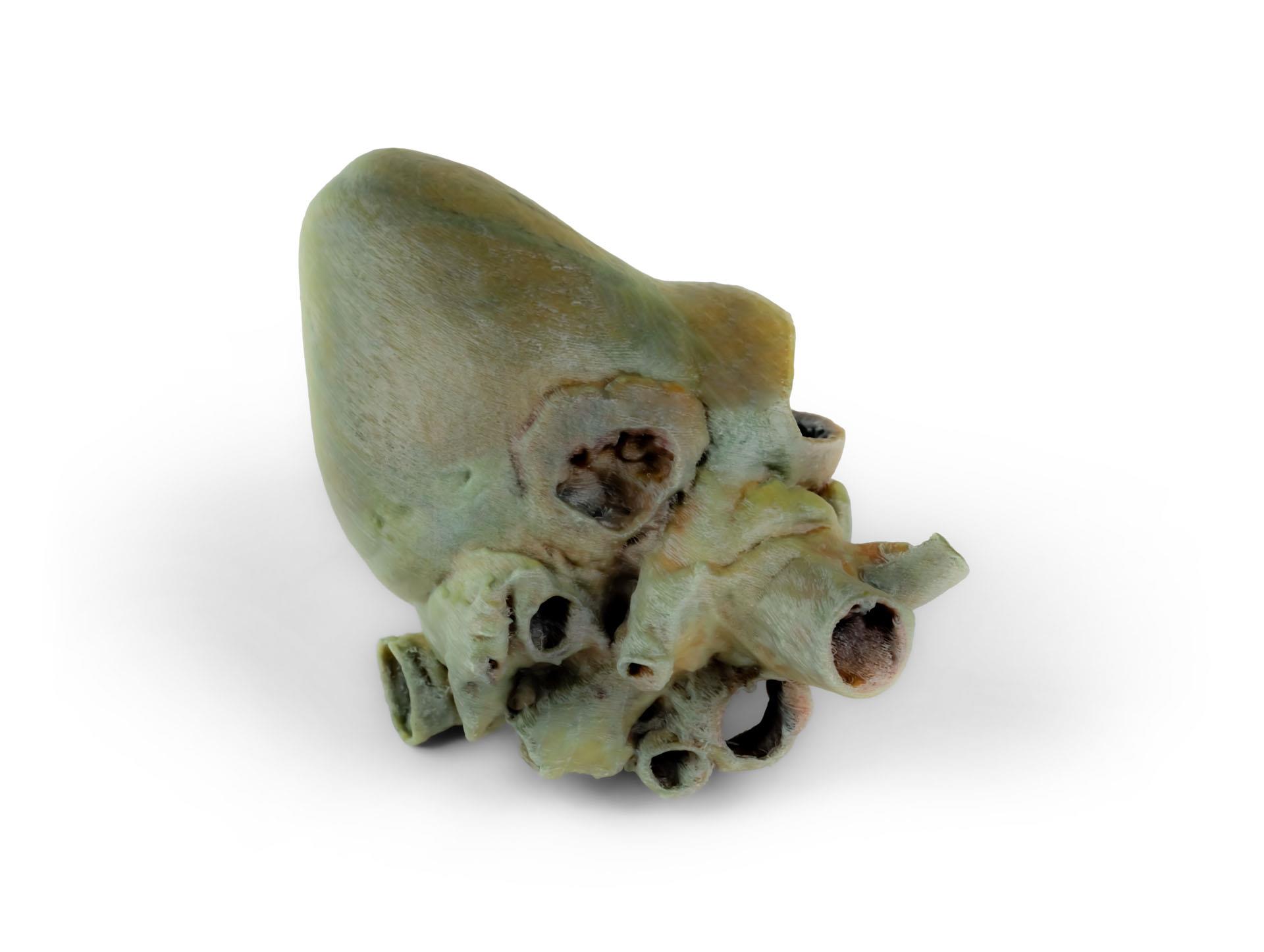
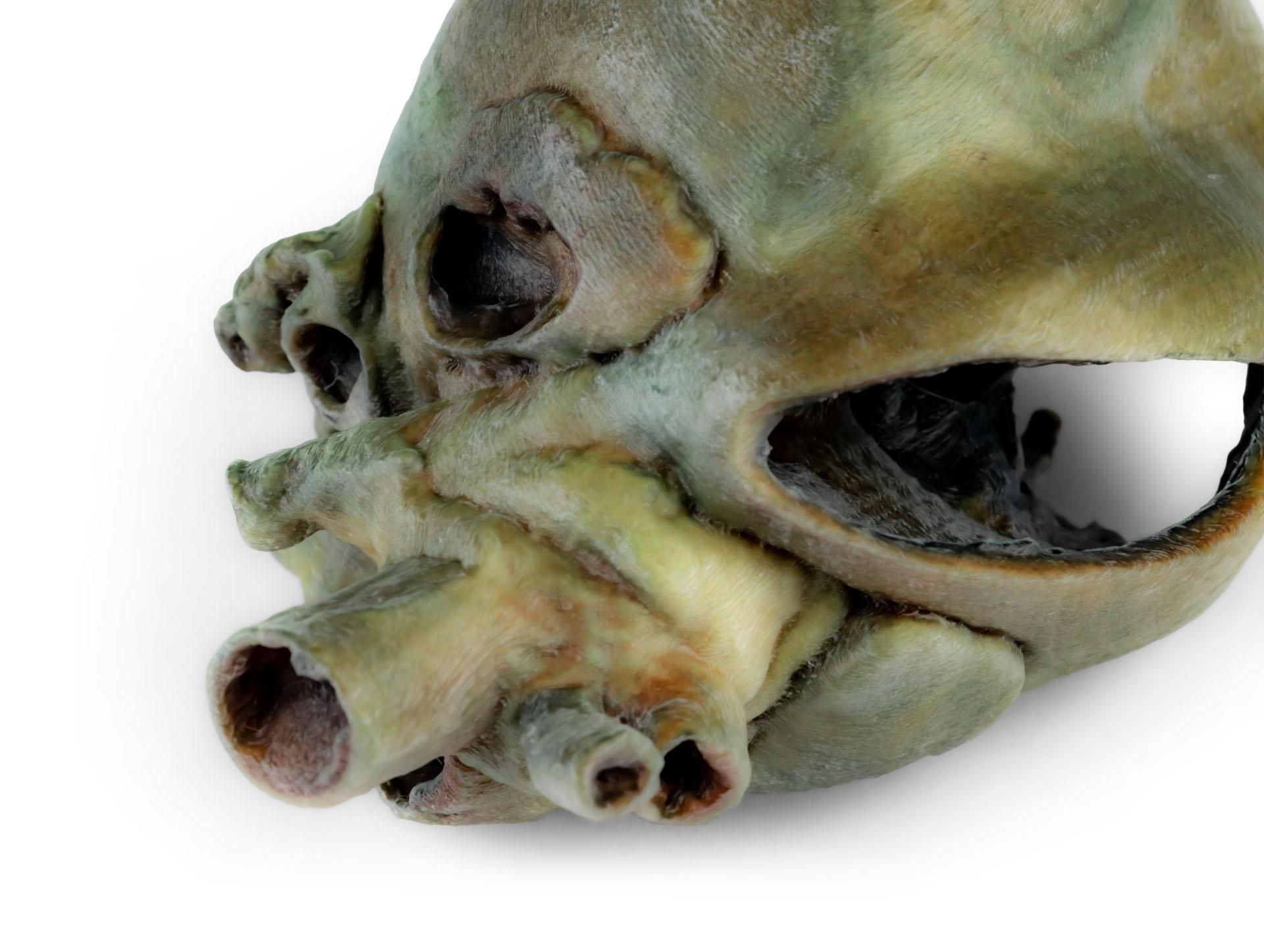
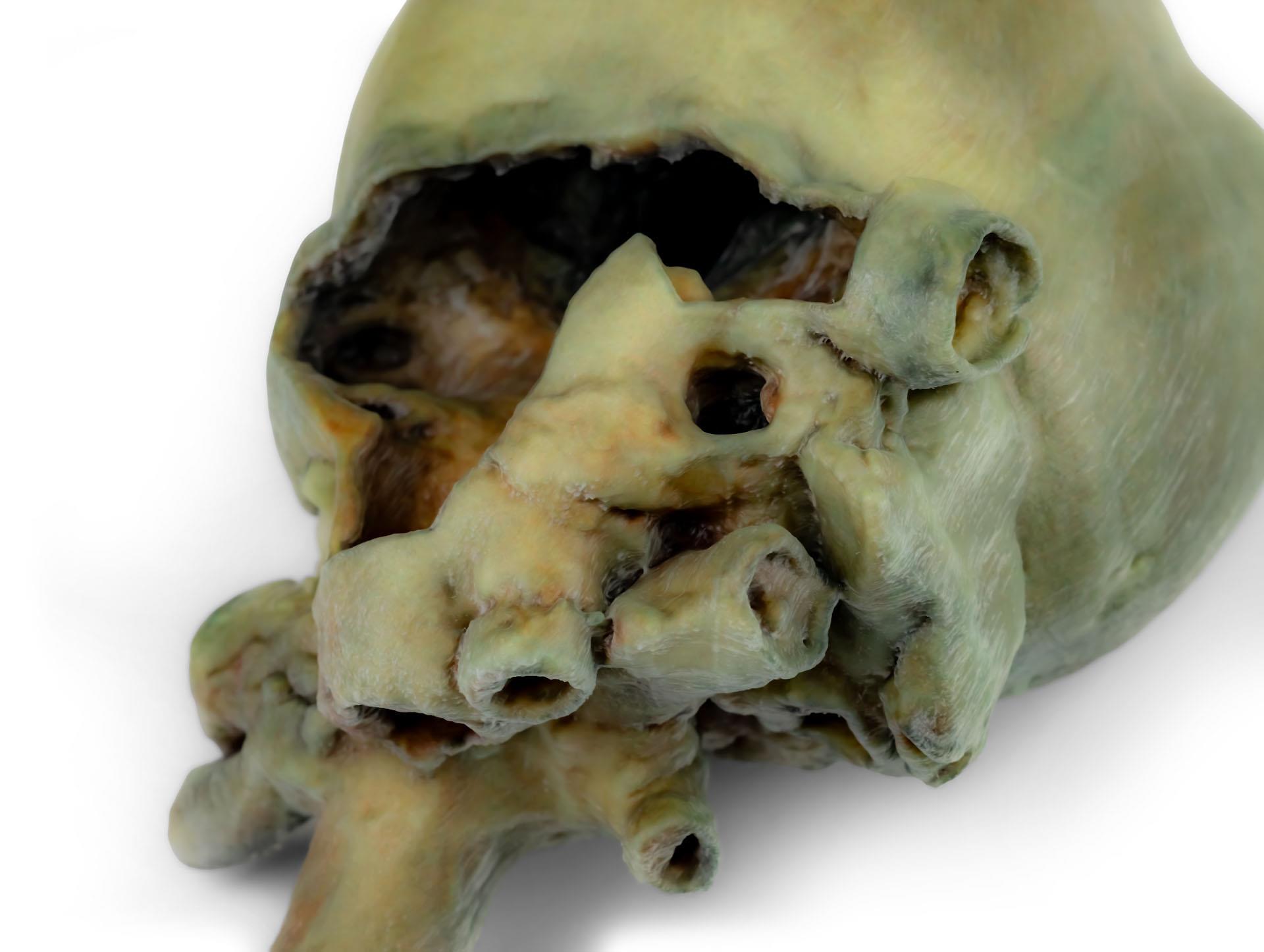
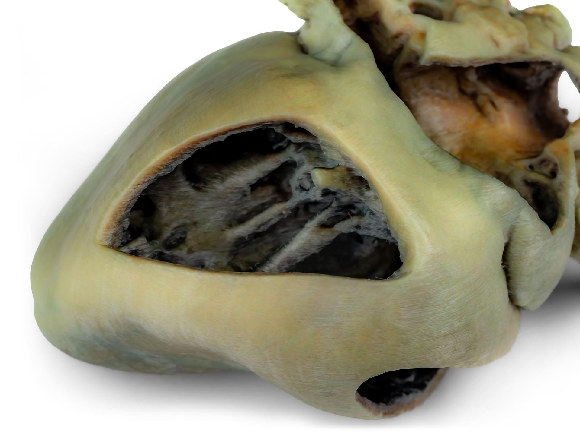
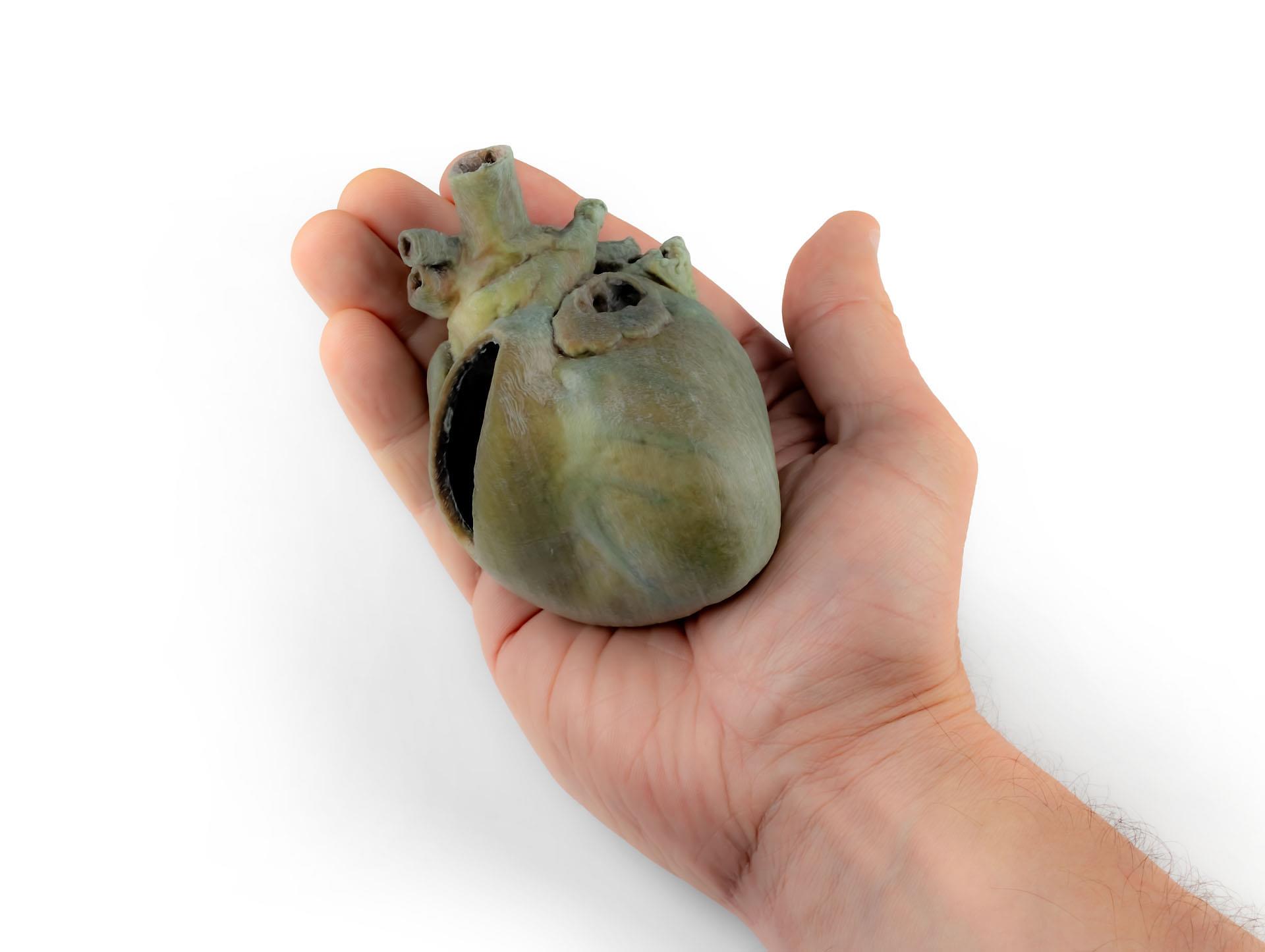
Dissected Dog Heart showing the Cardiac Cavities.

This specimen displays the anatomical features of the external surface of the heart, including both the atrial and auricular faces. At the base of the heart, the ascending aorta and its relationship with the pulmonary trunk are clearly visible, along with the sites of entry of the cranial and caudal venae cavae into the right atrium. The bifurcation of the pulmonary trunk into the pulmonary arteries can be observed, as well as the termination of the pulmonary veins into the left atrium. Both the interventricular and coronary grooves are visible.

A portion of the right atrial wall has been removed to expose the right atrioventricular (tricuspid) valve, including the chordae tendineae and papillary muscles.

Additionally, the septomarginal trabecula (moderator band) is visible within the right ventricle, as well as the trajectory from the ventricle to the pulmonary trunk. The left atrial wall is also open to display its lumen.
Documents
Anleitung / Manual VP9030 Download

0 of 0 reviews

Leave a review!

Share your experiences with other customers.


Documents
Anleitung / Manual VP9030 Download

Other customers also bought

Dog forelimb - muscles, tendons, ligaments, vessels and nerves distal to the elbow
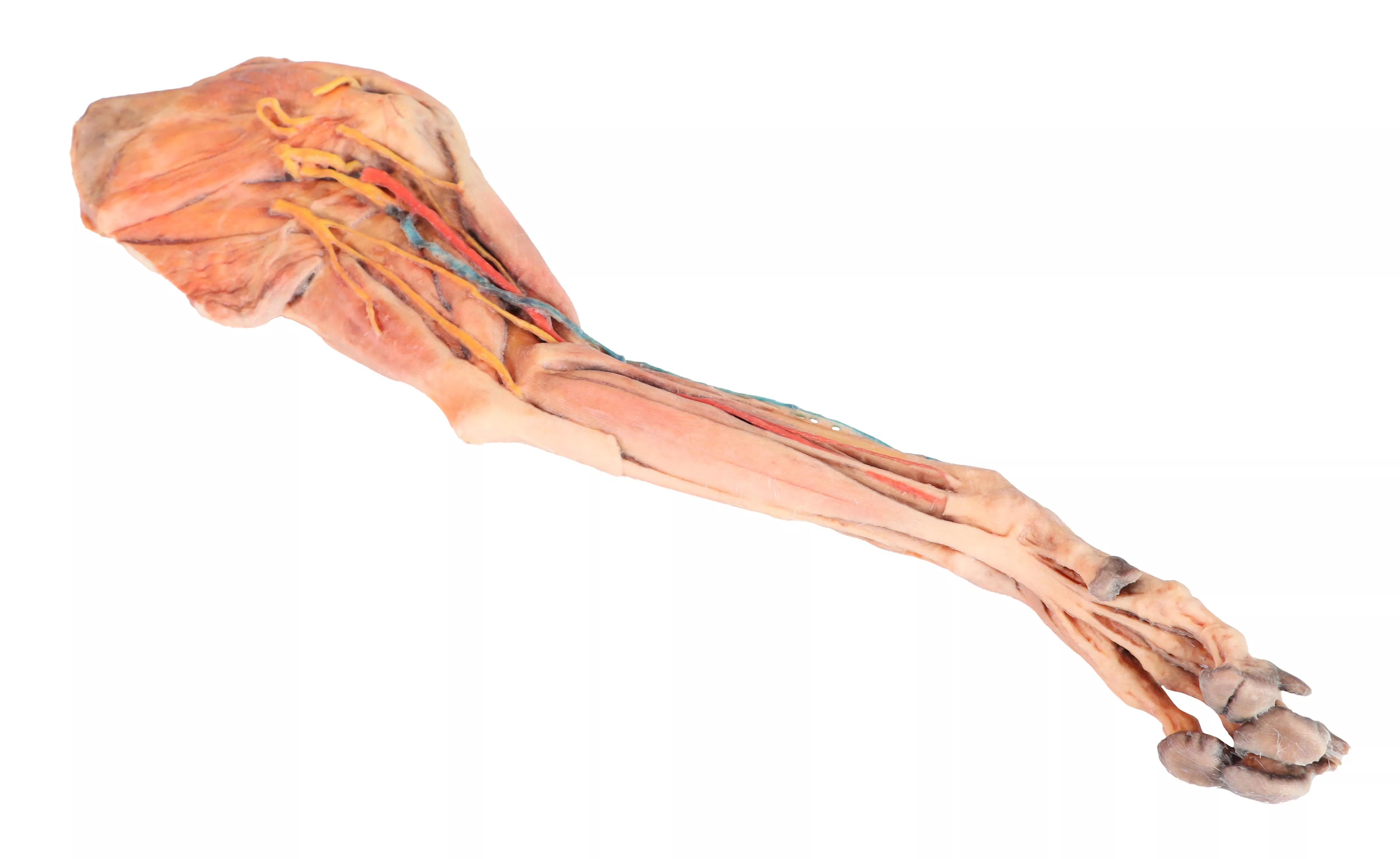
This specimen demonstrates the superficial anatomy of a dog's right thoracic limb from the scapula to the hand. The shoulder flexor and extensor muscles of the scapular region have been preserved, along with the arm flexor and extensor muscles of the elbow. In the forearm and hand are the flexor and extensor muscles of the carpus and fingers. On the medial side of the axillary region the main nerves of the brachial plexus have been preserved. Similarly, the paths of the brachial and median arteries and their respective veins are identified. In a superficial position, the cephalic vein has been preserved together with the antebrachialis lateral cutaneous nerve of the forearm. Detailed anatomical description on request.

€1,949.22*
Canine thoracic cavity left side
This specimen depicts the topography of the heart following theremoval of the left lung during a dissection of the thoracic cavityand cranial abdomen of a dog approached from the left side. The cranial and accessory lobes of the right lung remain in situ, servingas anatomical reference points. Within the cranial mediastinum, the brachiocephalic trunk and the left subclavian artery areidentifiable as major branches arising from the aortic arch. Thethoracic portion of the esophagus runs through the mediastinumin a craniocaudal direction. The diaphragm has been preserved to serve as a landmark forretrodiaphragmatic organs such as the liver and the stomach.The diaphragmatic crura and their relation to the aortic hiatus aremaintained, as are the initial visceral branches of the abdominalaorta—specifically, the celiac trunk and the cranial mesentericartery. Both the interventricular and coronary grooves are visible. A portion of the right atrial wall has been removed to exposethe right atrioventricular (tricuspid) valve, including the chordaetendineae and papillary muscles. Additionally, the septomarginaltrabecula (moderator band) is visible within the right ventricle, as well as the trajectory from the ventricle to the pulmonary trunk.The left atrial wall is also open to display its lumen.

€3,973.41*
Dog hindlimb- muscles, tendons, ligaments, vessels and nerves distal to the stifle
This specimen preserves a superficial dissection of the lower hindlimb musculature from the mid-thigh to the foot, as well as nerves and vessels of the femoral canal and popliteal fossa.The insertions of the muscles of the thigh in the stifle joint and tibial bone are visible. The capsule of the stifle joint has been opened to demonstrate the menisci and the collateralligaments. Extensor and flexor muscles of the tarsus and fingers are exposed. The dissection preserves the topography of the lateral saphenous vein on the foot surface and caudal aspect of the leg Dorsal and palmar anatomical structures of the foot are dissected. Detailed anatomical description on request.

€2,145.57*
Dog head - superficial and deep dissections
Superficial anatomical structures:Tip of the nose (Apez nasi)Right and left wings of the nose (Alae nasi)Nonglandular skin on the tip of the nose (Planun nasale)Superficial disscetion on the left side:On the left side, the skin has been removed to identify the main anatomical structures,which are described grouped below.Muscles of the facial neuromuscular system:• M. nasolabial levator (M. levator nasolabialis)• M. canine (M. caninus)• M. buccinator (M. buccinator)• M. Zygoma9c (M. zygoma:cus)• M. parodidoauricularisMuscles of the mandibular neuromuscular system (mas9cators):• M. masseter (M. masseter)• M. Temporary (M.temporalis)Nerves:• Facial nerve: (N . facialis)• Dorsal buccal Branch (Rami buccales)• Ventral buccal Branch (Rami buccales)• Bucolabial branches (Rami buccolabiales)• N. Auriculopalpebral (N. Auriculopalpebralis)Vascular:• Facial artery (Arteria facialis)• External jugular vein: (V. Jugularis externa)o Maxillary vein (V. Maxillaris)o Linguofacial vein (V. Linguofacialis)Salivary glands:• Parotid gland and paro9d duct (Glandula parotis) (Ductus parotideus)• Mandibular gland Glandula mandibularisLymph nodes:• N. L. mandibular (Lymphonodi mandubulares)Deep dissection on the right side:On this right side mandible has been removed to see deeper anatomical structures. You can see the surfaces of the temporal bone for the temporomandibular joint (Articulatio Temporomandibularis). The medial pterygoid muscle (M. pterygoideus medialis) is identified and sectioned in its insertion area to the ramus of the mandible (Ramus mandibulae). Next to this muscle, the branches of the mandibular nerve (N. mandibularis) are identified, as well as the maxillary nerve (nevus maxillaris) and the maxillary artery (arteria maxillaris). Near the external acoustic meatus (Meatus acusticus externus), the facial nerve (N . facialis) has been maintained, with one of its branches,the auriculopalpebral nerve (N. Auriculopalpebralis), running parallel to the zygomatic arch (Arcus zygomaticus), visible atier removing the parotd salivary gland (Glandula parotis). The tongue is identified in its entire caudal extension, and the styloglossus (M. stylogossus), genioglossus (M. genioglossus) and genihyoid (M. geniohydeus) muscles reach it. Next to these muscles, the hypoglossal nerve (N. hypoglossus) is identified. In relation to the pharynx (Pharynx), the constrictor muscles of the pharynx (Mm. constrictors phyngis caudalis) are identified. In the most caudal area, in relation to the neck, the course of the common caro9d artery (arteria caarotis communis) and the vagosympathetic trunk nerve (Truncus vagosympathicus) are identified.

€4,001.97*

Continuous innovation

Social responsibility

Active customer orientation

Understanding quality

Sustainable actions

ISO 9001 certification

Your last viewed products