


Product information "Heart and the distal trachea, carina and primary bronchi"
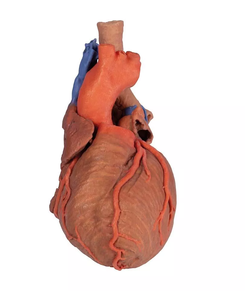
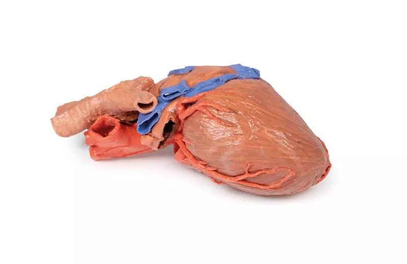
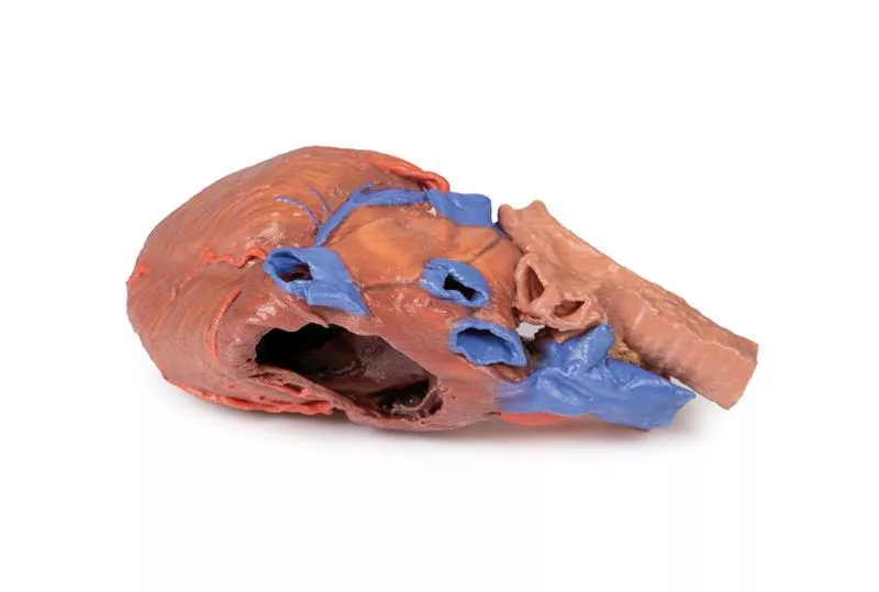
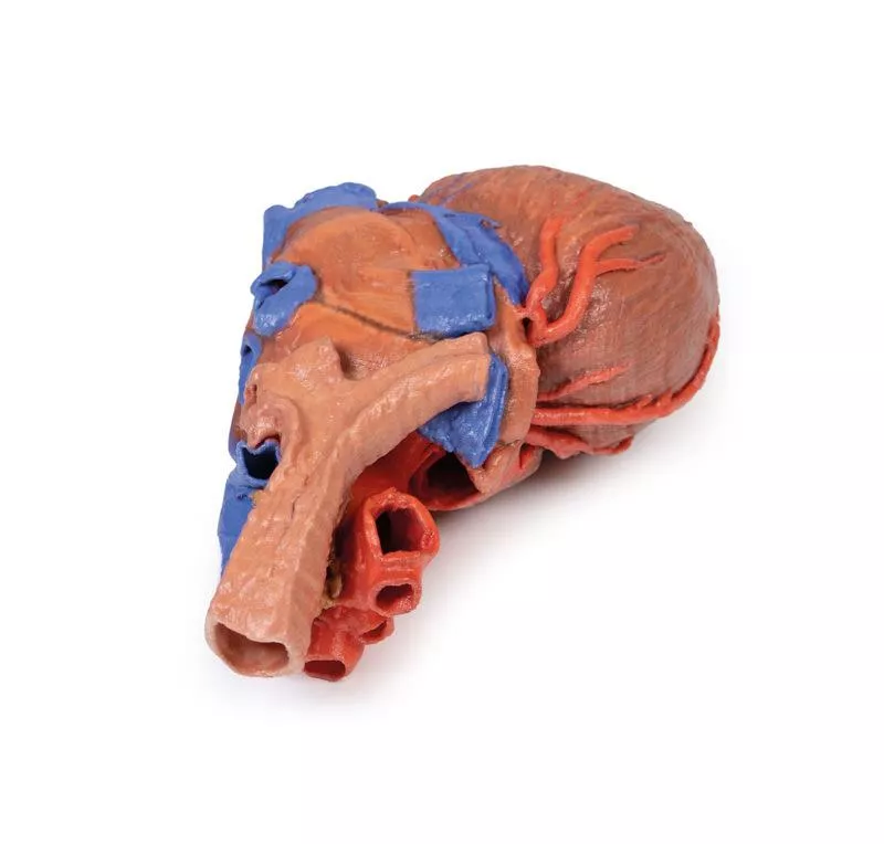
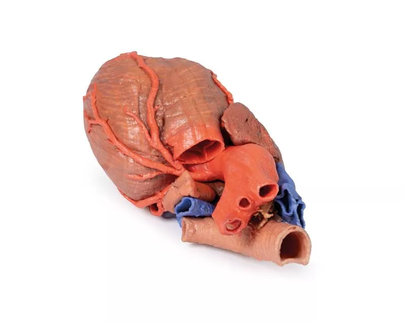
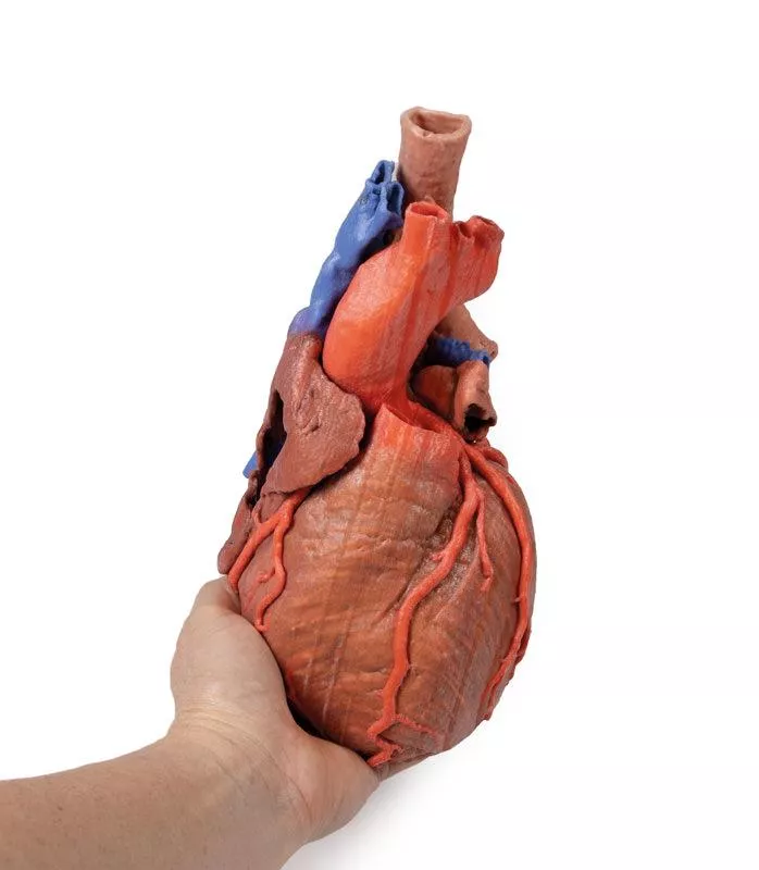
This 3D printed specimen demonstrates the external anatomy of the heart alongside the distal trachea, carina, and primary bronchi in the posterior mediastinum. The relationship of these structures to the great vessels and left atrium is clearly shown, with the pericardial reflections marking the transverse and oblique pericardial sinuses.
Heart Chambers and Valves
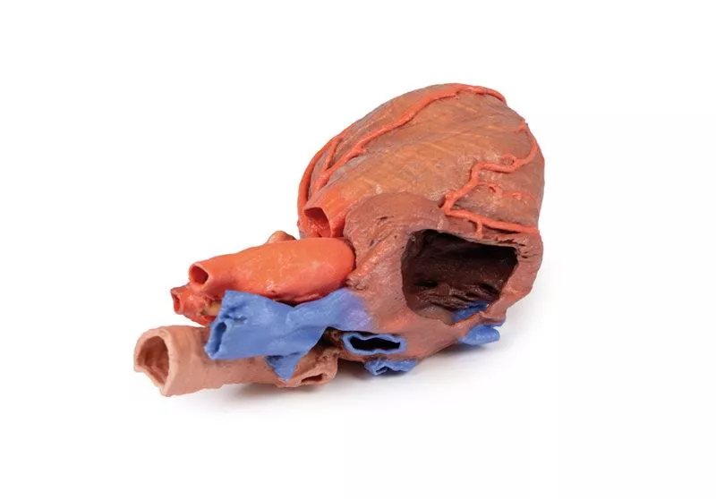
An anterior window has been dissected into the right atrium and base of the auricle, exposing the right atrioventricular (tricuspid) valve and passage into the right ventricle. The pulmonary trunk has been removed to reveal the open pulmonary semilunar valves.
Coronary Arteries and Sinus
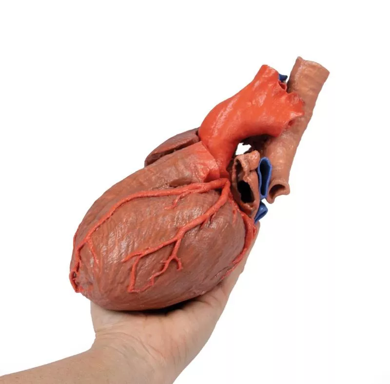
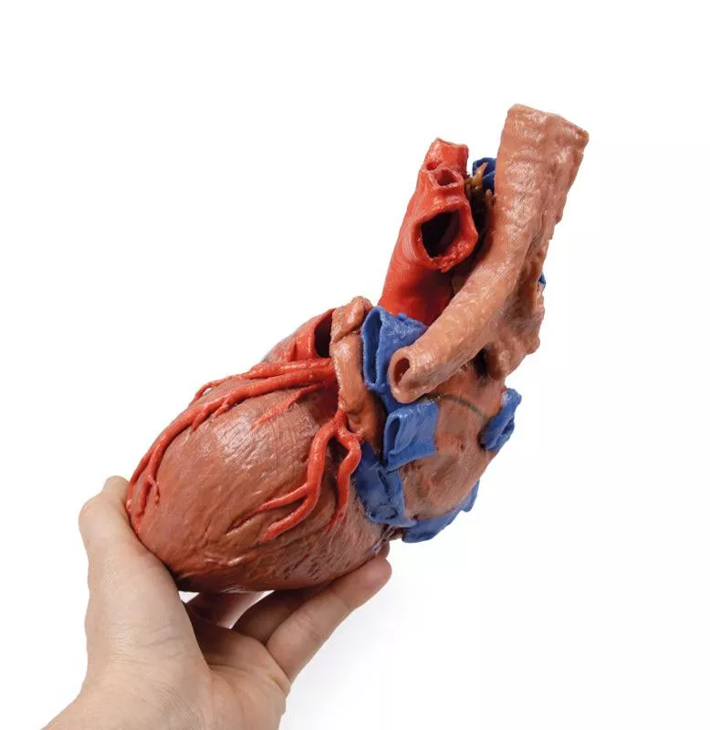
Both right and left coronary arteries with their named branches are visible. The posterior interventricular artery arises from the right coronary, while the circumflex artery is displayed in the coronary groove via a sectioned left auricle. The coronary sinus is retained inferior to the left atrium, although other cardiac veins have been removed.
Great Vessels
The arch of the aorta is intact, showing the origins of the brachiocephalic trunk, left common carotid, and left subclavian artery. Adjacent to the aorta, the termination of the left and right brachiocephalic veins and azygos vein into the superior vena cava is preserved, illustrating the venous return to the heart.
Heart Chambers and Valves
An anterior window has been dissected into the right atrium and base of the auricle, exposing the right atrioventricular (tricuspid) valve and passage into the right ventricle. The pulmonary trunk has been removed to reveal the open pulmonary semilunar valves.
Coronary Arteries and Sinus
Both right and left coronary arteries with their named branches are visible. The posterior interventricular artery arises from the right coronary, while the circumflex artery is displayed in the coronary groove via a sectioned left auricle. The coronary sinus is retained inferior to the left atrium, although other cardiac veins have been removed.
Great Vessels
The arch of the aorta is intact, showing the origins of the brachiocephalic trunk, left common carotid, and left subclavian artery. Adjacent to the aorta, the termination of the left and right brachiocephalic veins and azygos vein into the superior vena cava is preserved, illustrating the venous return to the heart.
Login
Erler-Zimmer
Erler-Zimmer Medical GmbH
Hauptstrasse 27
77886 Lauf
Germany
info@erler-zimmer.de
Achtung! Medizinisches Ausbildungsmaterial, kein Spielzeug. Nicht geeignet für Personen unter 14 Jahren.
Attention! Medical training material, not a toy. Not suitable for persons under 14 years of age.
Other customers also bought

Male left pelvis and proximal thigh
This 3D print displays a male left pelvis and proximal thigh, sectioned midsagittally and transversely through L3/4.It highlights superficial and deep structures of the pelvis, inguinal region, and proximal thigh. Muscles and FasciaThe transverse section shows the epaxial and abdominal wall muscles (rectus abdominis, obliques, transversus abdominis), psoas major, and quadratus lumborum, separated by rectus sheath and thoracolumbar fascia. The psoas major lies lateral to the external iliac artery, with testicular vessels on its surface. Lateral nerves—including ilioinguinal, lateral cutaneous thigh, and femoral—run over the iliacus.Arteries and VeinsThe left common iliac artery bifurcates into external and internal iliac arteries. Branches of the internal iliac—including inferior gluteal, internal pudendal, obturator, and umbilical arteries—are visible. The deep circumflex iliac artery passes posterior to the inguinal ligament, and the inferior epigastric artery perforates the rectus abdominis. The left common iliac vein, obturator, and external iliac veins are preserved.Pelvic Organs and Urogenital StructuresThe midline section reveals the bladder, left seminal vesicle, vas deferens, and rectum with the external anal sphincter. The urethra passes through the prostate, pelvic diaphragm, and penis. The scrotum shows the parietal tunica vaginalis after removal of the skin. Thigh and Femoral RegionOn the thigh, the fascia lata is removed to reveal muscles and neurovasculature. A window above the inguinal ligament exposes the transversus abdominis aponeurosis. The femoral nerve lies over iliopsoas, with the great saphenous vein running medially. Thigh muscles—including sartorius, rectus femoris, vasti muscles, and tensor fasciae latae—are visible, and a window shows gluteus medius attaching to the greater trochanter.

Bowel - Portion of Jejenum
This 3D printed small loop of jejunum illustrates the arterial arcades in the mesentery, featuring many long straight vasa rectae and fewer vascular arcades compared to the ileum. The mesentery has been dissected to reveal the fat and visceral peritoneum, along with lymph nodes (grey-light green), which are prominent near the root of the mesentery and larger vessels. Mesenteric Fat and VesselsTypically, the fat in the jejunal mesentery does not extend to the mesenteric border, allowing a clear view of the long straight vessels. In this specimen, however, a patient with a larger amount of abdominal fat shows fat extending further toward the mesenteric border, partially obscuring this window. Jejunal MucosaA small segment of the jejunum has been opened to display the mucosal folds, which are more numerous and smaller than in the ileum, providing a detailed view of the internal structure of the bowel.

Male Pelvis
This multipart 3D printed specimen displays the lower posterior abdominal wall, pelvic cavity, and proximal thigh, including the gluteal regions and femoral triangles.Lower Posterior Abdominal Wall & False PelvisTransected at L2/L3, it shows the common iliac veins forming the inferior vena cava and the common iliac arteries. The iliacus and psoas muscles, including the psoas minor tendon, are clearly visible under the inguinal ligament. The iliac fossa nerves—ilioinguinal, lateral cutaneous of thigh, femoral, and genitofemoral—and the ureters descending along the psoas are easily identified. External iliac vessels and the vas deferens are also preserved.True PelvisThe pelvis shows the bladder, dilated rectum, and ureters, with branches of the internal iliac artery, including the obturator artery and accessory vein, and the obliterated umbilical arteries ascending anteriorly.Femoral TriangleThe right femoral triangle reveals dissected floor muscles; the left side shows the femoral vein, artery, and nerve passing deep to the inguinal ligament. Gluteal RegionThe gluteus maximus is exposed on the right with perforating cutaneous nerves. Laterally, the tensor fascia lata and iliotibial tract are visible. A window on the left shows gluteus medius, piriformis, the sciatic nerve, and superior and inferior gluteal nerves and arteries. Small nerves like the inferior cluneal and posterior cutaneous thigh branch are also identifiable.

Cubital fossa - muscles, large nerves and the brachial artery
This 3D print shows the distal arm, partially flexed elbow, and proximal forearm, highlighting the muscles, major nerves, and brachial artery at the cubital fossa.All fat, superficial veins, and cutaneous nerves have been removed for a clear view of the underlying structures. Anterior ViewThe biceps muscle is prominent, with its flattened bicipital aponeurosis passing medially over the common flexor muscles and the tendon inserting into the radial tuberosity. Deep to the biceps, the brachialis is clearly visible. In the proximal forearm, the brachioradialis (partially reflected) and extensor carpi radialis longus are identifiable.The classic arrangement of biceps tendon, brachial artery, and median nerve runs from lateral to medial, partially covered by the bicipital aponeurosis. The ulnar nerve passes behind the medial epicondyle into the cubital tunnel, while the radial nerve is visible between brachialis and brachioradialis. Posterior ViewThe triceps tendon inserts into the olecranon, with the medial and lateral epicondyles clearly visible. The ulnar nerve is also seen passing behind the medial epicondyle before entering the cubital tunnel.Cross-SectionIn transverse view, the biceps lies anteriorly with the brachial artery, median nerve, and ulnar nerve medially. The three heads of triceps are visible posteriorly. In the forearm, the radius and ulna are clear, with the brachial artery medial to pronator teres and the median nerve deep to this muscle.

Heart internal structures
This 3D printed heart displays the internal structures of all chambers. The superior and inferior vena cava enter the right atrium, revealing the pectinate muscles of the auricle and the nearly translucent fossa ovalis.Right Atrium and VentricleThe right atrioventricular (tricuspid) valve with its three cusps is exposed, along with chordae tendineae and papillary muscles. The conus arteriosus leads to the pulmonary semilunar valve, and the right coronary artery continues posteriorly to the posterior interventricular artery. Left Atrium and VentricleThe pulmonary veins enter the left atrium, and the left atrioventricular (mitral) valve with associated chordae and papillary muscles is visible. The left ventricle shows well-developed trabeculae carneae, and the aortic semilunar valve sits at the base of the aorta. Coronary ArteriesThe left coronary artery gives rise to the left anterior descending, diagonal, ramus intermedius, and circumflex branches. The circumflex passes near the coronary sinus, while the left anterior descending penetrates the myocardium and emerges near the apex.

Bowel - Portion of Ileum
This 3D printed small loop of ileum highlights the arterial arcades in the mesentery. It demonstrates many short vasa rectae and a higher number of arcades compared to the jejunum. The mesentery is dissected to reveal the vessels and one or two large lymph nodes near the root of the mesentery. Mesenteric Fat and VesselsThe model clearly shows the fat in the mesentery extending up to and beyond the mesenteric border of the bowel, emphasizing the anatomical relationship between fat, vessels, and lymph nodes. Ileal MucosaA small segment of the ileum has been opened to display the mucosal folds, which are fewer but larger than those found in the jejunum, giving a detailed view of the bowel’s internal structure.

Female right pelvis superficial and deep structures
This 3D printed specimen preserves superficial and deep structures of the true and false pelvis, the inguinal ligament, obturator canal, and sciatic foramina, with ‘windows’ exposing extraperitoneal structures.Posterior Abdominal Wall & Major VesselsSectioned at L4, it shows the colon, psoas and quadratus lumborum, and abdominal wall muscles. The common iliac artery and vein, inferior vena cava, and the bifurcation into external and internal iliac arteries are clearly visible. The external iliac vessels pass along the pelvic brim, giving off inferior epigastric and deep circumflex branches, with the femoral nerve lateral to psoas and the lateral cutaneous nerve of the thigh running over iliacus.Internal Iliac & Obturator StructuresBranches of the internal iliac artery supply the bladder, uterus, and lateral pelvic wall, while the obturator nerve, artery, and vein pass through the obturator canal. Sacral nerves (S1–S3) form the sciatic nerve, which exits via the greater sciatic foramen alongside gluteal vessels.Pelvic OrgansThe ureter, bladder, uterus, vagina, and cervix are clearly visible in midsagittal view. The Fallopian tube, fimbria, and ovary with its ligaments are preserved, as well as small portions of the rectum and nearby pararectal lymph nodes. Femoral Triangle & Gluteal RegionThe inguinal ligament is intact, with femoral vessels and nerve passing beneath it. In the gluteal region, the sciatic nerve and gluteal vessels are visible, and the pudendal nerve and vessels pass into the lesser sciatic foramen toward the perineum.

Cubital Fossa
This 3D printed cubital fossa presents a superficial dissection of the right distal arm and proximal forearm. The skin and superficial fascia have been removed anteriorly, medially, and laterally to reveal the superficial veins—basilic, cephalic, and median cubital—as well as the cutaneous nerves (medial, lateral, and posterior antebrachial). Deep Fascia and Connective TissueThe deep fascia underlying the superficial structures has largely been removed, but the antebrachial fascia is retained medially to demonstrate the merging of connective tissue fibers with the tendon of the biceps brachii through the bicipital aponeurosis. Medially, the ulnar artery is visible entering the cubital tunnel proximal to the medial epicondyle of the humerus. Anteriorly, the median nerve, brachial artery, and accompanying veins run parallel to the biceps brachii. On the lateral aspect, the cephalic vein rests on the brachioradialis muscle, while the posterior antebrachial cutaneous nerve lies on the common origin of the forearm extensors, just anterior to the exposed origin of the triceps brachii. Cross-Sectional AnatomyThe proximal cross-section displays the anterior and posterior arm compartment muscles (biceps brachii, brachialis, triceps brachii), neurovascular bundles (median, ulnar, and radial nerves; brachial artery and veins), and superficial veins (basilic and cephalic) at the midshaft of the humerus.The distal cross-section reveals the anterior and posterior forearm compartment muscles separated by the interosseous membrane, along with superficial and deep neurovascular bundles (radial artery, vein, and superficial branch of the radial nerve; ulnar artery, vein, and nerve; median nerve; anterior and posterior interosseous arteries, veins, and nerves) and the distal continuations of the superficial veins and cutaneous nerves.

Heart
This 3D print preserves the superficial anatomy of an isolated human heart along with the bases of the great vessels. All four chambers—atria and ventricles—are clearly visible, with pericardial reflections on the left atrium marking the positions of the transverse and oblique pericardial sinuses.Coronary Arteries and VeinsThe right marginal branch of the right coronary artery is visible exiting the fat-filled coronary sulcus, along with the posterior interventricular (posterior descending) artery within its sulcus. On the anterior side, the anterior interventricular (left anterior descending) artery and diagonal branches from the left coronary artery are clearly displayed. The terminal portion of the circumflex branch can be seen deep to the left auricle, alongside the great cardiac vein. On the posterior aspect, the coronary sinus collects all cardiac veins—great, middle, and small—and includes a prominent posterior vein of the left ventricle. Heart ValvesAt the bases of the ascending aorta and pulmonary trunk, the aortic and pulmonary semilunar valves are visible, providing a detailed view of the outflow pathways from the ventricles.

Female right pelvis
This 3D printed female right pelvis is sectioned midsagittally and transversely at L4 and the proximal thigh, revealing the true and false pelvis, inguinal region, femoral triangle, and gluteal region.Muscles & NervesKey muscles include psoas, iliacus, quadratus lumborum, obturator internus, piriformis, coccygeus, and hamstrings. Major nerves are the femoral, obturator, lateral femoral cutaneous, genitofemoral, lumbosacral trunk (S1–S3), and the sciatic and pudendal nerves, with early divisions of the common peroneal and tibial nerves in the gluteal region.VesselsThe common iliac artery bifurcates into external and internal iliac arteries, giving rise to branches such as deep circumflex iliac, inferior epigastric, superior/inferior gluteal, internal pudendal, obturator, uterine, and vesical arteries. Veins include external/internal iliac veins, common iliac vein, and tributaries.Pelvic VisceraPreserved viscera include the bladder, ureter, urethra, uterus remnants, vagina, rectum, and anal canal, with surrounding levator ani and external anal sphincter fibers. Thigh & Femoral TriangleThe inguinal ligament and fascia lata are exposed, showing the femoral artery, vein, nerve, deep femoral artery, great saphenous vein, and anterior/medial thigh muscles including sartorius, rectus femoris, iliopsoas, gracilis, adductors, and obturator externus.Gluteal RegionA window in gluteus maximus exposes piriformis, superior and inferior gluteal vessels, sciatic nerve, internal pudendal vessels, obturator internus, gemelli muscles, and proximal hamstrings, illustrating deep gluteal anatomy.

Female left pelvis and proximal thigh
This 3D printed specimen displays superficial and deep structures of the pelvis, inguinal region, femoral triangle, and gluteal region, offering a detailed view of female pelvic anatomy.Posterior Abdominal Wall & SpineSectioned at L4, it shows the psoas, quadratus lumborum, epaxial muscles, and cauda equina, including ventral and dorsal roots exiting the intervertebral and sacral foramina.Major VesselsThe abdominal aorta and its branches—including common, external, and internal iliac arteries—are preserved. Internal iliac branches supply the bladder, uterus, vagina, and pelvic walls. The external iliac artery passes under the inguinal ligament, alongside the femoral nerve and lateral cutaneous nerve of the thigh.Pelvic OrgansThe midsagittal view shows the bladder, uterus, vagina, rectum, and clitoral body, as well as the anterior and posterior perineal triangles. Femoral Triangle & ThighThe femoral triangle is exposed with the femoral artery and vein, great saphenous vein, and femoral nerve branches. The proximal thigh reveals anterior, medial, and posterior muscles along with the sciatic nerve. Gluteal & Posterior ThighDissection of the gluteal region exposes the piriformis, gluteal vessels and nerves, sciatic nerve, and lateral rotators. The internal pudendal vessels and pudendal nerve are visible in the perineum alongside the levator ani and external anal sphincter.

Continuous innovation

Social responsibility

Active customer orientation

Understanding quality

Sustainable actions

ISO 9001 certification