


Product information "Ruptured Thoracic Aortic Aneurysm"
Clinical History
No clinical details are available for this specimen.
Pathology
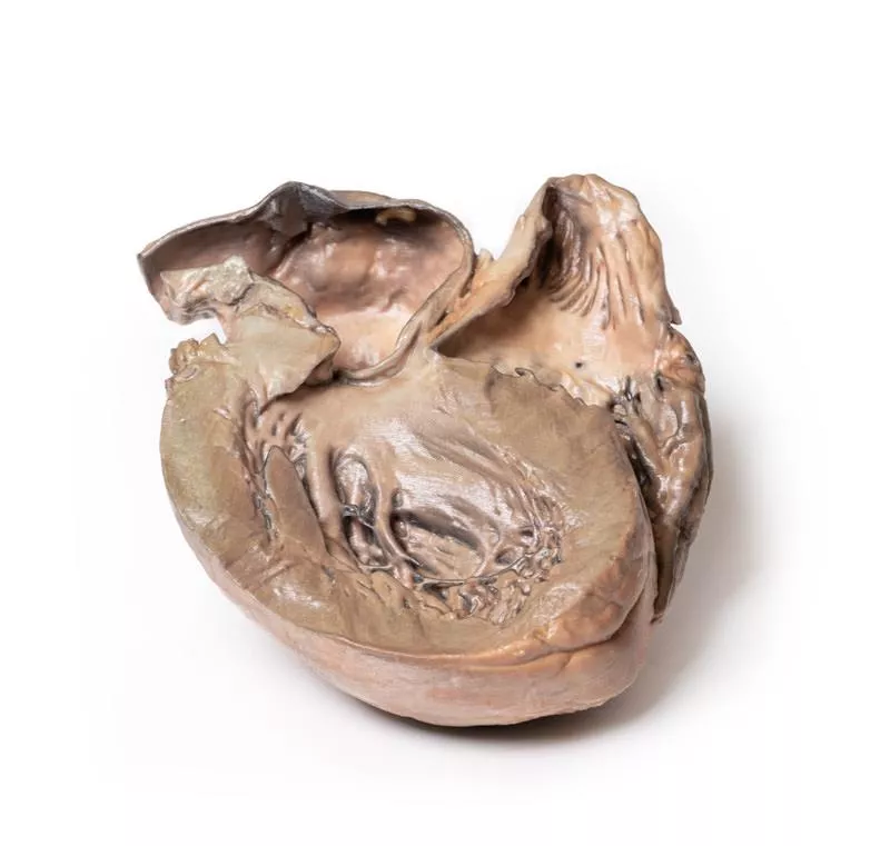
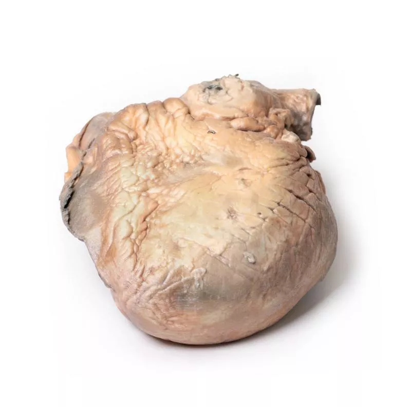
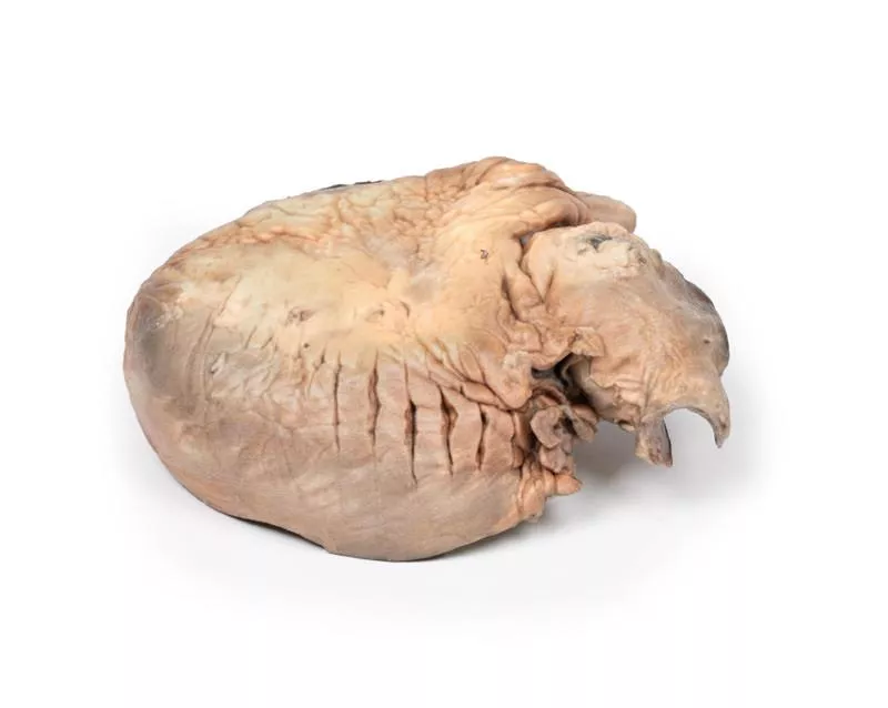
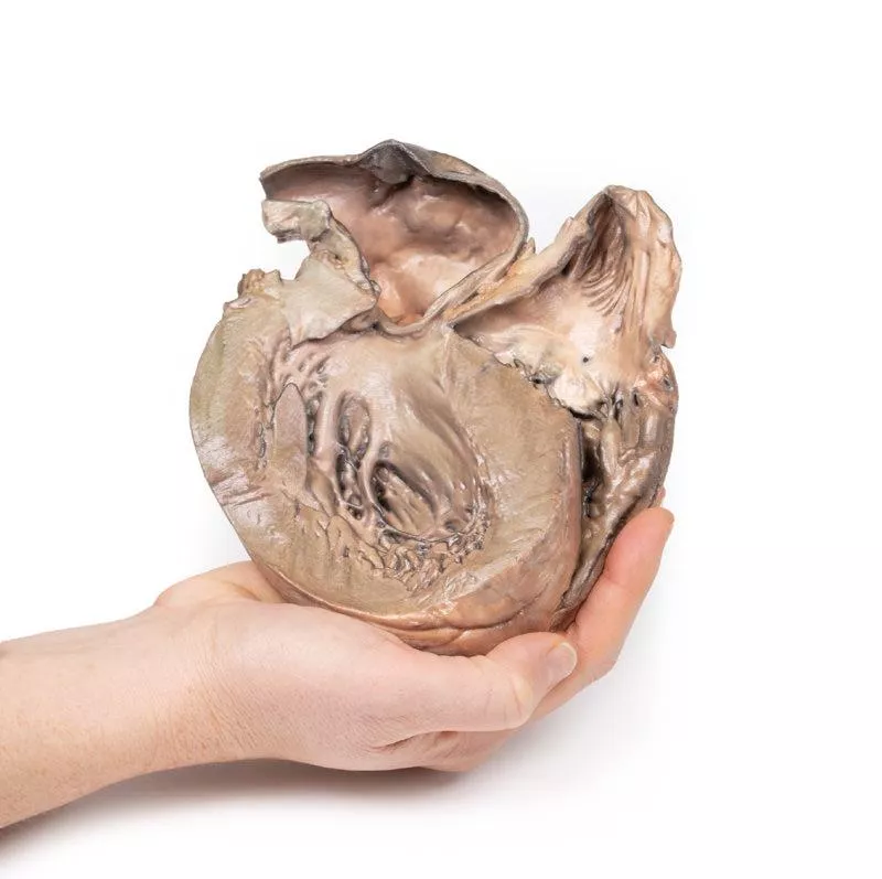
The heart is shown from the posterior aspect, displaying both ventricles. A prominent saccular dilatation of the ascending thoracic aorta is present, featuring several atherosclerotic plaques. The aorta is ruptured posteriorly, indicated by dark staining. Both ventricles are hypertrophied, while the coronary arteries and aortic and tricuspid valves appear normal. This represents a ruptured aneurysm of the ascending aorta.
Further Information
Dilation of the ascending aorta is often an incidental finding on transthoracic echocardiography. The thoracic aorta consists of three parts: ascending, arch, and descending. The ascending aorta begins just beyond the aortic valve and ends before the brachiocephalic trunk, measuring about 5 cm in length. It includes the aortic root (with coronary sinuses and sinotubular junction) and the tubular ascending aorta. Over 50% of thoracic aortic aneurysms occur here, affecting either the root or tubular segment. An aneurysm is a localized dilation of the aorta exceeding 50% of the expected diameter (observed/expected ratio = 1.5) and differs from ectasia, which is a diffuse dilation less than 50%. The incidence of ascending thoracic aortic aneurysms is about 10 per 100,000 person-years.
Reference: Saliba et al. (2015). Int J Cardiol Heart Vasc. 6: 91–100.
No clinical details are available for this specimen.
Pathology
The heart is shown from the posterior aspect, displaying both ventricles. A prominent saccular dilatation of the ascending thoracic aorta is present, featuring several atherosclerotic plaques. The aorta is ruptured posteriorly, indicated by dark staining. Both ventricles are hypertrophied, while the coronary arteries and aortic and tricuspid valves appear normal. This represents a ruptured aneurysm of the ascending aorta.
Further Information
Dilation of the ascending aorta is often an incidental finding on transthoracic echocardiography. The thoracic aorta consists of three parts: ascending, arch, and descending. The ascending aorta begins just beyond the aortic valve and ends before the brachiocephalic trunk, measuring about 5 cm in length. It includes the aortic root (with coronary sinuses and sinotubular junction) and the tubular ascending aorta. Over 50% of thoracic aortic aneurysms occur here, affecting either the root or tubular segment. An aneurysm is a localized dilation of the aorta exceeding 50% of the expected diameter (observed/expected ratio = 1.5) and differs from ectasia, which is a diffuse dilation less than 50%. The incidence of ascending thoracic aortic aneurysms is about 10 per 100,000 person-years.
Reference: Saliba et al. (2015). Int J Cardiol Heart Vasc. 6: 91–100.
Login
Erler-Zimmer
Erler-Zimmer Medical GmbH
Hauptstrasse 27
77886 Lauf
Germany
info@erler-zimmer.de
Achtung! Medizinisches Ausbildungsmaterial, kein Spielzeug. Nicht geeignet für Personen unter 14 Jahren.
Attention! Medical training material, not a toy. Not suitable for persons under 14 years of age.
Other customers also bought

Acute Bacterial Endocarditis
Clinical History A 15-year-old boy presented with cough and sputum, then developed a hectic fever and chest pain days before admission in a comatose state. Examination revealed an early diastolic murmur at the aortic area radiating down the left sternal edge. Despite antibiotic treatment, he deteriorated rapidly and died. Blood cultures grew Staphylococcus aureus.Pathology The small heart specimen shows the left ventricle and valves. The non-coronary cusp of the aortic valve is ulcerated, perforated, and covered with friable vegetations. A perforation extends below this cusp into the right atrium above the tricuspid valve. Another aortic cusp is thickened. This represents acute bacterial endocarditis with aortic cusp and atrioventricular perforations.Further Information Acute bacterial endocarditis is a severe infection of the heart valves or endocardium, usually requiring prior damage to the endothelial lining for bacteria or fungi to adhere. Staphylococcus aureus is highly virulent and can infect even normal valves. The infection leads to platelet-fibrin aggregation forming vegetations, which grow due to coagulation activation and inflammation. These vegetations can embolize, spreading infection to distant organs. Risk factors include valvular disease (e.g., rheumatic or congenital heart disease), prosthetic valves, or previous cardiac procedures. Diagnosis involves clinical exam, blood cultures, and echocardiography (transthoracic and transoesophageal). Treatment consists of antibiotics, anticoagulants, and sometimes surgery.

Syphilitic Aneurysm
Clinical History A 61-year-old man presented with exertional anginal chest pain and dyspnoea, worsening over 6 years. On examination, he was cyanotic and tachycardic with a collapsing pulse. A swelling and thrill were noted on the right side of his neck. The apex beat was displaced inferolaterally, and a loud systolic and diastolic murmur was heard over the aortic area. Chest X-rays revealed cardiomegaly and a large rounded lesion in the right upper mediastinum continuous with the heart shadow, along with signs of cardiac failure. Blood tests were positive for anti-treponemal antibodies. Despite treatment, his condition deteriorated and he died from cardiac failure.Pathology The enlarged heart specimen includes the aortic arch and descending aorta. The ascending aorta was dilated up to 7 cm, with a large aneurysmal bulge measuring 11 x 13 cm. The aneurysm was opened, showing a wrinkled, scarred intimal surface with marked atheroma. The innominate, left common carotid, and subclavian arteries were displaced by the aneurysm. A 5 mm ridge-like thickening on the internal aneurysm surface marks the pericardial sac attachment. Adventitial vessels showed marked congestion. This is a syphilitic aneurysm of the aortic arch.Further Information Syphilis is a chronic infection caused by the spirochete Treponema pallidum, mainly sexually transmitted but sometimes congenital. Risk groups include sexually active individuals, intravenous drug users, HIV patients, and men who have sex with men. Penicillin remains the main treatment. Syphilis progresses in three stages: - Primary syphilis appears about 3 weeks after infection, with a painless chancre that heals spontaneously. - Secondary syphilis follows in untreated patients with systemic symptoms and characteristic rashes. - Tertiary syphilis develops years later and can cause cardiovascular syphilis, neurosyphilis, and gummatous lesions.Cardiovascular syphilis involves aortitis of the ascending aorta, leading to dilation, aortic valve insufficiency, and aneurysm formation from vasa vasorum endarteritis. Symptoms typically appear 15-30 years after infection. Neurosyphilis can cause headaches, vision loss, strokes, and cognitive decline. Gummatous syphilis causes nodular lesions in skin, bone, and mucosa, especially in HIV patients.

Traumatic Oesophageal-aortic fistula
Clinical History A woman swallowed a chop bone during lunch and later collapsed, suffering a massive haematemesis. At laparotomy, the stomach was filled with fresh blood, but the cause was not identified. She died the next day. Necropsy revealed a communication between the aorta and the oesophagus.Pathology The specimen includes the distal trachea, aortic arch (opened coronally and viewed anteriorly), and oesophagus (opened longitudinally). The oesophageal mucosa is ulcerated and haemorrhagic. A small probe shows a fistula connecting the oesophagus with the posterior wall of the thoracic descending aorta.Further Information Though this case was caused traumatically, aorto-oesophageal fistulas can also arise non-traumatically from aortic aneurysms compressing the oesophagus, gastrointestinal cancers, or erosion of aortic grafts into the digestive tract. Such fistulas can occur anywhere along the aorta. They are life-threatening and commonly present as gastrointestinal bleeding, ranging from minor bleeding to massive haemorrhage causing haemodynamic collapse. Symptoms include melaena, frank bleeding in stools, or haematemesis as in this case. Smaller fistulas may cause malaise or limb ischaemia due to reduced blood flow. Diagnosis can be challenging and depends on patient stability. Stable patients may be evaluated by endoscopy or CT angiography, while unstable patients often require urgent laparotomy and blood transfusions.

Continuous innovation

Social responsibility

Active customer orientation

Understanding quality

Sustainable actions

ISO 9001 certification