Product information "Syphilitic Aneurysm"
Clinical History
A 61-year-old man presented with exertional anginal chest pain and dyspnoea, worsening over 6 years. On examination, he was cyanotic and tachycardic with a collapsing pulse. A swelling and thrill were noted on the right side of his neck. The apex beat was displaced inferolaterally, and a loud systolic and diastolic murmur was heard over the aortic area. Chest X-rays revealed cardiomegaly and a large rounded lesion in the right upper mediastinum continuous with the heart shadow, along with signs of cardiac failure. Blood tests were positive for anti-treponemal antibodies. Despite treatment, his condition deteriorated and he died from cardiac failure.
Pathology
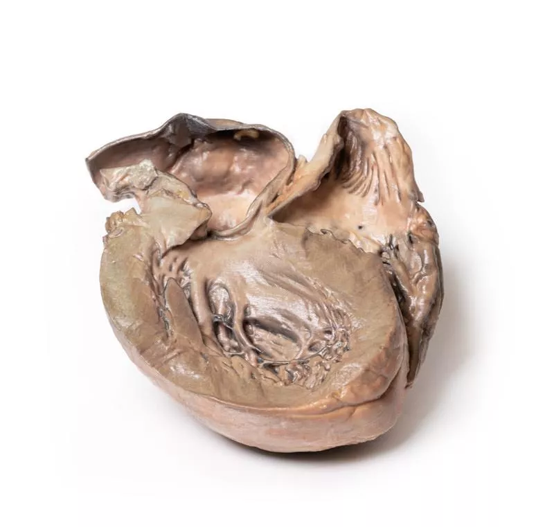
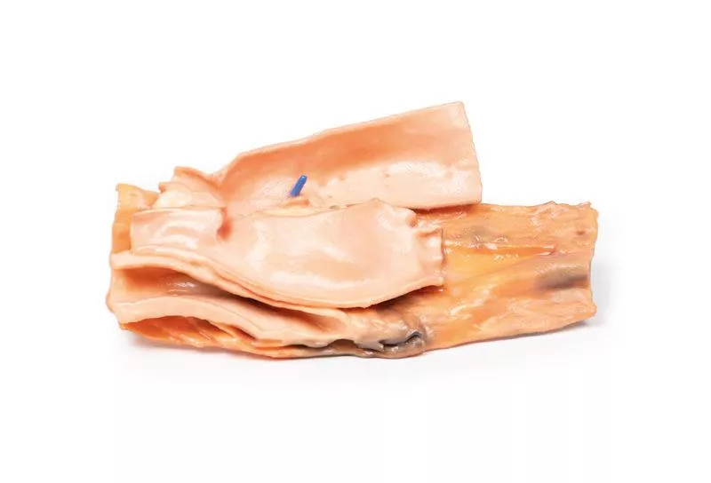
The enlarged heart specimen includes the aortic arch and descending aorta. The ascending aorta was dilated up to 7 cm, with a large aneurysmal bulge measuring 11 x 13 cm. The aneurysm was opened, showing a wrinkled, scarred intimal surface with marked atheroma. The innominate, left common carotid, and subclavian arteries were displaced by the aneurysm. A 5 mm ridge-like thickening on the internal aneurysm surface marks the pericardial sac attachment. Adventitial vessels showed marked congestion. This is a syphilitic aneurysm of the aortic arch.
Further Information
Syphilis is a chronic infection caused by the spirochete Treponema pallidum, mainly sexually transmitted but sometimes congenital. Risk groups include sexually active individuals, intravenous drug users, HIV patients, and men who have sex with men. Penicillin remains the main treatment. Syphilis progresses in three stages:
- Primary syphilis appears about 3 weeks after infection, with a painless chancre that heals spontaneously.
- Secondary syphilis follows in untreated patients with systemic symptoms and characteristic rashes.
- Tertiary syphilis develops years later and can cause cardiovascular syphilis, neurosyphilis, and gummatous lesions.
Cardiovascular syphilis involves aortitis of the ascending aorta, leading to dilation, aortic valve insufficiency, and aneurysm formation from vasa vasorum endarteritis. Symptoms typically appear 15-30 years after infection. Neurosyphilis can cause headaches, vision loss, strokes, and cognitive decline. Gummatous syphilis causes nodular lesions in skin, bone, and mucosa, especially in HIV patients.
Erler-Zimmer
Erler-Zimmer GmbH & Co.KG
Hauptstrasse 27
77886 Lauf
Germany
info@erler-zimmer.de
Achtung! Medizinisches Ausbildungsmaterial, kein Spielzeug. Nicht geeignet für Personen unter 14 Jahren.
Attention! Medical training material, not a toy. Not suitable for persons under 14 years of age.


