Product information "Upper Limb - biceps, bones and ligaments"
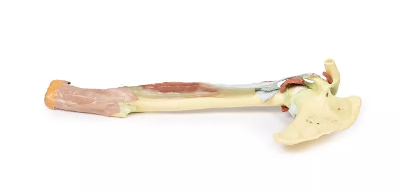
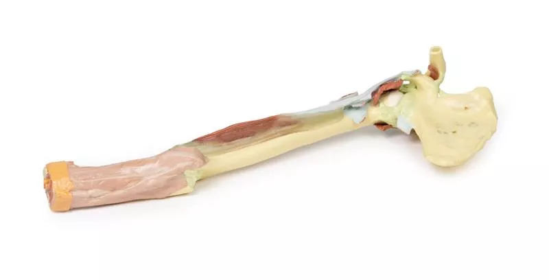
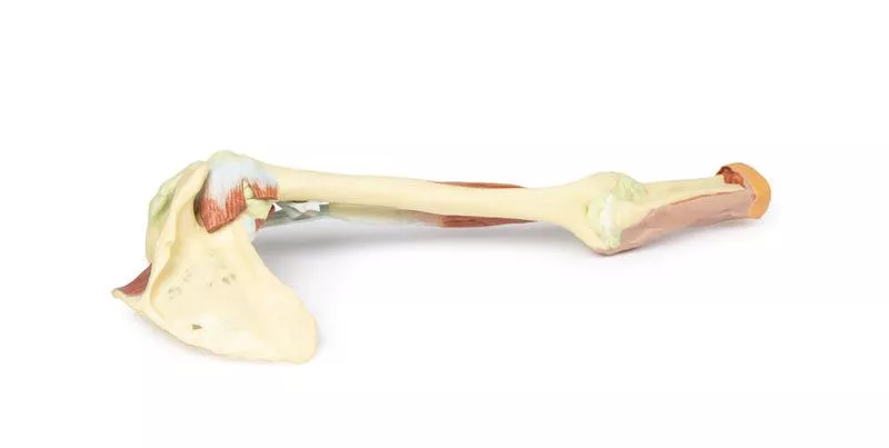
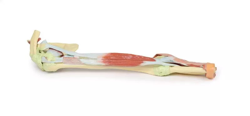
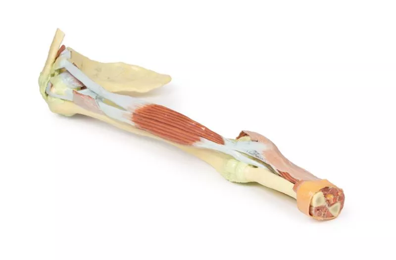
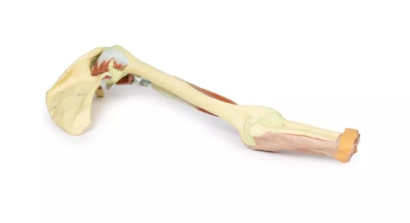
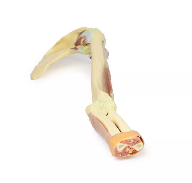
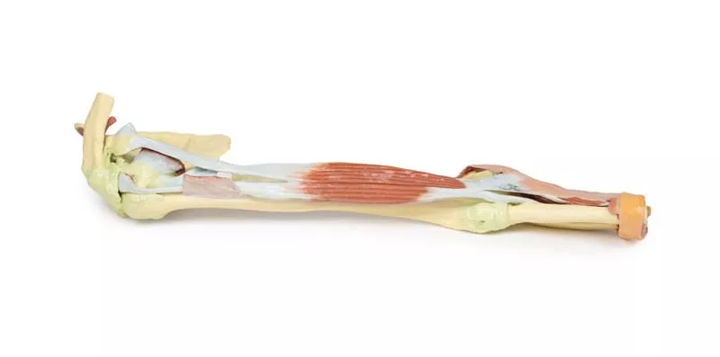
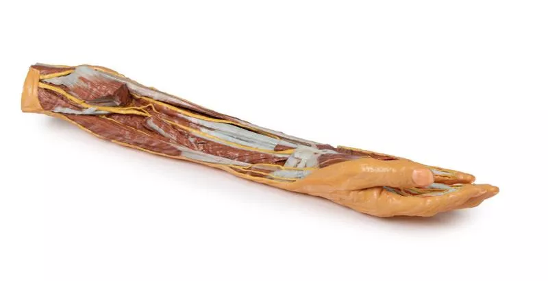
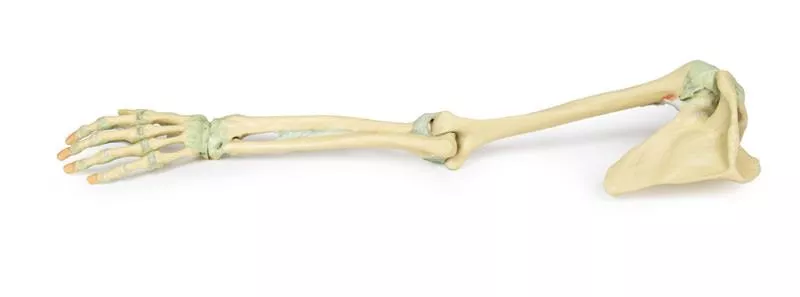
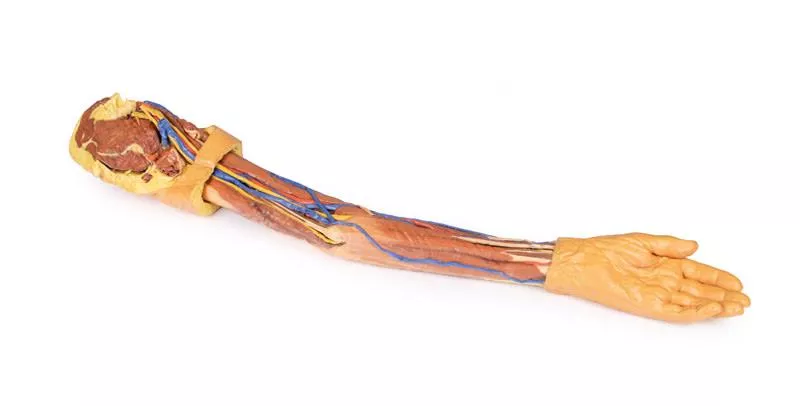
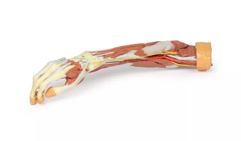
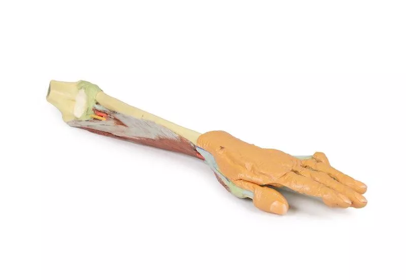
This 3D printed specimen highlights the origin, course, and insertion of the biceps brachii muscle, while most other arm and shoulder muscle bellies have been removed to enhance visibility.
Biceps Brachii: Origins and Insertion
The long head of the biceps originates from the supraglenoid tubercle (not visible) and travels through the bicipital groove, while the short head arises from the coracoid process. The bifid insertion is clearly shown: the bicipital aponeurosis and the round tendon wrapping around the radius to insert on the radial tuberosity.
Shoulder Region Structures
Several preserved elements are visible around the shoulder joint, including partial stumps or tendons of the subclavius, subscapularis, pectoralis major, teres minor, infraspinatus, long head of triceps, and latissimus dorsi. The teres major tendon lies along the medial lip, and the pectoralis major tendon along the lateral lip of the bicipital groove.
Also preserved are key shoulder ligaments:
- Coracoclavicular ligament
- Coracoacromial ligament
- Coracohumeral ligament
- Capsules of the shoulder and acromioclavicular joints
The supraspinatus is the only complete rotator cuff muscle retained, and the suprascapular ligament is seen bridging the suprascapular notch.
Elbow Joint Anatomy
At the elbow, the joint capsule, annular ligament of the radius, and radial collateral ligaments are visible. The ulnar collateral ligament is not shown due to retention of both heads of flexor carpi ulnaris.
This model is ideal for studying biceps anatomy, shoulder tendon insertions, and key ligamentous structures of the shoulder and elbow joints, making it a valuable tool for anatomical education and demonstration.
Erler-Zimmer
Erler-Zimmer GmbH & Co.KG
Hauptstrasse 27
77886 Lauf
Germany
info@erler-zimmer.de
Achtung! Medizinisches Ausbildungsmaterial, kein Spielzeug. Nicht geeignet für Personen unter 14 Jahren.
Attention! Medical training material, not a toy. Not suitable for persons under 14 years of age.


