


Product information "Upper Limb - elbow, forearm and hand"
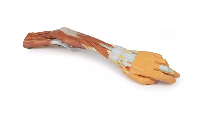
This detailed 3D print displays the left upper limb from the distal arm to the hand, highlighting key muscular, vascular, and neural structures through deep dissection.
Elbow Region & Distal Arm
The cubital fossa reveals the classic lateral-to-medial layout: biceps tendon, brachial artery, and median nerve, with the bicipital aponeurosis removed. The ulnar nerve passes behind the medial epicondyle, and the superficial radial nerve is visible between brachioradialis and brachialis.
Anterior & Posterior Forearm
Anteriorly, the superficial flexors—pronator teres, FCR, FDS, and FCU—are clearly shown (no palmaris longus present). The radial and ulnar arteries, as well as the superficial radial nerve, are identifiable.
Posteriorly, extensor muscles from the common extensor origin are preserved, including ECU, extensor digitorum, and ECRB/ECRL. The anatomical snuffbox is visible with the radial artery and cutaneous radial nerve.
Palm & Carpal Tunnel
The thenar/hypothenar muscles, flexor retinaculum, long tendons, and lumbricals are exposed. The median nerve runs beneath the retinaculum, and the superficial palmar arch (from the ulnar artery) is intact. Digital nerves and vessels are clearly seen, along with the recurrent median nerve branch and the extensor expansion on the middle finger.
This model is ideal for studying upper limb anatomy, with special focus on the elbow, forearm, and hand structures.
Login
Erler-Zimmer
Erler-Zimmer Medical GmbH
Hauptstrasse 27
77886 Lauf
Germany
info@erler-zimmer.de
Achtung! Medizinisches Ausbildungsmaterial, kein Spielzeug. Nicht geeignet für Personen unter 14 Jahren.
Attention! Medical training material, not a toy. Not suitable for persons under 14 years of age.
Other customers also bought

Posterior Body Wall / Ventral Deep Dissection
This 3D printed model provides a detailed ventral view of the head, neck, thorax, abdomen, and proximal thighs, offering insight into the central nervous system, nerve plexuses, and major vascular structures. It serves as a complementary piece to the dorsal dissection model (MP1400).Brain and Upper SpineThe facial skeleton is removed, exposing both cerebral hemispheres, parts of the Circle of Willis, and key arteries (vertebral, basilar, AICA, labyrinthine). Several cranial nerves (II, III, V, VI) and the right internal carotid artery are preserved. Below, the anterior spinal cord, vertebral arteries, and cervical and brachial plexus roots are visible, along with CN X, CN XII, and the sympathetic trunks. Axillae and Upper LimbsThe anterior thoracic wall, clavicles, and first ribs are removed to expose both axillae, with clear views of the brachial plexus, axillary arteries, and surrounding musculature. The proximal upper limbs are preserved to mid-arm level. Thorax and Lumbar RegionThe spinal cord extends to the conus medullaris, with ventral nerve roots and sympathetic chains visible throughout. Splanchnic nerves, intercostal nerves, and abdominal wall nerves (e.g., ilioinguinal, genitofemoral, femoral) are preserved. On the right, removal of the psoas muscle exposes the lumbar plexus.Pelvis and Proximal ThighThe pelvic viscera are removed, but the pelvic floor, rectum, and lumbosacral plexus remain. Obturator nerves are visible entering the obturator canals, and the femoral vessels are retained in both femoral triangles. This model is ideal for studying ventral CNS access, neurovascular pathways, and regional anatomical relationships across the entire torso.

Forearm and hand - deep dissection
This 3D printed specimen presents a deep dissection of the left upper limb, from the distal humerus to the palmar surface of the hand, offering detailed insights into the osseous, muscular, vascular, and neural structures.Elbow and ForearmAround the distal humerus, most musculature has been removed except the humeral origin of the flexor digitorum superficialis. The anterior joint capsule is opened, showing the articulation of the humerus, radius, and ulna.In the cubital fossa, the biceps brachii tendon, brachialis, brachial artery and vein, and median nerve are visible. Most surrounding forearm structures are removed to expose the anterior interosseous artery, vein, and nerve lying on the interosseous membrane, extending toward pronator quadratus. Wrist and Carpal TunnelThe flexor digitorum superficialis origin on the radius is exposed, with its tendons passing under the flexor retinaculum into the hand. Also visible at the wrist are the distal insertions of key forearm muscles (e.g. brachioradialis, FCR, FCU, ECRL, ECRB) and the radial artery. Palmar Hand AnatomyWithin the hand, tendons of the flexor digitorum superficialis and profundus continue into the fingers via fibrous digital sheaths. The flexor pollicis longus tendon runs to the thumb, while the sheath of the fifth digit is opened medially.The dissection reveals deep muscles including the adductor pollicis (both heads) and the first dorsal interosseous.Dorsal HandThe dorsal skin is largely intact but dissected medially to show the extensor digitorum, extensor digiti minimi, and the extensor expansion. This model offers a focused, high-resolution look at the deep anterior anatomy of the distal upper limb—ideal for anatomical education and demonstration.

Upper Limb
This 3D printed model presents a detailed anatomical view of the left upper limb, extending from the scapula to the hand. Most of the skin and fascia have been removed to expose the muscles, vessels, and nerves, while select regions (such as the dorsum of the scapula, proximal arm, and hand) retain superficial coverings for reference. Veins and Superficial StructuresThe cephalic, basilic, and median cubital veins are preserved, clearly demonstrating the superficial venous system from the wrist to the deltopectoral groove and brachial vein. Axilla and ShoulderIn the axillary region, the deltoid, supraspinatus, infraspinatus, teres minor and major, and subscapularis muscles are shown in cross-section. Key tendons — including those of the latissimus dorsi, coracobrachialis, and pectoralis major — are also retained.Portions of the axillary artery and vein are visible, along with the lateral cords of the brachial plexus. Several terminal branches are displayed, including the ulnar, median, musculocutaneous, axillary, radial, and upper subscapular nerves. Arm and Forearm DissectionThe model continues distally with a full view of the anterior and posterior compartments of the arm, displaying both muscle structure and the deep neurovascular pathways. In the cubital fossa, part of the bicipital aponeurosis remains intact.In the forearm, superficial flexor and extensor muscles are dissected from their origins to their tendinous insertions. A section of deep forearm fascia over the extensor compartment is retained for structural orientation. At the distal forearm, the radial and ulnar arteries and the median nerve are clearly visible. This model is ideal for teaching and studying upper limb anatomy, offering detailed views of musculature, vascular structures, and nerve pathways with a focus on both surface and deep anatomical relationships.
 - Superficial muscles and axillary/brachial artery")
Shoulder (left) - Superficial muscles and axillary/brachial artery
This 3D printed model of the left shoulder region provides a detailed view of the superficial musculature, rotator cuff muscles, and the axillary artery as it transitions into the brachial artery. The skeletal elements include the scapula, humerus (cut at midshaft), and a sectioned clavicle (cut at mid-length).Superficial and Deep Muscles of the ShoulderAnteriorly, the deltoid covers the lateral shoulder, concealing the long head of biceps brachii and lateral head of triceps brachii. The clavicular head of pectoralis major, subclavius, and pectoralis minor (inserting on the coracoid process) are preserved.Posteriorly, visible muscles include the trapezius (attached to the lateral third of the clavicle, acromion, and scapular spine), teres major, teres minor, and supraspinatus. The infraspinatus has been partially removed to reveal the suprascapular artery as it moves from the supraspinous to the infraspinous fossa. A remnant of the omohyoid and the suprascapular nerve passing beneath the suprascapular ligament are also visible. Subscapular and Serratus Anterior ViewsOn the costal (anterior) surface of the scapula, the subscapularis and serratus anterior muscles are preserved, offering insight into their relationship with the thoracic wall and axilla. Axillary and Brachial ArteriesThe axillary artery, just below the clavicle, gives rise to the thoracoacromial branch, suprascapular artery, subscapular artery, and the circumflex scapular artery. The posterior circumflex humeral artery is partly visible through the quadrangular space, beneath the deltoid. Beyond the inferior border of teres major, the artery becomes the brachial artery, which gives off a visible radial collateral branch.Neurovascular and Muscular Cross SectionA transverse cut at the humeral midshaft reveals the anatomical relationships between neurovascular bundles and the muscles of the anterior and posterior compartments, offering a clear sectional anatomy reference.

Hand
This high-detail 3D printed model presents a superficial dissection of the human hand and wrist, offering an exceptional overview of both anterior and posterior anatomical structures, including muscles, tendons, nerves, and vascular components.Anterior View: Carpal Tunnel, Tendons & ArteriesOn the anterior side, the transverse carpal and palmar carpal ligaments have been removed to expose the structures within the carpal tunnel and Canal of Guyon, including tendons and nerves. The palmar aponeurosis is also removed, revealing:- Tendons passing through the palm- Thenar and hypothenar muscles (mainly abductors and flexors)- Lumbrical muscles originating from the flexor digitorum tendonsIn the fingers, the fibrous tendon sheaths have been removed, clearly showing the insertions of the flexor digitorum superficialis and profundus, as well as the flexor pollicis longus tendon into the intermediate and distal phalanges.The superficial palmar arch, formed by branches of the ulnar and radial arteries, is visible, along with its common and proper palmar digital branches reaching the fingertips. These are accompanied by the corresponding median and ulnar digital nerves.Also preserved are the flexor carpi radialis and flexor carpi ulnaris tendons, plus the radial and ulnar arteries at the wrist. Posterior View: Extensor Tendons & Anatomical SnuffboxOn the posterior side, the radial artery is shown in the anatomical snuffbox, giving off the deep branch (piercing the first dorsal interosseous muscle) and dorsal carpal branch.The extensor retinaculum and superficial fascia have been removed to expose the:- Tendons of extensor digitorum, extensor carpi radialis, and ulnaris- Extensor pollicis longus, extensor pollicis brevis, and abductor pollicis longus- Intertendinous connections and extensor expansions, including insertions from the lumbricals and first dorsal interosseous muscle

Shoulder - deep dissection of a right shoulder girdle, preserving a complete scapula, lateral clavicle, and proximal humerus
This high-resolution 3D printed anatomical model displays a deep dissection of the right shoulder girdle, including a complete scapula, lateral clavicle, and proximal humerus. It offers detailed insight into key muscular, ligamentous, and joint structures, ideal for advanced anatomical study.Anterior View: Muscle Layers and Joint CapsuleOn the anterior side, the subscapularis muscle is preserved but partially sectioned to demonstrate the cross-sectional thickness within the subscapular fossa. The coracoclavicular and coracoacromial ligaments are intact and visible medial to the insertions of coracobrachialis and pectoralis minor on the coracoid process.The latissimus dorsi tendon is also retained, partially covered by the long head of the biceps brachii tendon, which travels through the bicipital groove toward the shoulder joint. The glenohumeral joint capsule has been opened anteriorly to reveal this passage, along with the supraspinatus muscle, partially covered by the collapsed subdeltoid bursa. Posterior View: Deep Joint StructuresFrom the posterior view, the supraspinatus muscle remains intact, while the infraspinatus and teres minor have been removed to expose the posterior glenohumeral joint capsule. Key tendon insertions—including those of the long head of the triceps brachii, infraspinatus, and teres minor—are preserved.

Shoulder - deep dissection of the left shoulder joint, musculature, and associated nerves and vessels
This 3D printed anatomical model presents a detailed deep dissection of the left shoulder, including the scapula, proximal humerus (up to midshaft), and surrounding muscles, nerves, and vessels. It offers a comprehensive view of the glenohumeral joint and associated neurovascular structures, ideal for teaching and clinical reference.Muscular Structures and Tendon AnatomyAnteriorly, the deltoid muscle has been removed from its origin to reveal deeper layers. Its insertion remains intact, overlying the long head of the biceps brachii, which travels through the bicipital groove toward the shoulder joint capsule.The subscapularis, a multipennate muscle, is fully exposed, including its tendinous insertion beneath the short head of the biceps brachii. Posteriorly, both the supraspinatus and infraspinatus muscles are visible from their scapular origins to their insertions on the proximal humerus. Nerve and Vascular AnatomyKey neurovascular structures are clearly depicted:- The suprascapular nerve and artery pass beneath and above the superior transverse scapular ligament, respectively.- A major neurovascular bundle—containing the brachial artery, brachial vein, and terminal branches of the brachial plexus (radial, ulnar, median, and medial antebrachial cutaneous nerves)—runs adjacent to the short head of the biceps.- The axillary nerve is shown entering the quadrangular space, while the profunda brachii artery originates nearby, following the radial nerve. Additional Muscle and Ligament FeaturesThe tendons of latissimus dorsi, teres major, and long head of triceps brachii have been transected to improve visibility of the medial humerus and deeper neurovascular pathways.The glenohumeral joint capsule remains intact, with all major extracapsular ligaments preserved, including:- Acromioclavicular ligament- Coracoacromial ligament- Coracoclavicular ligament (both conoid and trapezoid parts)

Right thoracic wall - axilla, and the root of the neck
This high-resolution 3D printed specimen displays a detailed dissection of the right thoracic wall, axilla, and root of the neck, offering exceptional anatomical insight from the parasagittal section to the proximal arm.Thoracic Wall & Intercostal RegionWith the viscera removed, the model exposes ribs, intercostal muscles, and the neurovascular bundles deep to the parietal pleura. The serratus anterior shows clear digitations on the lateral chest wall. Pectoral Region & AxillaThe pectoralis major is reflected to reveal pectoralis minor, which divides the axillary artery into three parts. The clavicle is partially removed, retaining the subclavius muscle. The cephalic vein runs in the deltopectoral groove, passing through the clavipectoral fascia. Brachial Plexus & NervesThe brachial plexus is preserved from C5–T1 roots to major terminal branches:- Musculocutaneous, Median, Ulnar, Radial, Axillary- Lateral and medial pectoral nerves- Long thoracic nerve (on serratus anterior)- Thoracodorsal nerve (with accompanying artery)- Dorsal scapular and suprascapular nerves- Axillary nerve with posterior circumflex humeral arteryAlso visible: the phrenic nerve crossing the scalenus anterior, plus a thin accessory phrenic nerve.Arteries & VeinsClearly shown arterial branches:- Thoracoacromial, Lateral thoracic, Thoracodorsal, Circumflex humeral arteries- Transverse cervical and suprascapular arteries (from the subclavian artery)Most deep veins are removed for visibility, except the cephalic vein. Posterior Neck & Shoulder MusclesVisible muscles include:- Latissimus dorsi, teres major, trapezius, rhomboid major- Infraspinatus, teres minor, triceps brachii- Scalene muscles, sternocleidomastoid, and omohyoid

Forearm and hand - superficial and deep dissection
This high-detail 3D printed specimen presents a combined superficial and deep dissection of the anterior right distal arm, forearm, and hand, offering a comprehensive view of key muscular, neural, and tendinous structures.Proximal Forearm & Cubital RegionMedially, the ulnar nerve is shown passing through the cubital tunnel and deep to the flexor carpi ulnaris. The cubital fossa has been opened by removing most flexor muscles, clearly exposing the biceps brachii and brachialis insertions, as well as the courses of the median, ulnar, and superficial radial nerves in the anterior compartment. Forearm: Muscle and Nerve DetailMuscular branches of the median nerve are visible entering the pronator teres and flexor digitorum profundus. The pronator teres insertion lies between the partially exposed supinator and flexor pollicis longus.Near the wrist, tendons of flexor digitorum superficialis, flexor digitorum profundus, and flexor pollicis longus are preserved, passing through the carpal tunnel alongside the median nerve. The pronator quadratus is partially visible deep to these structures.The flexor retinaculum is covered by the palmaris longus tendon, palmar aponeurosis, and palmaris brevis muscle. Wrist & Anatomical SnuffboxLaterally, the superficial radial nerve crosses over the extensor pollicis brevis and abductor pollicis longus tendons, entering the roof of the anatomical snuffbox, aiding in topographic orientation.Palm & Digital StructuresThe hand is superficially dissected between the thenar and hypothenar eminences, removing only skin and fascia. The tendons of flexor digitorum superficialis and profundus extend through the palm, surrounded by lumbricals and terminal branches of the median and ulnar nerves, forming common and proper digital nerves.Although most arterial structures have been removed for clarity, one common palmar digital artery and several proper palmar digital arteries are retained along the fingers.This model is ideal for detailed anatomical study of anterior forearm structures, neurovascular paths, and hand anatomy, particularly the carpal tunnel and digital innervation.

Upper Limb Ligaments
This 3D printed model presents the entire upper limb skeleton, from the pectoral girdle to the hand, with detailed ligamentous anatomy and select tendon and muscle insertions.Shoulder and Pectoral GirdleVisible ligaments include the acromioclavicular, coracoclavicular, coracoacromial, and superior transverse scapular ligament. A small portion of the supraspinatus muscle and tendon demonstrates its path beneath the coracoacromial ligament. Key preserved structures:- Subscapularis tendon (reflected to expose the glenohumeral capsule)- Tendons of teres major, latissimus dorsi, and long head of triceps- Long head of biceps tendon within the intertubercular groove and capsule Elbow JointThe elbow capsule is opened to show the articulation between the humerus, radius, and ulna.Preserved:- Ulnar and radial collateral ligaments- Anular ligament of radius- Biceps tendon insertion on the radial tuberosity Wrist and HandDistally, the model includes key wrist ligaments:- Palmar/dorsal radiocarpal and ulnocarpal ligaments- Radial/ulnar collateral ligaments, pisohamate, carpometacarpal, and othersIn the hand:- MCP and IP joint capsules with collateral ligaments and volar plates- Flexor digitorum superficialis/profundus and flexor pollicis longus tendons are preserved and shown at their insertions

Deep upper limb and hand
This 3D printed model presents a superficial dissection of the right distal arm, forearm, and hand, showcasing key vascular, nervous, and muscular anatomy.Distal Arm & Cubital FossaThe arrangement of the biceps tendon, brachial artery, and median nerve is visible from lateral to medial. The bicipital aponeurosis has been removed to expose deeper structures. The ulnar nerve is reflected from the cubital tunnel, and the radial nerve with its branches is visible near the supinator muscle. Forearm AnatomyOn the anterior forearm, superficial flexors (pronator teres, FCR, FDS, FCU) are preserved; palmaris longus is absent. The radial artery is exposed; the ulnar artery is not visible. Posteriorly, extensors from the common origin are visible, including ECRB, ED, EDM, and ECU. The APL, EPB, and EPL are also shown wrapping around the radius. Hand & SnuffboxThe anatomical snuffbox reveals the radial artery in its floor and cutaneous branches of the radial nerve. The palmar side displays thenar/hypothenar muscles, lumbricals, flexor tendons, and the median nerve beneath the flexor retinaculum. A superficial branch of the radial artery crosses the retinaculum.Ideal for anatomical education, this print offers a clear view of key structures in the distal upper limb.

Upper Limb - biceps, bones and ligaments
This 3D printed specimen highlights the origin, course, and insertion of the biceps brachii muscle, while most other arm and shoulder muscle bellies have been removed to enhance visibility.Biceps Brachii: Origins and InsertionThe long head of the biceps originates from the supraglenoid tubercle (not visible) and travels through the bicipital groove, while the short head arises from the coracoid process. The bifid insertion is clearly shown: the bicipital aponeurosis and the round tendon wrapping around the radius to insert on the radial tuberosity. Shoulder Region StructuresSeveral preserved elements are visible around the shoulder joint, including partial stumps or tendons of the subclavius, subscapularis, pectoralis major, teres minor, infraspinatus, long head of triceps, and latissimus dorsi. The teres major tendon lies along the medial lip, and the pectoralis major tendon along the lateral lip of the bicipital groove. Also preserved are key shoulder ligaments:- Coracoclavicular ligament- Coracoacromial ligament- Coracohumeral ligament- Capsules of the shoulder and acromioclavicular jointsThe supraspinatus is the only complete rotator cuff muscle retained, and the suprascapular ligament is seen bridging the suprascapular notch. Elbow Joint AnatomyAt the elbow, the joint capsule, annular ligament of the radius, and radial collateral ligaments are visible. The ulnar collateral ligament is not shown due to retention of both heads of flexor carpi ulnaris.This model is ideal for studying biceps anatomy, shoulder tendon insertions, and key ligamentous structures of the shoulder and elbow joints, making it a valuable tool for anatomical education and demonstration.

Continuous innovation

Social responsibility

Active customer orientation

Understanding quality

Sustainable actions

ISO 9001 certification