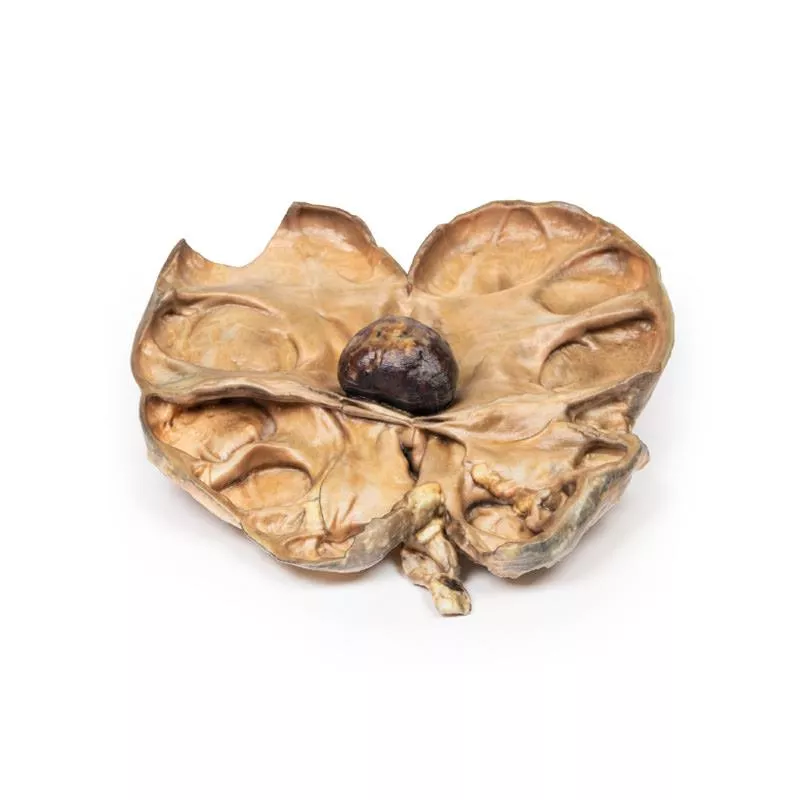
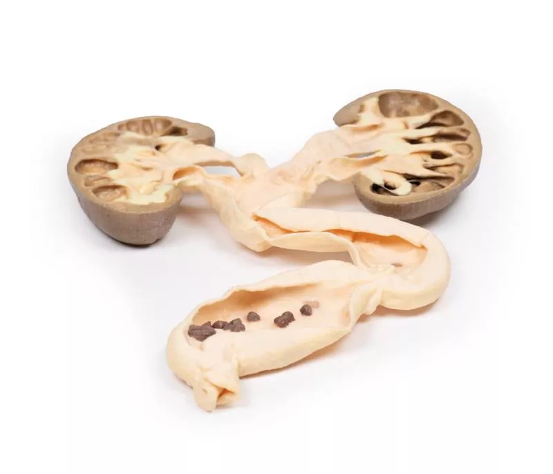
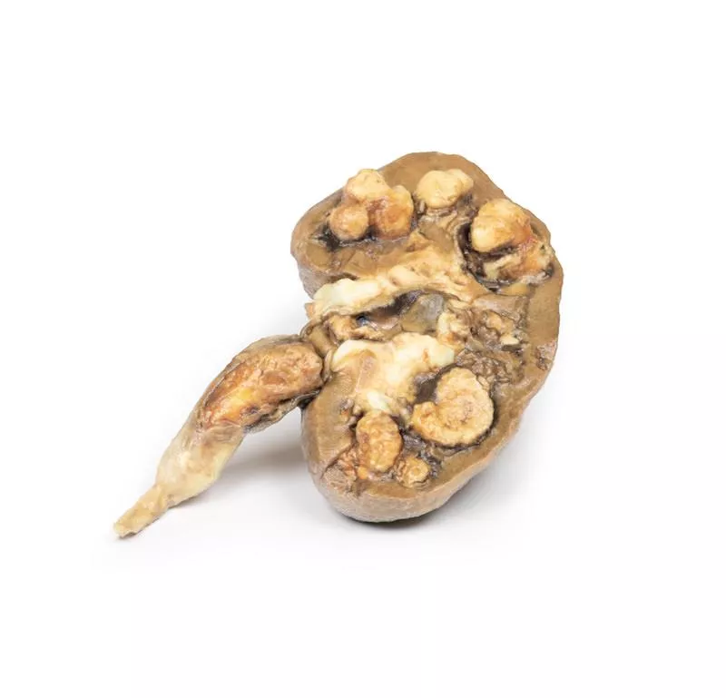
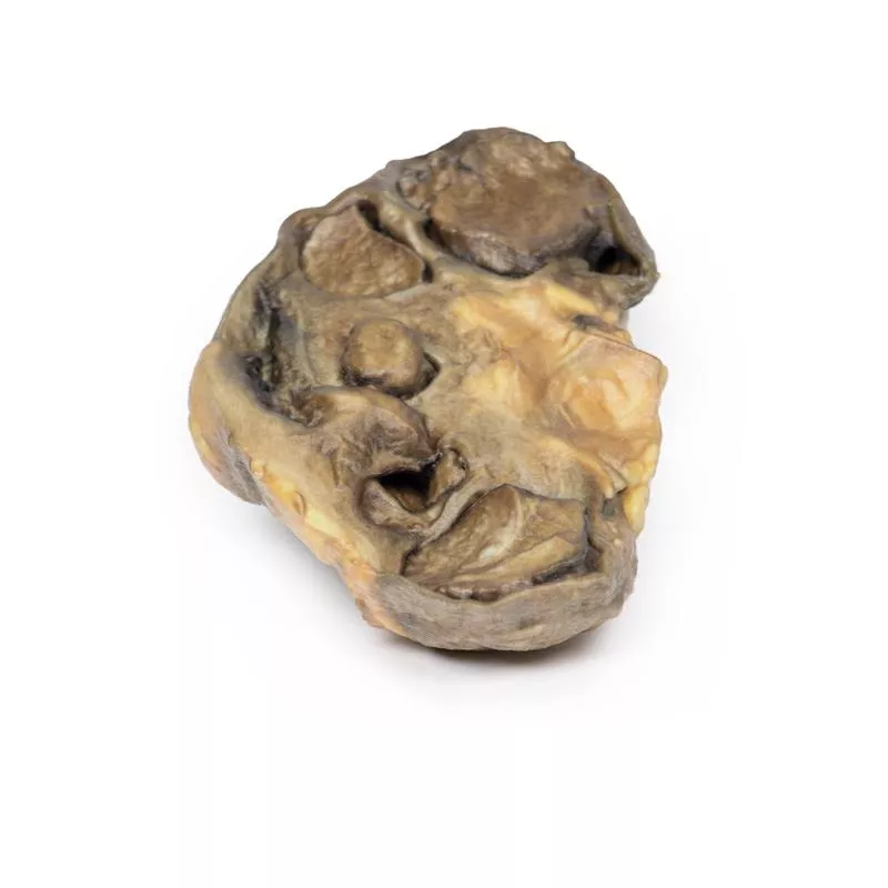
Product information "Renal Cell Carcinoma"
Clinical History
A 64-year-old male presented with 5 months of malaise, weight loss, and dull right flank pain. Examination revealed a palpable right abdominal mass and hypertension. Urinalysis showed microscopic haematuria. He underwent right nephrectomy.
Pathology
The kidney specimen shows a 5 cm irregular mass replacing the lower pole, compressing the renal parenchyma. The tumour surface shows haemorrhage and necrosis. Several small tumour nodules represent intrarenal metastases. The renal pelvis is slightly dilated, indicating mild hydronephrosis. Histology confirmed renal cell carcinoma (RCC).
Further Information
RCC accounts for 85% of primary kidney cancers, mostly arising in the renal cortex. It affects men twice as often, typically in their 60s. Risk factors include smoking, obesity, hypertension, certain toxins, and familial syndromes (e.g., Von Hippel Lindau).
Major RCC types are clear cell (70–80%), papillary (10–15%), chromophobe (5–10%), oncocytic (3–7%), and rare collecting duct carcinoma (less than 1%). Clear cell carcinoma is linked to chromosome 3p deletion and may be sporadic or hereditary. Papillary carcinoma often shows genetic trisomies and is multifocal. Chromophobe and oncocytic variants usually have better prognosis. Collecting duct carcinoma is aggressive with poor response to treatment.
Clinical signs include flank pain, palpable mass, and haematuria, with possible systemic symptoms. RCC metastasizes early, mainly to lungs and bones. Tumours can invade the renal vein and inferior vena cava.
Diagnosis uses ultrasound and CT; biopsy may be needed. Many RCC cases are incidental findings.
The 5-year survival is ~70%. Treatment involves radical nephrectomy, and for metastases, chemotherapy, VEGF inhibitors, and tyrosine kinase inhibitors.
Erler-Zimmer
Erler-Zimmer GmbH & Co.KG
Hauptstrasse 27
77886 Lauf
Germany
info@erler-zimmer.de
Achtung! Medizinisches Ausbildungsmaterial, kein Spielzeug. Nicht geeignet für Personen unter 14 Jahren.
Attention! Medical training material, not a toy. Not suitable for persons under 14 years of age.


