Tertiary Syphilis
Artikel in Produktion, lieferbar vorauss. in 2-3 Wochen
Produktnummer:
MP2113
Artikelnummer: MP2113
Produktinformationen "Tertiary Syphilis"
Clinical History
A 66-year old male presents with postprandial epigastric pain. Of note, he is deaf and non-verbal. On examination, he has a tender epigastrium and several nodular tender lesions over his forehead and scalp. Blood tests show a low haemoglobin, impaired hepatic function and are positive for anti-treponemal antibodies. After admission, he has a large gastrointestinal bleed, and dies despite intervention.
Pathology
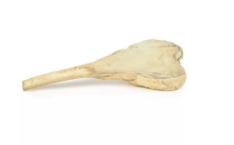
This specimen is the vault of the patient‘s skull. On the external surface, there are multiple circumscribed necrotic lesions in the parasagittal area to the left of the midline. The lesions are brown in colour and measure up to 3-4 cm in maximum diameter. The lesions have eroded the outer table of the skull and the adjacent periosteum is thickened with a fibrinous inflammation. These lesions are chronic syphilitic lesions or gumma of the skull, which are characteristic of benign tertiary syphilis.
Further Information
Syphilis is a chronic infection caused by the spirochete bacterium Treponema pallidum. Sexually transmitted infection is most common, but it may also be congenitally-acquired by transplacental transmission of the bacteria. Those who have the higher risk of infection, include those of a sexually active age, intravenous drug users, HIV-infected patients and homosexual males. Syphilis infection rates decreased significantly with the introduction of penicillin in 1943, which remains the main treatment today. However, the infection rate has been increasing since the early 2000’s.
Syphilis is divided into three stages with distinct clinical and pathological features with characteristic proliferative endarteritis affecting small vessels. Primary syphilis occurs usually 3 weeks after initial infection. This manifests typically as a single, painless and erythematous lesion called a chancre at the site of inoculation. The syphilis spreads throughout the body from this chancre, which then heals spontaneously after 3 to 6 weeks.
Secondary syphilis occurs weeks to a few months after the primary chancre resolves in 75% of untreated patients. During this stage patients commonly have generalised symptoms, such as malaise, lymphadenopathy and skin rashes. Palmar/plantar rashes are the most frequent site but rashes can also be diffuse. These rashes can be maculopapular, scaly or pustular. Condylomata lata are elevated gray plaques that arise on the moist mucous membranes, such as oral or genital regions. Other less common manifestations include hepatitis, gastrointestinal invasion or ulceration and neurosyphilis - discussed below.
Tertiary syphilis has three main characteristics: cardiovascular syphilis, neurosyphilis and gummatous syphilis. These occur after a latent period of 5 years or more in one third of untreated patients.
Cardiovascular syphilis involves an aortitis for which the exact pathophysiology is unclear. The vasculitis involves the ascending thoracic aorta leading to progressive dilation of the aortic root which can cause aortic valve insufficiency and aneurysms. Clinical manifestation usually happens 15-30 years post initial infection.
Neurosyphilis can be symptomatic or asymptomatic. It occurs in 10% of untreated patients. Early clinical manifestations include headaches, meningitis, hearing loss and ocular involvement, most commonly uveitis, causing vision loss. Late manifestations can occur up to 25 years post initial infection. Main features are meningovascular neurosyphilis, paretic neurosyphilis and tabes dorsalis. Meningovascular involvement involves chronic meningitis and endarteritis which can lead to
strokes. Tabes dorsalis is caused from degeneration of the posterior columns within the spinal cord. This causes loss of proprioception, ataxia, loss of pain sensation, and loss of reflexes. Paretic neurosyphilis is caused by invasion and damage of the brain parenchyma, most commonly the frontal lobes. This leads to progressive cognitive impairment and mood disturbance.
Gummatous syphilis is characterised by the formation of nodular lesions most commonly bone, skin and mucosa of the upper airway and mouth called gummas. Gummas can occur anywhere including viscera. The formation of gummas is rare but occurs more frequently in HIV-infected patients. Skeletal involvement causes pain and pathological fractures.
Erler-Zimmer
Erler-Zimmer GmbH & Co.KG
Hauptstrasse 27
77886 Lauf
Germany
info@erler-zimmer.de
Achtung! Medizinisches Ausbildungsmaterial, kein Spielzeug. Nicht geeignet für Personen unter 14 Jahren.
Attention! Medical training material, not a toy. Not suitable for persons under 14 years of age.