Pelvis
3
Products
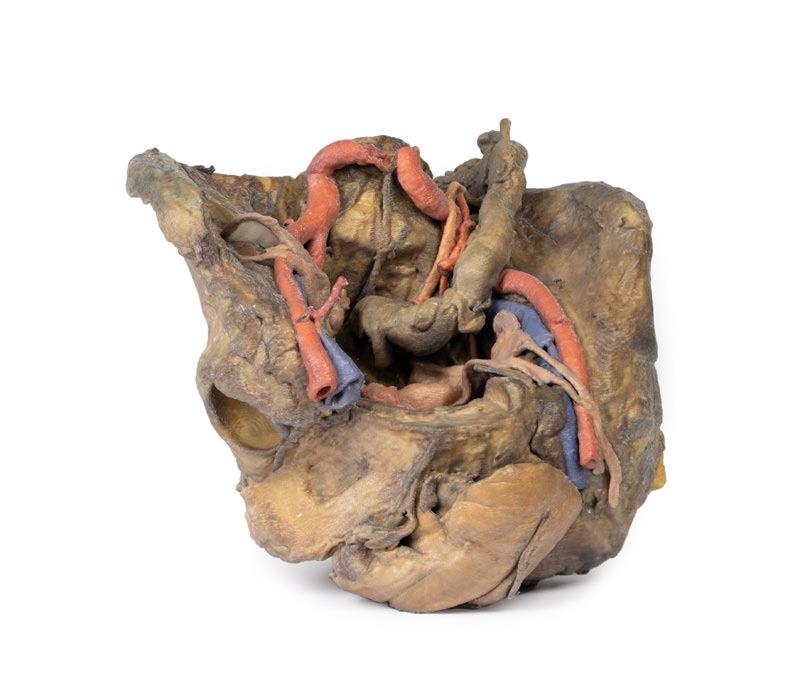
Female pelvis deep dissection
€5,472.81*
This 3D model presents a deep dissection and isolation of the pelvis from surrounding regions, particularly demonstrating visceral and neurovascular structures relative to deep ligaments and osseous features.Within the false pelvis, the sigmoid colon descends on the left side of the specimen to the rectum, passing superficially across the pelvic brim and the passage of the common and external iliac artery and vein. Adjacent to the sigmoid colon are parts of the sigmoid arteries and superior rectal artery, resting superficial to the common iliac vessels and near the descending ureter. Anterior in the true pelvis is the collapsed urinary bladder, and between the bladder and rectum rests the uterus. The organ is partially covered in the broad ligament, with both the suspensory ligament of the ovary and round ligament have been separated and pulled away from the peritoneum on both sides to expose surrounding blood vessels. While the ovarian ligaments, round ligaments, uterine tubes and ovaries are trapped within the peritoneal fold of the broad ligament, the reduction in ovary size (common with advanced age) has rendered these indistinguishable in the model.Lateral to these organs, branches of the internal iliac artery can be identified – as well as a retained median sacral artery in the midline between the two common iliac arteries. On the left side only the uterine artery can be seen laterally. On the right side, the obturator, superior vesical, and uterine arteries can be observed. In addition, the origins of the inferior epigastric artery and vein can be seen arising from the external iliac vessels just prior to exiting the inferior abdominal cavity.On the right side of the preserved pelvis, the entire femur and thigh musculature has been removed to demonstrate the obturator membrane, the articular cartilage of the acetabulum and the transverse ligament of the acetabulum. Posteriorly the entire gluteal region has been dissected to expose the superior gluteal foramen and the origin of the superior gluteal artery. The sacrotuberous ligament has been removed to demonstrate the sacrospinous ligament, with some branches of the inferior rectal artery retained within the exposed ischoanal fossa.On the left side of the preserved pelvis the sciatic nerve has been maintained within the greater sciatic foramen, as has the sacrotuberous ligament. The ischioanal fossa mirrors that of the right side, where branches of the inferior rectal artery have been retained relative to the fibres of the pelvic diaphragm, and the integration of the external anal sphincter on the projecting external rectal surface.
Male hemipelvis and thigh
€5,853.61*
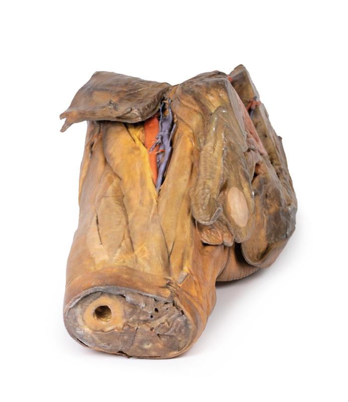
This 3D model preserves a right male pelvis sectioned just superior to the L5 vertebra and sectioned at the midsagittal plane, with the thigh preserved to near the midshaft of the femur. This specimen compliments our LW 91 female hemipelvic specimen and thigh.The common iliac artery is preserved with several key branches visible, particularly the distribution of the internal iliac within the true pelvis. Several major vessels including the obturator artery and the partially obliterated umbilical artery passes towards the anterior abdominal wall (to form the medial umbilical ligament) and gives off the superior vesicle artery; while the roots of the iliolumbar, superior gluteal, inferior gluteal and internal pudendal artery are visible lateral to the urinary bladder. The ureter descends superficial to these vessels to approach the urinary bladder which is covered with peritoneum in this model. The ductus deferens is exposed from the entry into the space via the deep inguinal ring and passing posteriorly (though sectioned from its normal insertion pathway and resting on the internal iliac artery). Adjacent to the ureter and on the superficial surface of the psoas major muscle is an enlarged iliac lymph node and part of the lymphatic vasculature ascending along the external iliac artery. The majority of the pelvis has been left undissected, allowing for an appreciation of the rectovesicular pouch and the exposed superior rectal artery and vein approaching the preserved portion of rectum. In cross section, the rectum, seminal vesicle and prostate are visible (the section plane preserves parts of both the prostatic urethra and ejaculatory duct).In the anterior thigh the borders and contents of the femoral triangle are well-preserved, with partial coverage by the flap of the anterior abdominal wall. Posteriorly the skin over the gluteal region and the gluteus maximus muscle have been removed as sequential windows to expose the gluteus medius and minimum muscles, the piriformis, the obturator internus with gemelli muscles, and the quadratus femoris muscle. The superior and inferior gluteal arteries are maintained superior and inferior to the piriformis, respectively; with the sciatic nerve exiting inferior to piriformis before passing deep to the retained portion of the gluteus maximus.
Female hemipelvis and thigh
€3,292.73*
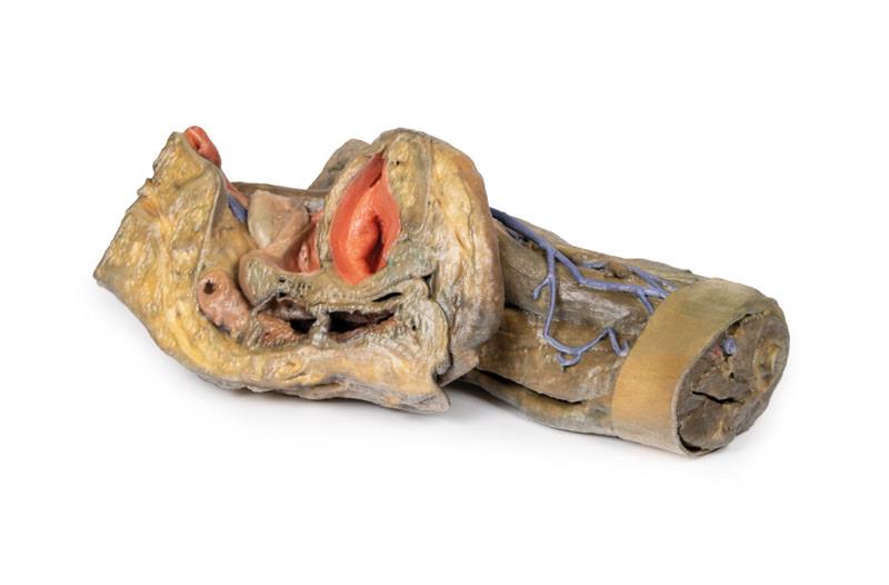
This 3D model preserves a left pelvis divided at the midsagittal plane, and the proximal thigh to approximately the midthigh.
Human body replicas to improve teaching!
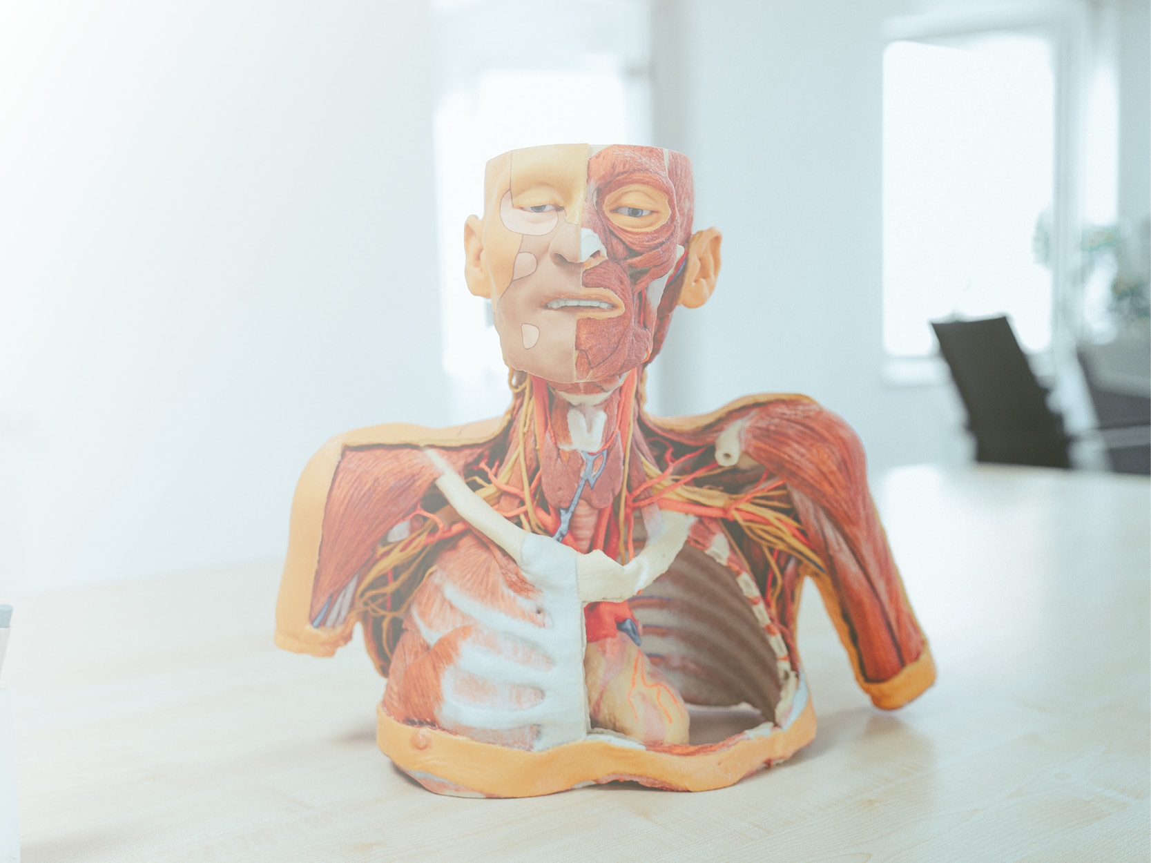
Erler-Zimmer's groundbreaking Anatomy Series features a unique and unrivalled collection of colourised human body replicas specifically designed to enhance teaching and learning. This premium collection of highly accurate human anatomy has been created directly from radiological data or real specimens using the latest imaging techniques. The 3D Human Anatomy Series offers a cost-effective way to meet your specific teaching and demonstration needs across the curriculum in medicine, health sciences and biology. A detailed description of the anatomy represented in each 3D printed specimen is included. What are the advantages of the Monash 3D Anatomy Series compared to plastic models or real human plastinates?
Each body replica has been carefully developed from selected radiological patient data or dissected human bodies of the highest quality, chosen by a highly skilled team of anatomists at Monash University's Human Anatomy Teaching Centre, to represent clinically important areas of anatomy in a quality and detail not possible with conventional models - it is real anatomy, not stylised. Each body replica has been rigorously checked by the highly qualified team of anatomists at Monash University's Human Anatomy Teaching Centre to ensure the anatomical accuracy of the final product. The body replicas are not real human tissue and are therefore not subject to any restrictions on transport, import or use in educational institutions that do not have permission to use cadavers. The
exclusive 3D Anatomy Series avoids these and other ethical issues that arise when dealing with plastinated human remains.
