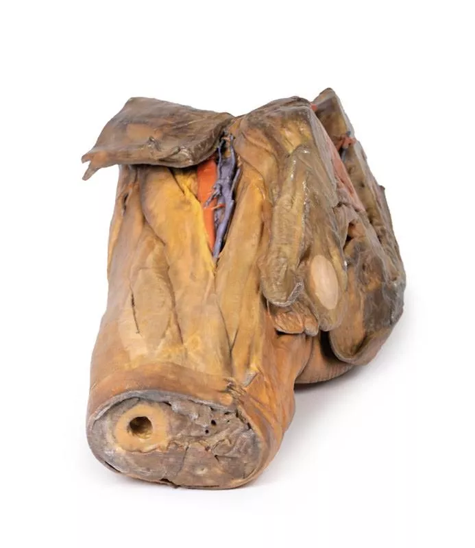
Product information "Female pelvis deep dissection"
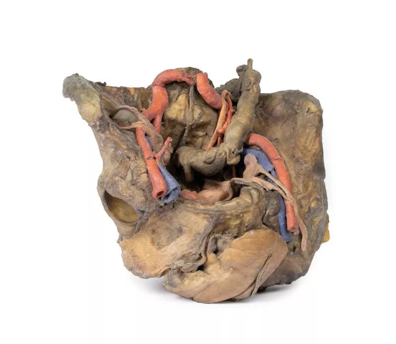
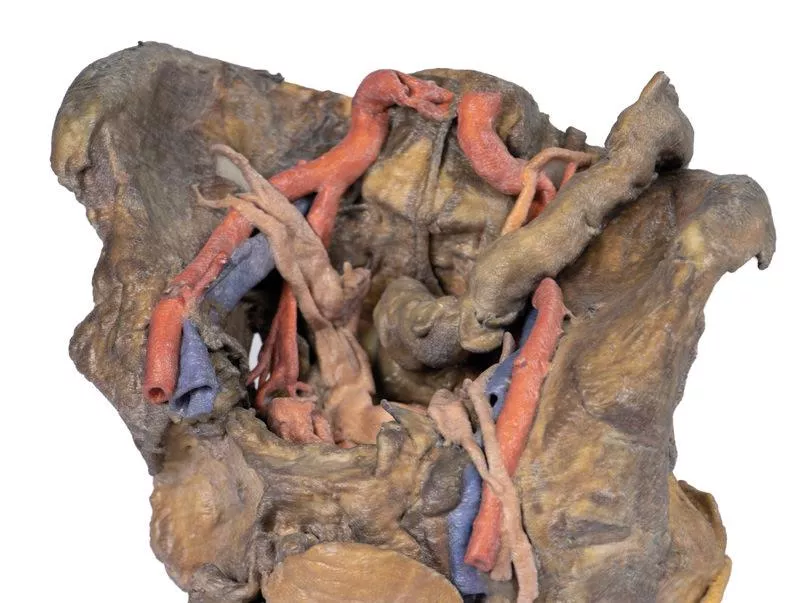
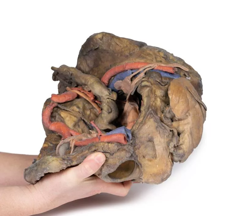
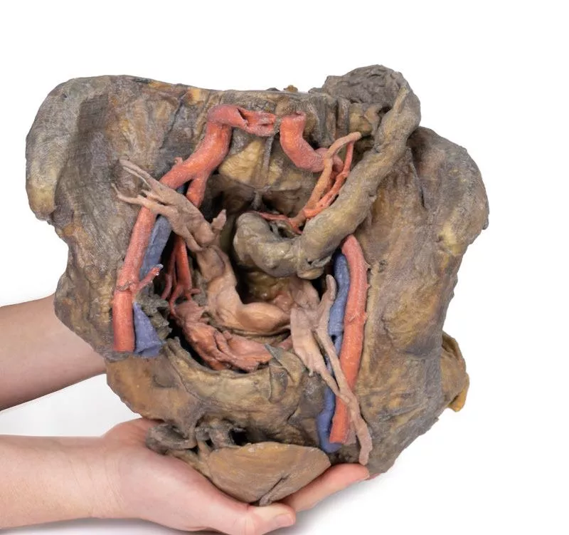
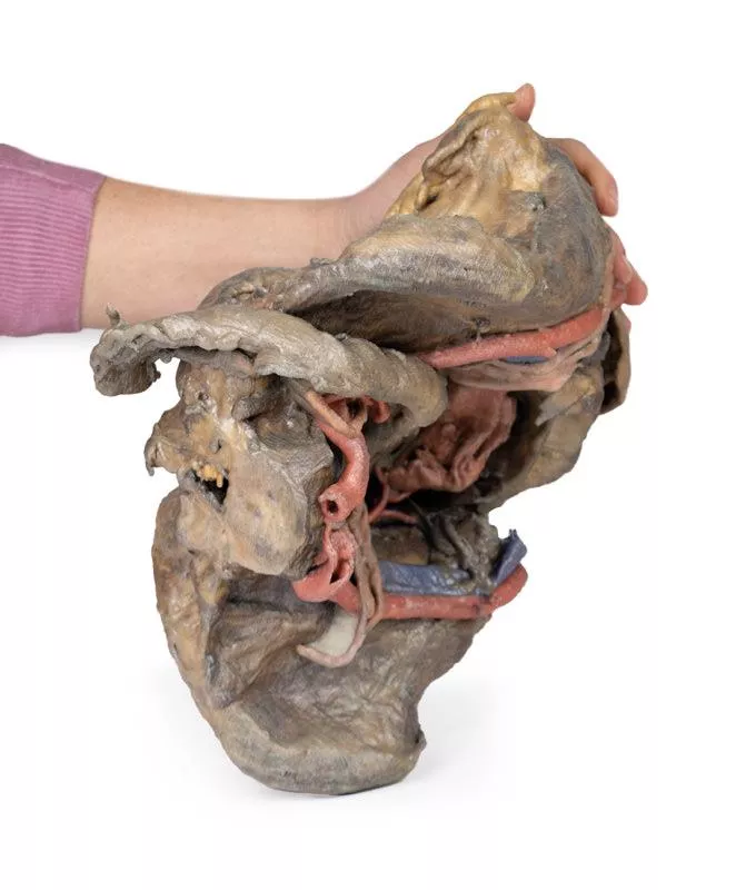
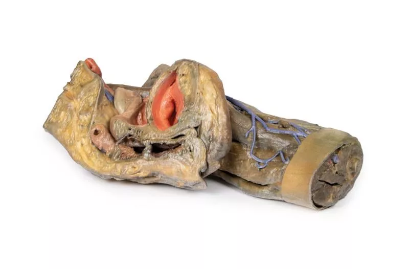
This high-detail 3D model showcases a deep dissection of the female pelvis, isolated from surrounding regions, with emphasis on visceral, vascular, and ligamentous structures in relation to bony landmarks.
Pelvic Organs & Peritoneal Structures
- Sigmoid colon descends into the rectum over the pelvic brim, crossing the common and external iliac vessels.
- Nearby: Sigmoid and superior rectal arteries, and the descending ureter.
- Urinary bladder (collapsed) and uterus are positioned anteriorly in the true pelvis.
- The broad ligament is retained, though ovaries, uterine tubes, ovarian and round ligaments are present but indistinct due to age-related atrophy.
- Suspensory and round ligaments are detached from the peritoneum to expose surrounding vessels.
Arteries & Veins
- Internal iliac artery branches are visible bilaterally.
- Median sacral artery is seen in the midline between the common iliac arteries.
- Left side: Uterine artery only.
- Right side: Uterine, superior vesical, and obturator arteries.
- Inferior epigastric artery and vein arise from the external iliac vessels, visible near the inferior abdominal wall.
Musculoskeletal Features
- Right side: Entire femur and thigh muscles removed to expose:
- Posterior dissection reveals:
Nerves & Ligaments
- Left sciatic nerve preserved within the greater sciatic foramen
- Sacrotuberous ligament retained on the left
- Ischioanal fossae on both sides show:
Pelvic Organs & Peritoneal Structures
- Sigmoid colon descends into the rectum over the pelvic brim, crossing the common and external iliac vessels.
- Nearby: Sigmoid and superior rectal arteries, and the descending ureter.
- Urinary bladder (collapsed) and uterus are positioned anteriorly in the true pelvis.
- The broad ligament is retained, though ovaries, uterine tubes, ovarian and round ligaments are present but indistinct due to age-related atrophy.
- Suspensory and round ligaments are detached from the peritoneum to expose surrounding vessels.
Arteries & Veins
- Internal iliac artery branches are visible bilaterally.
- Median sacral artery is seen in the midline between the common iliac arteries.
- Left side: Uterine artery only.
- Right side: Uterine, superior vesical, and obturator arteries.
- Inferior epigastric artery and vein arise from the external iliac vessels, visible near the inferior abdominal wall.
Musculoskeletal Features
- Right side: Entire femur and thigh muscles removed to expose:
- Obturator membrane
- Acetabular cartilage
- Transverse acetabular ligament
- Acetabular cartilage
- Transverse acetabular ligament
- Posterior dissection reveals:
- Superior gluteal foramen and artery
- Sacrospinous ligament (with sacrotuberous ligament removed)
- Inferior rectal artery branches within the ischioanal fossa
- Sacrospinous ligament (with sacrotuberous ligament removed)
- Inferior rectal artery branches within the ischioanal fossa
Nerves & Ligaments
- Left sciatic nerve preserved within the greater sciatic foramen
- Sacrotuberous ligament retained on the left
- Ischioanal fossae on both sides show:
- Inferior rectal artery branches
- Pelvic diaphragm fibers
- External anal sphincter integration with the rectal wall
- Pelvic diaphragm fibers
- External anal sphincter integration with the rectal wall
Erler-Zimmer
Erler-Zimmer GmbH & Co.KG
Hauptstrasse 27
77886 Lauf
Germany
info@erler-zimmer.de
Achtung! Medizinisches Ausbildungsmaterial, kein Spielzeug. Nicht geeignet für Personen unter 14 Jahren.
Attention! Medical training material, not a toy. Not suitable for persons under 14 years of age.


