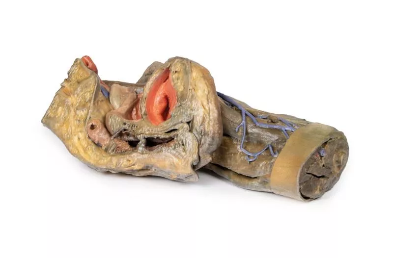
Product information "Internal abdominal wall"
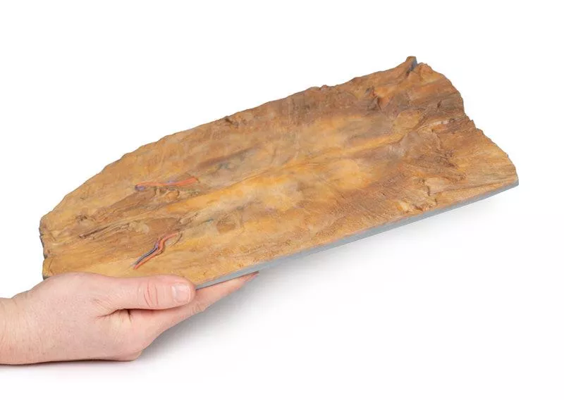
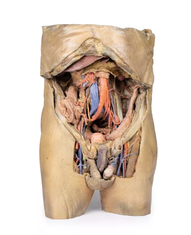
This detailed 3D model captures the internal surface of the anterior abdominal wall—a region often removed or damaged during dissections. It complements our MP1130 abdominal specimen, where the anterior wall has been removed, providing a clear view of key muscle and connective tissue structures.
Key Features:
Muscle Fibers & Aponeurosis:
The horizontally oriented transversus abdominus muscle fibers converge toward their aponeurosis (tendon sheet), visible especially along the specimen’s superior margins.
Arcuate Line:
Located in the lower third of the model, this landmark marks where the aponeurosis shifts relative to the rectus abdominus muscle.
- Above the arcuate line: Aponeurosis fibers split evenly around the rectus abdominus.
- Below the arcuate line: All aponeurotic fibers pass anteriorly to the rectus abdominus, reflecting a change in abdominal wall structure.
Vascular Structures:
Inferior Epigastric Arteries & Veins:
These vessels originate from the external iliac arteries and veins, ascending superiorly through the anterior abdominal wall.
Hesselbach’s Triangle:
On the right side of the model, the orientation of the inferior epigastric artery relative to the rectus abdominus muscle defines the apex of the inguinal (Hesselbach’s) triangle—a critical anatomical region often associated with direct inguinal hernias. (Note: The inguinal ligament forming the base of this triangle is not present in this specimen.)
Embryological Remnant:
Median Abdominal Ligament:
Positioned midline between the two rectus abdominus muscles, this fold of parietal peritoneum covers the urachus, a fibrous remnant from embryological development extending from the bladder to the umbilicus.
Erler-Zimmer
Erler-Zimmer GmbH & Co.KG
Hauptstrasse 27
77886 Lauf
Germany
info@erler-zimmer.de
Achtung! Medizinisches Ausbildungsmaterial, kein Spielzeug. Nicht geeignet für Personen unter 14 Jahren.
Attention! Medical training material, not a toy. Not suitable for persons under 14 years of age.


