


Product information "Abdomen with bilateral Hernias"
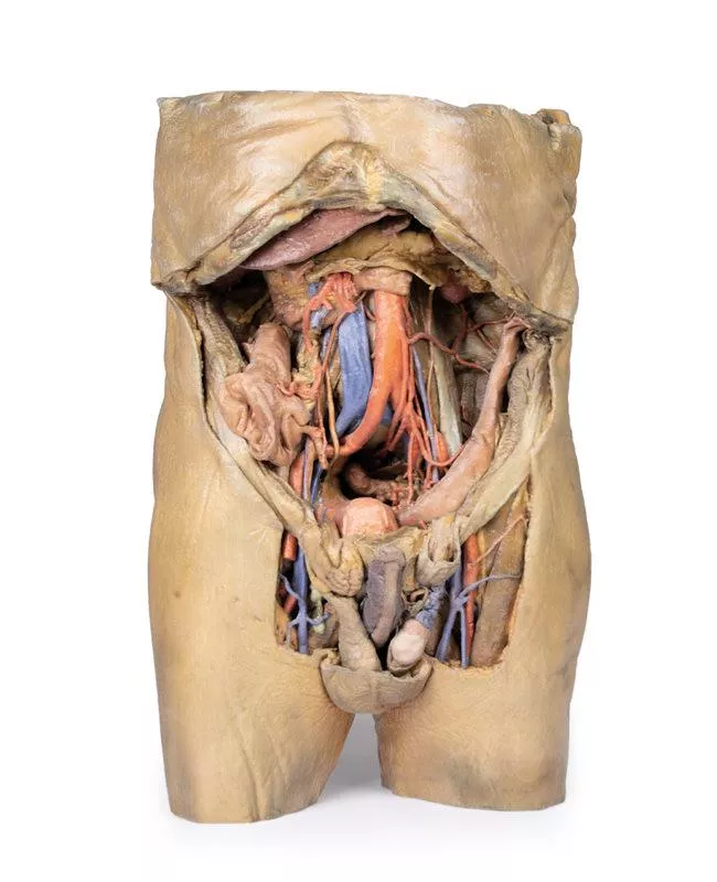
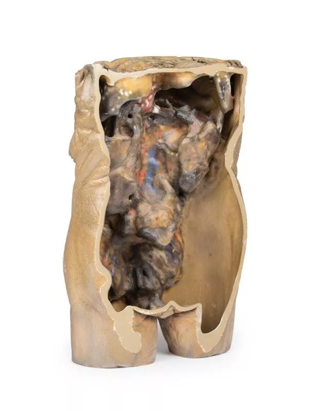
This 3D model represents one of the largest and most complex in the series, consisting of a partial torso from the diaphragm to the proximal thigh with a complete abdominal cavity preserving varying levels of dissection. This 3D model also records the rare, simultaneous occurrence of indirect and direct inguinal hernias allowing for a consideration of the anatomical underpinnings for both conditions. Given the scale of the dissection this 3D model description is divided into discrete parts based on views and regions.
The diaphragm
The diaphragm is preserved on the model’s superior aspect, with both domes and costodiaphragmatic recesses visible despite some distortion from rib removal. The fibrous pericardium rests on the central tendon, with the terminal inferior vena cava seen in the caval foramen. Lateral to this lies the oesophagus in the oesophageal hiatus, and the descending thoracic aorta approaching the aortic hiatus near the vertebrae.
The epigastric and hypochondriac regions
In the abdomen, removal of the anterior wall, greater omentum, and much of the GI tract reveals retroperitoneal structures. The terminal oesophagus enters just left of the liver. With the stomach removed, the pancreas is fully exposed from head to tail, reaching the spleen in the left hypochondrium. Above it, the splenic and common hepatic arteries span the narrow space between pancreas, diaphragm, and liver. The tortuous splenic artery divides near the splenic vein; the common hepatic gives rise to the gastroduodenal and right gastric arteries, superficial to the portal vein. The superior mesenteric vessels pass near the pancreatic head, and the ileocolic artery leads to the caecum. The inferior mesenteric vein arises from the superior rectal vein and crosses the descending aorta.
Below the liver, the gallbladder lies between the lobes. On the left, renal vessels pass deep to the pancreas, with ureters descending across the psoas muscles.
The umbilical and lumbar regions
Most abdominal organs in the umbilical and lumbar regions have been removed to reveal the posterior abdominal wall. Centrally, the descending aorta and inferior vena cava are prominent, with testicular vessels traceable toward the inguinal region. Two right lumbar arteries branch from the aorta, and the inferior mesenteric artery gives rise to the left colic, sigmoid, and superior rectal arteries. On the right, subcostal, iliohypogastric, and ilioinguinal nerves are visible, along with the circumflex iliac artery.
The hypogastrium and iliac regions
The abdominal aorta bifurcates into the common, internal, and external iliac arteries, with matching iliac veins merging into the inferior vena cava. The obturator artery, ureters, and testicular vessels are visible. In the true pelvis, the peritoneum covers the bladder, while the rectum remains obscured. The right iliac fossa contains the terminal ileum, caecum, and appendix, with nearby vessels and nerves. On the left, the sigmoid colon crosses the iliac fossa, where an epiploic appendage extends into an indirect hernia near the inferior epigastric artery.
The inguinal region and perineum
This model uniquely preserves both direct (right) and indirect (left) inguinal hernias, with the inferior epigastric vessels retained for anatomical orientation. The right hernia lies medial to these vessels; the left hernia sac extends laterally into the spermatic cord, containing an epiploic appendage. The perineum reveals the penis, testes, and spermatic cords. On the right, the cord remains intact; on the left, it’s opened, showing a varicose testicular vein linked to the indirect hernia.
The thigh
The femoral triangle has been dissected on both thighs. On the right, the femoral sheath was removed to reveal the femoral artery, vein, deep inguinal lymph nodes, and femoral nerve. On the left, a broader view exposes anterior and medial thigh muscles, with the femoral artery, profunda femoris, and circumflex iliac artery visible. The model ends mid-thigh, showing cross-sectional anatomy including the femoral shaft, vessels, and muscles in the subsartorial canal.
Login
🔬 3D Anatomy Series - Human Body Replicas to Enhance Teaching!
August 26, 2025
Discover exclusive 3D-printed models of the human body – created directly from radiological data or real specimens.
Erler-Zimmer
Erler-Zimmer Medical GmbH
Hauptstrasse 27
77886 Lauf
Germany
info@erler-zimmer.de
Achtung! Medizinisches Ausbildungsmaterial, kein Spielzeug. Nicht geeignet für Personen unter 14 Jahren.
Attention! Medical training material, not a toy. Not suitable for persons under 14 years of age.
Other customers also bought

Female hemipelvis and thigh
This detailed 3D model displays the left half of a female pelvis, sectioned midsagittally, and extending to the proximal mid-thigh.Pelvic Organs & Peritoneum- Visible structures: Urinary bladder, uterus, vagina, and rectum (from anterior to posterior).- The peritoneum is preserved, showing the vesicouterine and rectouterine pouches.- The broad ligament, uterine tube, fimbriae, and left ovary are identifiable near the pelvic brim. Vessels & Nerves- Common and external iliac arteries pass toward the subinguinal space, alongside the common iliac vein and psoas major.- The ureter crosses over these vessels. - The femoral nerve is visible between psoas major and iliacus muscles. Anterior Thigh & Inguinal Region- Superficial fascia removed, exposing thigh structures up to the perineal edge.- Femoral triangle dissected to show:- Femoral artery and vein, with the vein receiving tributaries from the great saphenous, superficial circumflex iliac, external pudendal, and deep pudendal veins.- Femoral nerve lateral to the artery.- Anterior cutaneous nerves and part of the lateral cutaneous nerve over the sartorius muscle. - Inguinal lymph nodes beneath the inguinal ligament. Posterior Gluteal Region- Gluteus maximus removed to reveal deeper gluteal muscles.- Piriformis reflected, exposing:- Sciatic nerve, formed by tibial and common peroneal nerves.- Superior and inferior gluteal arteries. - Posterior cutaneous nerve of the thigh running parallel to the sciatic nerve.- Obturator internus, gemelli, and quadratus femoris muscles exposed.- Internal pudendal artery and pudendal nerve track toward the ischioanal fossa.- Their branches, including the inferior rectal nerve, are visible near the pelvic diaphragm and external anal sphincter.

Abdomen vasculature
Coeliac TrunkThe celiac trunk, arising at the T12 level, supplies the embryological foregut. Visible branches include the left gastric artery from the left side, the splenic artery passing to the left hypochondrium, and the common hepatic artery on the right. The common hepatic gives rise to the gastroduodenal artery, which connects to the superior mesenteric artery, and the proper hepatic artery, which branches into the left and right hepatic arteries. The right hepatic artery eventually forms the cystic artery supplying the gallbladder. Superior Mesenteric Artery and Inferior Mesenteric ArteryThe superior and inferior mesenteric arteries arise at L1 and L3, supplying the midgut and hindgut. While not fully preserved, the superior mesenteric artery is visible exiting below the pancreas and branching out, and the inferior mesenteric artery descends along the left side of the aorta. The left colic artery branches from the inferior mesenteric artery, supplying the colon via the marginal arteries. Venous System of the AbdomenThe superior mesenteric vein lies posterior to the artery and appears less tubular. Removal of the liver’s left lobe exposes portal vein branches, which carry nutrients from the gut to hepatocytes before draining via hepatic veins into the inferior vena cava.Hilum of the KidneyThe right kidney shows typical anatomy with the renal vein superior, artery inferior, and ureter descending. The left kidney displays variation: the renal vein is inferior and subdivided, the artery is superior, and the ureter descends medially. Muscles, Nerves and Other VasculatureThe psoas major and iliacus muscles are visible on both sides, with key lumbar plexus nerves around them—especially on the left—including the iliohypogastric, ilioinguinal, femoral, and genitofemoral nerves. Medial to the psoas major, the left testicular artery (from the aorta) and vein (draining to the left renal vein) are seen. The right testicular vein drains directly into the IVC. A branch of the iliolumbar artery passes beneath the testicular vessels and ureter. GallbladderJust inferior to the liver, the gallbladder can be observed with the cystic artery moving inferiorly to meet it. The cystic duct can also be seen moving from the gallbladder, meeting the common hepatic duct moving from the liver to form the common bile duct.

Spleen and pancreas
This detailed 3D anatomical model preserves the deep foregut organs, including the descending, horizontal, and ascending parts of the duodenum, the pancreas, and the spleen. It offers a unique and insightful view into the complex anatomy of this region. DuodenumA small window is opened in the duodenum to reveal the plicae circularis, the characteristic circular folds of the proximal small intestine. This contrasts with the prominent rugae of the stomach, providing an educational comparison of mucosal patterns in the upper gastrointestinal tract. PancreasThe pancreas is preserved in its natural anatomical position, nestled within the curvature of the duodenum. The head of the pancreas is clearly visible, including the distinct uncus located at its distal margin, adjacent to the origin of the superior mesenteric artery (SMA). In this model, the SMA is already divided into its major named branches, highlighting its vascular complexity.The body of the pancreas features the superior margin where the celiac trunk, sectioned from the descending abdominal aorta, is located. The complete splenic artery is shown following its tortuous path from the celiac trunk to the spleen. The model also displays the origins of the left gastric artery and the common hepatic artery branching from the celiac trunk.Adjacent to the celiac trunk, a segment of the splenic vein is visible emerging from the pancreas’ capsule. This vein runs alongside the splenic artery en route to the spleen. Additionally, a portion of the superior mesenteric vein is seen adhering to the posterior pancreas, representing its path before converging with the splenic vein to form the hepatic portal vein.The tail of the pancreas is embedded within the splenic capsule, partially obscuring the splenic artery branches before entering the spleen. For more detailed views of this region, refer to our other spleen models (MP1130 and MP1134), which illustrate further anatomical and spatial relationships.

Female pelvis deep dissection
This high-detail 3D model showcases a deep dissection of the female pelvis, isolated from surrounding regions, with emphasis on visceral, vascular, and ligamentous structures in relation to bony landmarks.Pelvic Organs & Peritoneal Structures- Sigmoid colon descends into the rectum over the pelvic brim, crossing the common and external iliac vessels.- Nearby: Sigmoid and superior rectal arteries, and the descending ureter.- Urinary bladder (collapsed) and uterus are positioned anteriorly in the true pelvis.- The broad ligament is retained, though ovaries, uterine tubes, ovarian and round ligaments are present but indistinct due to age-related atrophy.- Suspensory and round ligaments are detached from the peritoneum to expose surrounding vessels. Arteries & Veins- Internal iliac artery branches are visible bilaterally.- Median sacral artery is seen in the midline between the common iliac arteries.- Left side: Uterine artery only.- Right side: Uterine, superior vesical, and obturator arteries.- Inferior epigastric artery and vein arise from the external iliac vessels, visible near the inferior abdominal wall. Musculoskeletal Features- Right side: Entire femur and thigh muscles removed to expose:- Obturator membrane- Acetabular cartilage- Transverse acetabular ligament- Posterior dissection reveals:- Superior gluteal foramen and artery- Sacrospinous ligament (with sacrotuberous ligament removed)- Inferior rectal artery branches within the ischioanal fossa Nerves & Ligaments- Left sciatic nerve preserved within the greater sciatic foramen- Sacrotuberous ligament retained on the left- Ischioanal fossae on both sides show:- Inferior rectal artery branches- Pelvic diaphragm fibers- External anal sphincter integration with the rectal wall

Vasculature of the spleen
This anatomical model vividly displays the splenic hilum, focusing on the critical vascular structures supplying and draining the spleen.Key Features:Splenic Artery and Vein:Both vessels enter the spleen at the hilum. The splenic vein’s opening is kept patent using inserted silicon tubing, allowing clear visualization of venous drainage. The model shows the most superior branch of the splenic vein carefully sectioned to reveal its course. Tortuous Splenic Artery:The model highlights the distinctive twisted and curled shape of the splenic artery as it branches at the hilum, reflecting its natural, winding path from the coeliac trunk to the spleen. Branching Vessels:The splenic artery and vein give rise to the short gastric arteries and the left gastro-omental artery. In this specimen, these branches are cut beyond their origin, so they are not fully visible, providing a focused view of the main vessels at the hilum.Ligament Attachments (Not Present):- Splenorenal Ligament: Connects the spleen to the left kidney and contains the splenic artery, vein, and tail of the pancreas. Formed embryologically from the dorsal mesentery’s peritoneum, this ligament is removed in the model to expose the splenic vessels clearly.- Gastrosplenic Ligament: Connects the stomach to the spleen, containing the short gastric arteries and part of the left gastro-omental artery. This ligament is also absent in the model due to dissection beyond the splenic artery branch. Spleen Capsule:The outer surface of the spleen is covered by a thin fibrous capsule. This delicate layer is prone to rupture because of the spleen’s high blood content, an important clinical consideration highlighted by the model.

Abdomen with inguinal hernia
Diaphragm and Xyphoid ProcessThe diaphragm has been secured to the superior border of the dissected specimen with sutures to ensure an unobstructed view of the abdomen. The xyphoid process is in the middle of this sutured border. Liver and GallbladderThe liver in the right hypochondrium is pushed laterally to reveal the kidney behind it. The falciform ligament divides the liver’s right and left lobes and contains the ligamentum teres, a remnant of the fetal umbilical vein. Below the ligamentum teres, the gallbladder sits between the liver lobes. Stomach and Splenic VasculatureThe deflated stomach is pushed up to reveal the twisted splenic artery and vein, which branch into the spleen’s hilum.Spleen and PancreasThe spleen sits in the left hypochondrium, with its gastric impression marking the stomach’s greater curvature. The pancreas tail, intraperitoneal and fused to the spleen’s hilum, lies near its inferior pole. KidneysThe kidneys are mostly retroperitoneal, but the covering peritoneum is removed in this specimen. Normally, the right kidney lies lower due to the liver, but here it is smaller and higher than the left. The left kidney is enlarged, with two accessory renal arteries from the aorta supplying its hilum and lower pole.Adrenal GlandsThe left adrenal gland is detached from its usual position on the superior pole of the kidney. The middle adrenal artery originates directly from the aorta, left of the coeliac trunk, whilst the inferior adrenal artery is derived from the left renal artery: both supply the adrenal gland. The superior adrenal artery has been obscured by connective tissue.Rectum and BladderAlthough the majority of the peritoneum in the abdomen has been removed below the level of the sacral prominence (S1), a layer of peritoneum remains intact, which overlays the rectum and bladder. Notably this the first part of the rectum, which is intraperitoneal. Gastrointestinal TractThe final part of the ascending duodenum and the descending colon at the left colic flexure has been ligated with twine, with the intestine in between being removed in order to provide a better view of the abdomen. Pelvic RegionIn this specimen, the sigmoid colon has herniated indirectly through the inguinal canal. On the right, the vas deferens exits the superficial inguinal ring toward the right testicle, while other spermatic cord contents are removed. Sutures below the vas deferens mark the embalming entry via the right femoral artery.Abdominal VasculatureThe coeliac trunk, located just below the stomach, typically branches into the left gastric, splenic, and common hepatic arteries to supply the foregut.In this 3D model, the coeliac trunk gives off right and left gastric arteries, the splenic artery, and a gastroduodenal branch that splits into two superior pancreaticoduodenal arteries. The proper hepatic artery arises independently from the abdominal aorta and gives off the right inferior phrenic artery. The iliolumbar artery emerges deep to the right psoas, connecting with branches of the right deep circumflex iliac artery along the iliac crest.

Stomach
This 3D model is an isolated stomach with two dissection windows to expose the rugae and pylorus. A small portion of the terminal oesophagus is preserved at the cardiac region, and a small portion of the proximal duodenum beyond the pyloric sphincter. The large window within the body of the stomach allows for a clear view into the fundus and the well-developed rugae on the posterior aspect of the wall of the organ. The smaller window, opened just at the pyloric region, allows for an appreciation of the thickening of the organ wall at the pyloric sphincter just proximal to the start of the duodenum.

Internal abdominal wall
This detailed 3D model captures the internal surface of the anterior abdominal wall—a region often removed or damaged during dissections. It complements our MP1130 abdominal specimen, where the anterior wall has been removed, providing a clear view of key muscle and connective tissue structures. Key Features:Muscle Fibers & Aponeurosis:The horizontally oriented transversus abdominus muscle fibers converge toward their aponeurosis (tendon sheet), visible especially along the specimen’s superior margins. Arcuate Line:Located in the lower third of the model, this landmark marks where the aponeurosis shifts relative to the rectus abdominus muscle.- Above the arcuate line: Aponeurosis fibers split evenly around the rectus abdominus.- Below the arcuate line: All aponeurotic fibers pass anteriorly to the rectus abdominus, reflecting a change in abdominal wall structure. Vascular Structures:Inferior Epigastric Arteries & Veins:These vessels originate from the external iliac arteries and veins, ascending superiorly through the anterior abdominal wall. Hesselbach’s Triangle:On the right side of the model, the orientation of the inferior epigastric artery relative to the rectus abdominus muscle defines the apex of the inguinal (Hesselbach’s) triangle—a critical anatomical region often associated with direct inguinal hernias. (Note: The inguinal ligament forming the base of this triangle is not present in this specimen.) Embryological Remnant: Median Abdominal Ligament:Positioned midline between the two rectus abdominus muscles, this fold of parietal peritoneum covers the urachus, a fibrous remnant from embryological development extending from the bladder to the umbilicus.

Male hemipelvis and thigh
This 3D model preserves a right male pelvis sectioned just superior to the L5 vertebra and sectioned at the midsagittal plane, with the thigh preserved to near the midshaft of the femur. This specimen compliments our LW 91 female hemipelvic specimen and thigh. The common iliac artery is preserved with several key branches visible, particularly the distribution of the internal iliac within the true pelvis. Several major vessels including the obturator artery and the partially obliterated umbilical artery passes towards the anterior abdominal wall (to form the medial umbilical ligament) and gives off the superior vesicle artery; while the roots of the iliolumbar, superior gluteal, inferior gluteal and internal pudendal artery are visible lateral to the urinary bladder. The ureter descends superficial to these vessels to approach the urinary bladder which is covered with peritoneum in this model. The ductus deferens is exposed from the entry into the space via the deep inguinal ring and passing posteriorly (though sectioned from its normal insertion pathway and resting on the internal iliac artery). Adjacent to the ureter and on the superficial surface of the psoas major muscle is an enlarged iliac lymph node and part of the lymphatic vasculature ascending along the external iliac artery. The majority of the pelvis has been left undissected, allowing for an appreciation of the rectovesicular pouch and the exposed superior rectal artery and vein approaching the preserved portion of rectum. In cross section, the rectum, seminal vesicle and prostate are visible (the section plane preserves parts of both the prostatic urethra and ejaculatory duct).In the anterior thigh the borders and contents of the femoral triangle are well-preserved, with partial coverage by the flap of the anterior abdominal wall. Posteriorly the skin over the gluteal region and the gluteus maximus muscle have been removed as sequential windows to expose the gluteus medius and minimum muscles, the piriformis, the obturator internus with gemelli muscles, and the quadratus femoris muscle. The superior and inferior gluteal arteries are maintained superior and inferior to the piriformis, respectively; with the sciatic nerve exiting inferior to piriformis before passing deep to the retained portion of the gluteus maximus.

Liver with vessels and gallbladder
This liver specimen displays notable differences compared to a typical liver. It is less wedge-shaped and elongated in the superoinferior dimension, resulting in a greater vertical height when viewed from the posterior. Size- Measures approximately 18 cm along the midclavicular line.- Typical livers measure under 16 cm in this dimension.- The increased length suggests mild hepatomegaly (enlargement). Important Notes- Size estimates may be affected by specimen preservation and fixing, which can cause some distortion.- Diagnosing hepatomegaly based on a single measurement is limited and varies with individual anatomy, measurement technique, sex, and body mass index (BMI). Anatomical VariationsThis specimen does not match common anatomical variations often confused with hepatomegaly such as:- Riedel’s lobe: a downward projection of the right lobe- Beaver tail liver: elongated left lobe- Papillary process from the caudate lobe

Continuous innovation

Social responsibility

Active customer orientation

Understanding quality

Sustainable actions

ISO 9001 certification