Product information "Vasculature of the spleen"
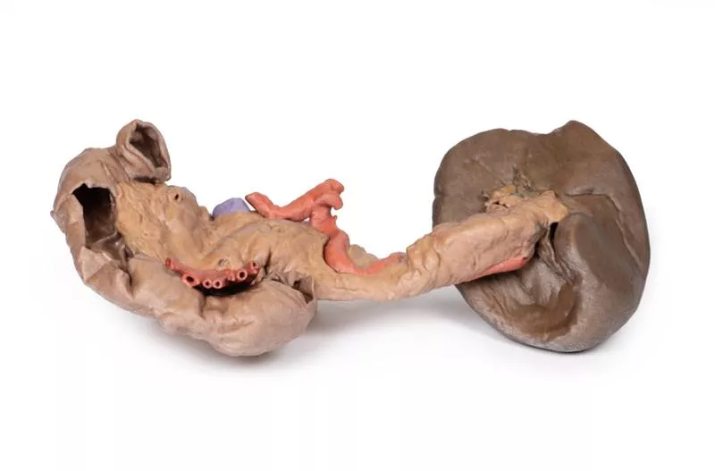
This anatomical model vividly displays the splenic hilum, focusing on the critical vascular structures supplying and draining the spleen.
Key Features:
Splenic Artery and Vein:
Both vessels enter the spleen at the hilum. The splenic vein’s opening is kept patent using inserted silicon tubing, allowing clear visualization of venous drainage. The model shows the most superior branch of the splenic vein carefully sectioned to reveal its course.
Tortuous Splenic Artery:
The model highlights the distinctive twisted and curled shape of the splenic artery as it branches at the hilum, reflecting its natural, winding path from the coeliac trunk to the spleen.
Branching Vessels:
The splenic artery and vein give rise to the short gastric arteries and the left gastro-omental artery. In this specimen, these branches are cut beyond their origin, so they are not fully visible, providing a focused view of the main vessels at the hilum.
Ligament Attachments (Not Present):
- Splenorenal Ligament: Connects the spleen to the left kidney and contains the splenic artery, vein, and tail of the pancreas. Formed embryologically from the dorsal mesentery’s peritoneum, this ligament is removed in the model to expose the splenic vessels clearly.
- Gastrosplenic Ligament: Connects the stomach to the spleen, containing the short gastric arteries and part of the left gastro-omental artery. This ligament is also absent in the model due to dissection beyond the splenic artery branch.
Spleen Capsule:
The outer surface of the spleen is covered by a thin fibrous capsule. This delicate layer is prone to rupture because of the spleen’s high blood content, an important clinical consideration highlighted by the model.
Erler-Zimmer
Erler-Zimmer GmbH & Co.KG
Hauptstrasse 27
77886 Lauf
Germany
info@erler-zimmer.de
Achtung! Medizinisches Ausbildungsmaterial, kein Spielzeug. Nicht geeignet für Personen unter 14 Jahren.
Attention! Medical training material, not a toy. Not suitable for persons under 14 years of age.


