


Product information "Male hemipelvis and thigh"
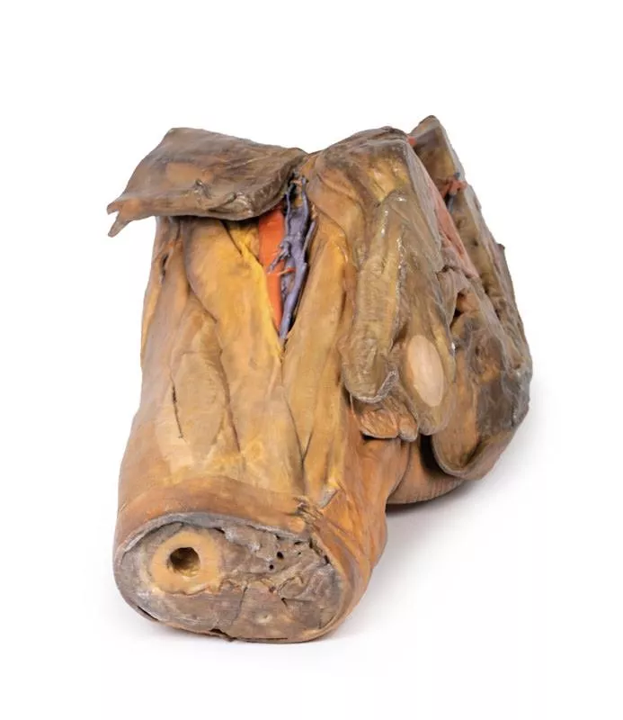
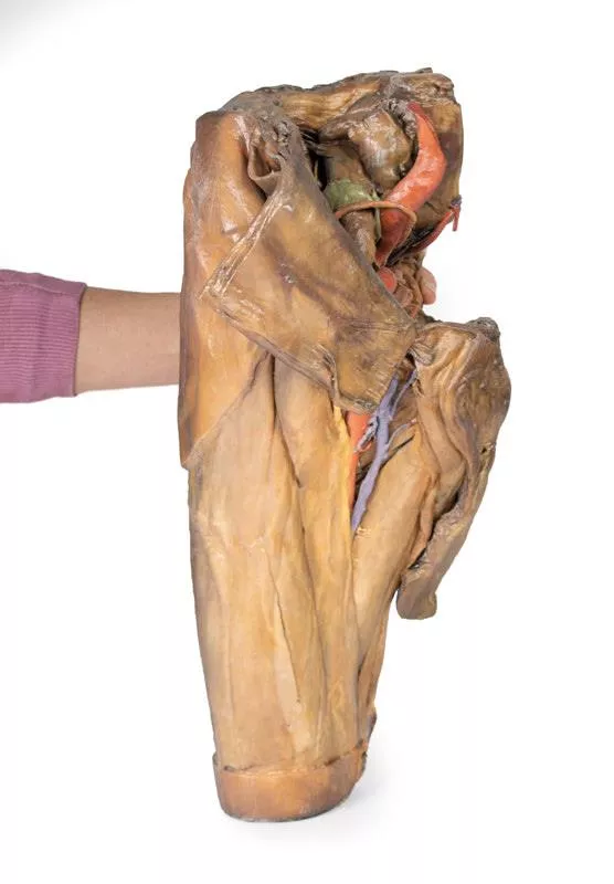
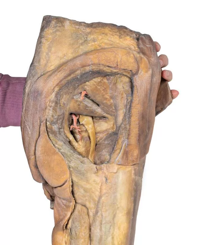
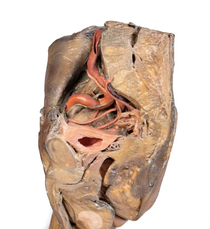
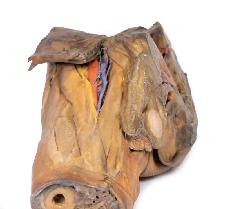
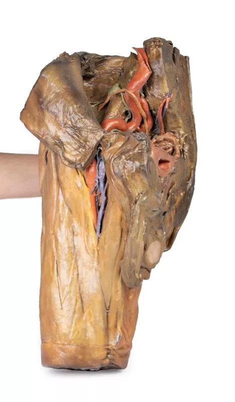
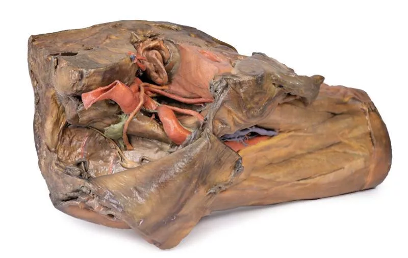
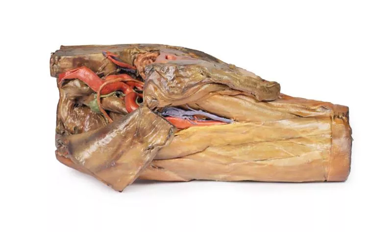
This 3D model preserves a right male pelvis sectioned just superior to the L5 vertebra and sectioned at the midsagittal plane, with the thigh preserved to near the midshaft of the femur. This specimen compliments our LW 91 female hemipelvic specimen and thigh.
The common iliac artery is preserved with several key branches visible, particularly the distribution of the internal iliac within the true pelvis. Several major vessels including the obturator artery and the partially obliterated umbilical artery passes towards the anterior abdominal wall (to form the medial umbilical ligament) and gives off the superior vesicle artery; while the roots of the iliolumbar, superior gluteal, inferior gluteal and internal pudendal artery are visible lateral to the urinary bladder. The ureter descends superficial to these vessels to approach the urinary bladder which is covered with peritoneum in this model. The ductus deferens is exposed from the entry into the space via the deep inguinal ring and passing posteriorly (though sectioned from its normal insertion pathway and resting on the internal iliac artery). Adjacent to the ureter and on the superficial surface of the psoas major muscle is an enlarged iliac lymph node and part of the lymphatic vasculature ascending along the external iliac artery. The majority of the pelvis has been left undissected, allowing for an appreciation of the rectovesicular pouch and the exposed superior rectal artery and vein approaching the preserved portion of rectum. In cross section, the rectum, seminal vesicle and prostate are visible (the section plane preserves parts of both the prostatic urethra and ejaculatory duct).
In the anterior thigh the borders and contents of the femoral triangle are well-preserved, with partial coverage by the flap of the anterior abdominal wall. Posteriorly the skin over the gluteal region and the gluteus maximus muscle have been removed as sequential windows to expose the gluteus medius and minimum muscles, the piriformis, the obturator internus with gemelli muscles, and the quadratus femoris muscle. The superior and inferior gluteal arteries are maintained superior and inferior to the piriformis, respectively; with the sciatic nerve exiting inferior to piriformis before passing deep to the retained portion of the gluteus maximus.
Login
Erler-Zimmer
Erler-Zimmer Medical GmbH
Hauptstrasse 27
77886 Lauf
Germany
info@erler-zimmer.de
Achtung! Medizinisches Ausbildungsmaterial, kein Spielzeug. Nicht geeignet für Personen unter 14 Jahren.
Attention! Medical training material, not a toy. Not suitable for persons under 14 years of age.
Other customers also bought

Female hemipelvis and thigh
This detailed 3D model displays the left half of a female pelvis, sectioned midsagittally, and extending to the proximal mid-thigh.Pelvic Organs & Peritoneum- Visible structures: Urinary bladder, uterus, vagina, and rectum (from anterior to posterior).- The peritoneum is preserved, showing the vesicouterine and rectouterine pouches.- The broad ligament, uterine tube, fimbriae, and left ovary are identifiable near the pelvic brim. Vessels & Nerves- Common and external iliac arteries pass toward the subinguinal space, alongside the common iliac vein and psoas major.- The ureter crosses over these vessels. - The femoral nerve is visible between psoas major and iliacus muscles. Anterior Thigh & Inguinal Region- Superficial fascia removed, exposing thigh structures up to the perineal edge.- Femoral triangle dissected to show:- Femoral artery and vein, with the vein receiving tributaries from the great saphenous, superficial circumflex iliac, external pudendal, and deep pudendal veins.- Femoral nerve lateral to the artery.- Anterior cutaneous nerves and part of the lateral cutaneous nerve over the sartorius muscle. - Inguinal lymph nodes beneath the inguinal ligament. Posterior Gluteal Region- Gluteus maximus removed to reveal deeper gluteal muscles.- Piriformis reflected, exposing:- Sciatic nerve, formed by tibial and common peroneal nerves.- Superior and inferior gluteal arteries. - Posterior cutaneous nerve of the thigh running parallel to the sciatic nerve.- Obturator internus, gemelli, and quadratus femoris muscles exposed.- Internal pudendal artery and pudendal nerve track toward the ischioanal fossa.- Their branches, including the inferior rectal nerve, are visible near the pelvic diaphragm and external anal sphincter.

Posterior Abdominal wall
This detailed 3D printed model presents the male posterior abdominal wall from the diaphragm down to the pelvic brim, including the pelvis and proximal thigh.A focused pelvic and thigh version is also available (MP1770).Muscular Anatomy & DiaphragmThe parietal peritoneum is removed to expose the key muscles of the posterior abdominal wall: psoas major, quadratus lumborum, transversus abdominis, and iliacus below the iliac crest. The diaphragm shows distinct muscular fibers originating from the thoracic cage and lumbar vertebrae (right crus L1–L3, left crus L1–L2), connected by the median arcuate ligament. Major diaphragmatic openings for the esophagus, aorta, and inferior vena cava are visible, although the aorta is removed. NervesSomatic nerves are clearly identifiable, including the subcostal, iliohypogastric, and ilioinguinal nerves (which arise together in this specimen), the lateral cutaneous nerve of the thigh, the genitofemoral nerve (on psoas), and the femoral nerve situated between psoas and iliacus. The sympathetic trunks run alongside the lumbar vertebrae. Vessels & KidneysThe aorta and inferior vena cava are transected at L3, with the aortic bifurcation positioned slightly higher than usual. The renal arteries and veins are preserved, though their full origin is partly obscured by the absence of the great vessels. Both kidneys are dissected free from surrounding fat, showing the typical lower position of the right kidney. The ureters are visible descending from the renal hilum, passing medial to the psoas before crossing the pelvic brim into the true pelvis. This model offers an exceptional view of the complex anatomy of the posterior abdominal wall, pelvis, and proximal thigh, making it ideal for advanced anatomical study, surgical planning, and clinical reference.

Liver with vessels and gallbladder
This liver specimen displays notable differences compared to a typical liver. It is less wedge-shaped and elongated in the superoinferior dimension, resulting in a greater vertical height when viewed from the posterior. Size- Measures approximately 18 cm along the midclavicular line.- Typical livers measure under 16 cm in this dimension.- The increased length suggests mild hepatomegaly (enlargement). Important Notes- Size estimates may be affected by specimen preservation and fixing, which can cause some distortion.- Diagnosing hepatomegaly based on a single measurement is limited and varies with individual anatomy, measurement technique, sex, and body mass index (BMI). Anatomical VariationsThis specimen does not match common anatomical variations often confused with hepatomegaly such as:- Riedel’s lobe: a downward projection of the right lobe- Beaver tail liver: elongated left lobe- Papillary process from the caudate lobe

Internal abdominal wall
This detailed 3D model captures the internal surface of the anterior abdominal wall—a region often removed or damaged during dissections. It complements our MP1130 abdominal specimen, where the anterior wall has been removed, providing a clear view of key muscle and connective tissue structures. Key Features:Muscle Fibers & Aponeurosis:The horizontally oriented transversus abdominus muscle fibers converge toward their aponeurosis (tendon sheet), visible especially along the specimen’s superior margins. Arcuate Line:Located in the lower third of the model, this landmark marks where the aponeurosis shifts relative to the rectus abdominus muscle.- Above the arcuate line: Aponeurosis fibers split evenly around the rectus abdominus.- Below the arcuate line: All aponeurotic fibers pass anteriorly to the rectus abdominus, reflecting a change in abdominal wall structure. Vascular Structures:Inferior Epigastric Arteries & Veins:These vessels originate from the external iliac arteries and veins, ascending superiorly through the anterior abdominal wall. Hesselbach’s Triangle:On the right side of the model, the orientation of the inferior epigastric artery relative to the rectus abdominus muscle defines the apex of the inguinal (Hesselbach’s) triangle—a critical anatomical region often associated with direct inguinal hernias. (Note: The inguinal ligament forming the base of this triangle is not present in this specimen.) Embryological Remnant: Median Abdominal Ligament:Positioned midline between the two rectus abdominus muscles, this fold of parietal peritoneum covers the urachus, a fibrous remnant from embryological development extending from the bladder to the umbilicus.

Head, Neck and Shoulder with angiosomes
This large, multipart 3D printed specimen showcases detailed anatomy of the head, neck, thorax, axillae, and upper limbs.Head and Neck:The calotte has been removed ~2?cm above the orbits to expose the brain and endocranial cavity. A transverse cerebral section shows grey and white matter, lateral ventricles, and choroid plexus. The right side retains skin and fascia, false-coloured to highlight facial and neck angiosomes. The left side reveals facial expression and mastication muscles, and infratemporal structures including the lingual nerve and terminal branches of the external carotid artery. The carotid sheaths are opened bilaterally, exposing the common, internal, and external carotid arteries, and vagus nerves. The sternocleidomastoid and internal jugular veins are mostly removed. On the right, the great auricular and hypoglossal nerves are visible, along with the stylohyoid ligament and supra-/infrahyoid muscles. The thyroid gland is prominent, with preserved superior and inferior thyroid vessels.Root of the Neck and Axilla:On the left, partial clavicle removal reveals the first rib, anterior scalene, and brachial plexus roots (C5–T1) forming trunks between scalene muscles. The subclavian artery passes posterior to scalenus anterior, transitioning to the axillary artery, closely related to the brachial plexus cords.The left axilla displays brachial plexus divisions and cords. The formation of the median nerve around the axillary artery is distinct. The ulnar, musculocutaneous, axillary, thoracodorsal, and long thoracic nerves are clearly identified with their courses and muscular targets.On the right, the clavicle and subclavius muscle are intact, showing the cervico-axillary canal. Pectoralis major and minor have been reflected, exposing deeper structures.Thorax:A window in the left thoracic wall reveals the mediastinum. The left lung has been removed. Intercostal spaces are visible beneath the parietal pleura; neurovascular bundles are identifiable posteriorly. The heart is exposed without pericardium, showing the left atrium and ventricle, pulmonary vessels, aorta, and both left vagus and recurrent laryngeal nerves. The right thoracic wall remains intact, displaying intercostal and upper limb muscles. From below, the right lung, pleural cavities, and diaphragmatic heart surface are visible. Posterior thoracic skin and fascia are intact, showing cutaneous nerve distribution.Size: 50 x 20 x 41 cm

Female pelvis deep dissection
This high-detail 3D model showcases a deep dissection of the female pelvis, isolated from surrounding regions, with emphasis on visceral, vascular, and ligamentous structures in relation to bony landmarks.Pelvic Organs & Peritoneal Structures- Sigmoid colon descends into the rectum over the pelvic brim, crossing the common and external iliac vessels.- Nearby: Sigmoid and superior rectal arteries, and the descending ureter.- Urinary bladder (collapsed) and uterus are positioned anteriorly in the true pelvis.- The broad ligament is retained, though ovaries, uterine tubes, ovarian and round ligaments are present but indistinct due to age-related atrophy.- Suspensory and round ligaments are detached from the peritoneum to expose surrounding vessels. Arteries & Veins- Internal iliac artery branches are visible bilaterally.- Median sacral artery is seen in the midline between the common iliac arteries.- Left side: Uterine artery only.- Right side: Uterine, superior vesical, and obturator arteries.- Inferior epigastric artery and vein arise from the external iliac vessels, visible near the inferior abdominal wall. Musculoskeletal Features- Right side: Entire femur and thigh muscles removed to expose:- Obturator membrane- Acetabular cartilage- Transverse acetabular ligament- Posterior dissection reveals:- Superior gluteal foramen and artery- Sacrospinous ligament (with sacrotuberous ligament removed)- Inferior rectal artery branches within the ischioanal fossa Nerves & Ligaments- Left sciatic nerve preserved within the greater sciatic foramen- Sacrotuberous ligament retained on the left- Ischioanal fossae on both sides show:- Inferior rectal artery branches- Pelvic diaphragm fibers- External anal sphincter integration with the rectal wall

Continuous innovation

Social responsibility

Active customer orientation

Understanding quality

Sustainable actions

ISO 9001 certification