


Product information "Head, Neck and Shoulder with angiosomes"
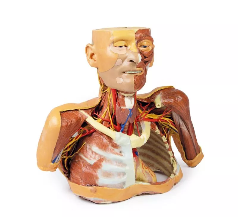
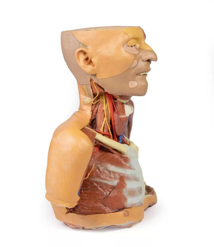
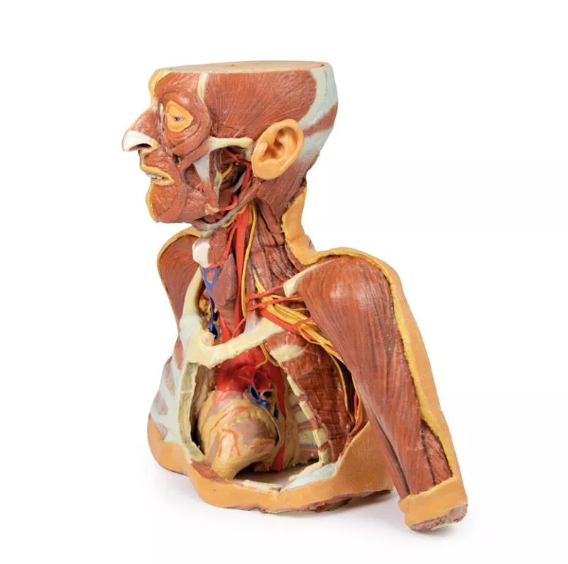
This large, multipart 3D printed specimen showcases detailed anatomy of the head, neck, thorax, axillae, and upper limbs.
Head and Neck:
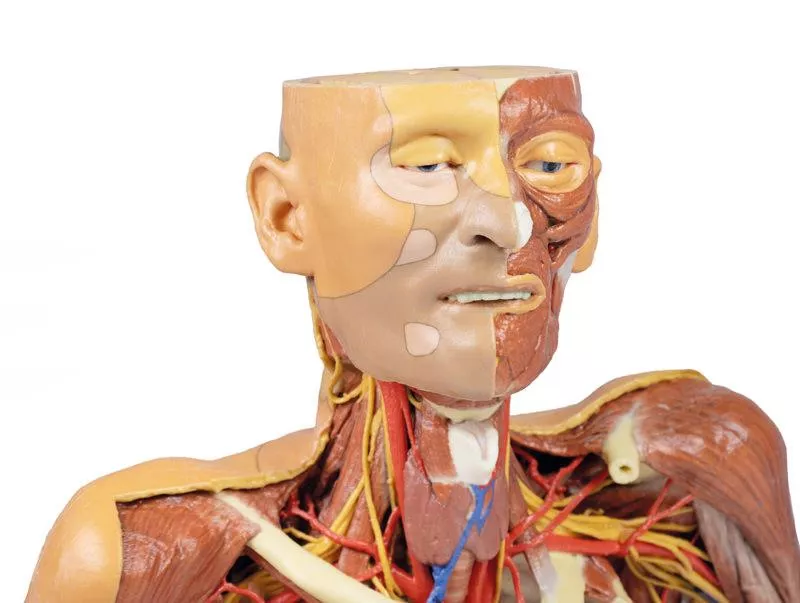
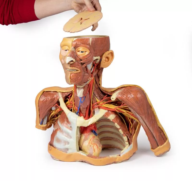
The calotte has been removed ~2?cm above the orbits to expose the brain and endocranial cavity. A transverse cerebral section shows grey and white matter, lateral ventricles, and choroid plexus. The right side retains skin and fascia, false-coloured to highlight facial and neck angiosomes. The left side reveals facial expression and mastication muscles, and infratemporal structures including the lingual nerve and terminal branches of the external carotid artery. The carotid sheaths are opened bilaterally, exposing the common, internal, and external carotid arteries, and vagus nerves. The sternocleidomastoid and internal jugular veins are mostly removed. On the right, the great auricular and hypoglossal nerves are visible, along with the stylohyoid ligament and supra-/infrahyoid muscles. The thyroid gland is prominent, with preserved superior and inferior thyroid vessels.
Root of the Neck and Axilla:
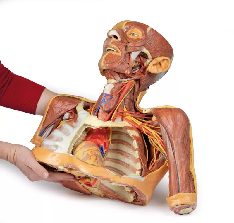
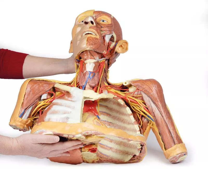
On the left, partial clavicle removal reveals the first rib, anterior scalene, and brachial plexus roots (C5–T1) forming trunks between scalene muscles. The subclavian artery passes posterior to scalenus anterior, transitioning to the axillary artery, closely related to the brachial plexus cords.
The left axilla displays brachial plexus divisions and cords. The formation of the median nerve around the axillary artery is distinct. The ulnar, musculocutaneous, axillary, thoracodorsal, and long thoracic nerves are clearly identified with their courses and muscular targets.
On the right, the clavicle and subclavius muscle are intact, showing the cervico-axillary canal. Pectoralis major and minor have been reflected, exposing deeper structures.
Thorax:
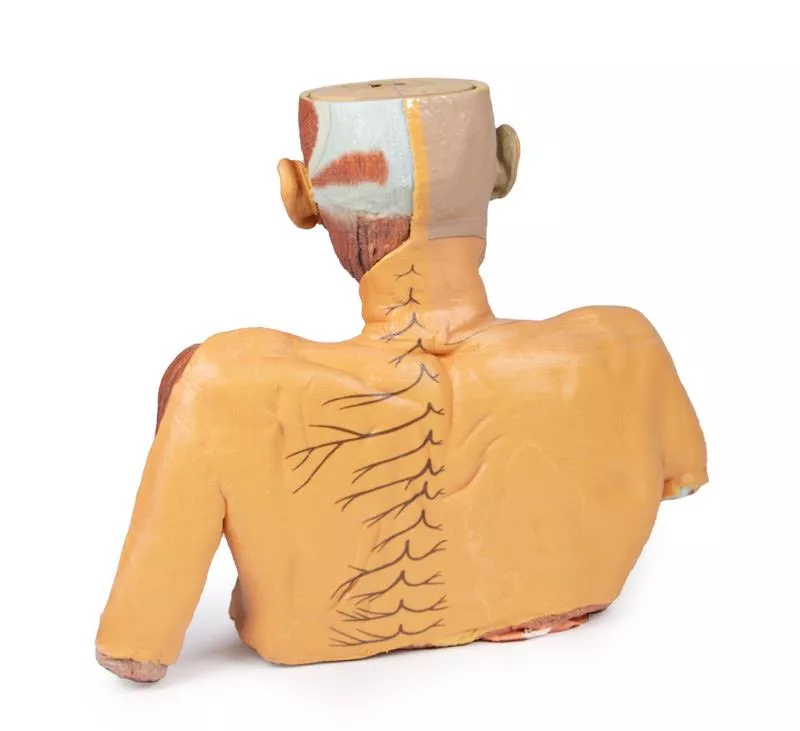
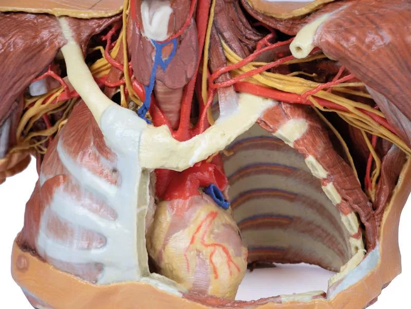
A window in the left thoracic wall reveals the mediastinum. The left lung has been removed. Intercostal spaces are visible beneath the parietal pleura; neurovascular bundles are identifiable posteriorly. The heart is exposed without pericardium, showing the left atrium and ventricle, pulmonary vessels, aorta, and both left vagus and recurrent laryngeal nerves. The right thoracic wall remains intact, displaying intercostal and upper limb muscles. From below, the right lung, pleural cavities, and diaphragmatic heart surface are visible. Posterior thoracic skin and fascia are intact, showing cutaneous nerve distribution.
Size: 50 x 20 x 41 cm
Head and Neck:
The calotte has been removed ~2?cm above the orbits to expose the brain and endocranial cavity. A transverse cerebral section shows grey and white matter, lateral ventricles, and choroid plexus. The right side retains skin and fascia, false-coloured to highlight facial and neck angiosomes. The left side reveals facial expression and mastication muscles, and infratemporal structures including the lingual nerve and terminal branches of the external carotid artery. The carotid sheaths are opened bilaterally, exposing the common, internal, and external carotid arteries, and vagus nerves. The sternocleidomastoid and internal jugular veins are mostly removed. On the right, the great auricular and hypoglossal nerves are visible, along with the stylohyoid ligament and supra-/infrahyoid muscles. The thyroid gland is prominent, with preserved superior and inferior thyroid vessels.
Root of the Neck and Axilla:
On the left, partial clavicle removal reveals the first rib, anterior scalene, and brachial plexus roots (C5–T1) forming trunks between scalene muscles. The subclavian artery passes posterior to scalenus anterior, transitioning to the axillary artery, closely related to the brachial plexus cords.
The left axilla displays brachial plexus divisions and cords. The formation of the median nerve around the axillary artery is distinct. The ulnar, musculocutaneous, axillary, thoracodorsal, and long thoracic nerves are clearly identified with their courses and muscular targets.
On the right, the clavicle and subclavius muscle are intact, showing the cervico-axillary canal. Pectoralis major and minor have been reflected, exposing deeper structures.
Thorax:
A window in the left thoracic wall reveals the mediastinum. The left lung has been removed. Intercostal spaces are visible beneath the parietal pleura; neurovascular bundles are identifiable posteriorly. The heart is exposed without pericardium, showing the left atrium and ventricle, pulmonary vessels, aorta, and both left vagus and recurrent laryngeal nerves. The right thoracic wall remains intact, displaying intercostal and upper limb muscles. From below, the right lung, pleural cavities, and diaphragmatic heart surface are visible. Posterior thoracic skin and fascia are intact, showing cutaneous nerve distribution.
Size: 50 x 20 x 41 cm
Login
September 29, 2021 07:56
Ausgezeichnete Qualität, perfekte Darstellung kleinster Strukturen!
Ausgezeichnete Qualität, perfekte Darstellung kleinster Strukturen!
🔬 3D Anatomy Series - Human Body Replicas to Enhance Teaching!
August 26, 2025
Discover exclusive 3D-printed models of the human body – created directly from radiological data or real specimens.
Erler-Zimmer
Erler-Zimmer Medical GmbH
Hauptstrasse 27
77886 Lauf
Germany
info@erler-zimmer.de
Achtung! Medizinisches Ausbildungsmaterial, kein Spielzeug. Nicht geeignet für Personen unter 14 Jahren.
Attention! Medical training material, not a toy. Not suitable for persons under 14 years of age.
Other customers also bought

Male hemipelvis and thigh
This 3D model preserves a right male pelvis sectioned just superior to the L5 vertebra and sectioned at the midsagittal plane, with the thigh preserved to near the midshaft of the femur. This specimen compliments our LW 91 female hemipelvic specimen and thigh. The common iliac artery is preserved with several key branches visible, particularly the distribution of the internal iliac within the true pelvis. Several major vessels including the obturator artery and the partially obliterated umbilical artery passes towards the anterior abdominal wall (to form the medial umbilical ligament) and gives off the superior vesicle artery; while the roots of the iliolumbar, superior gluteal, inferior gluteal and internal pudendal artery are visible lateral to the urinary bladder. The ureter descends superficial to these vessels to approach the urinary bladder which is covered with peritoneum in this model. The ductus deferens is exposed from the entry into the space via the deep inguinal ring and passing posteriorly (though sectioned from its normal insertion pathway and resting on the internal iliac artery). Adjacent to the ureter and on the superficial surface of the psoas major muscle is an enlarged iliac lymph node and part of the lymphatic vasculature ascending along the external iliac artery. The majority of the pelvis has been left undissected, allowing for an appreciation of the rectovesicular pouch and the exposed superior rectal artery and vein approaching the preserved portion of rectum. In cross section, the rectum, seminal vesicle and prostate are visible (the section plane preserves parts of both the prostatic urethra and ejaculatory duct).In the anterior thigh the borders and contents of the femoral triangle are well-preserved, with partial coverage by the flap of the anterior abdominal wall. Posteriorly the skin over the gluteal region and the gluteus maximus muscle have been removed as sequential windows to expose the gluteus medius and minimum muscles, the piriformis, the obturator internus with gemelli muscles, and the quadratus femoris muscle. The superior and inferior gluteal arteries are maintained superior and inferior to the piriformis, respectively; with the sciatic nerve exiting inferior to piriformis before passing deep to the retained portion of the gluteus maximus.

Female hemipelvis and thigh
This detailed 3D model displays the left half of a female pelvis, sectioned midsagittally, and extending to the proximal mid-thigh.Pelvic Organs & Peritoneum- Visible structures: Urinary bladder, uterus, vagina, and rectum (from anterior to posterior).- The peritoneum is preserved, showing the vesicouterine and rectouterine pouches.- The broad ligament, uterine tube, fimbriae, and left ovary are identifiable near the pelvic brim. Vessels & Nerves- Common and external iliac arteries pass toward the subinguinal space, alongside the common iliac vein and psoas major.- The ureter crosses over these vessels. - The femoral nerve is visible between psoas major and iliacus muscles. Anterior Thigh & Inguinal Region- Superficial fascia removed, exposing thigh structures up to the perineal edge.- Femoral triangle dissected to show:- Femoral artery and vein, with the vein receiving tributaries from the great saphenous, superficial circumflex iliac, external pudendal, and deep pudendal veins.- Femoral nerve lateral to the artery.- Anterior cutaneous nerves and part of the lateral cutaneous nerve over the sartorius muscle. - Inguinal lymph nodes beneath the inguinal ligament. Posterior Gluteal Region- Gluteus maximus removed to reveal deeper gluteal muscles.- Piriformis reflected, exposing:- Sciatic nerve, formed by tibial and common peroneal nerves.- Superior and inferior gluteal arteries. - Posterior cutaneous nerve of the thigh running parallel to the sciatic nerve.- Obturator internus, gemelli, and quadratus femoris muscles exposed.- Internal pudendal artery and pudendal nerve track toward the ischioanal fossa.- Their branches, including the inferior rectal nerve, are visible near the pelvic diaphragm and external anal sphincter.

Hilum of the right lung
This high-quality 3D model presents a sagittal section of the right lung, focused on the hilum, where key anatomical structures enter and exit the lung. It serves as an essential tool for understanding pulmonary vascular and bronchial anatomy, with clear orientation from apex to base and medial to lateral surfaces.Key Features:Hilum Structure:- The hilum marks the transition between visceral and parietal pleura and serves as the lung’s only anatomical connection to the body via the pulmonary ligament.- Major structures entering the lung at this point include:- Pulmonary artery (superior in the hilum) – carrying deoxygenated blood from the heart.- Superior and inferior pulmonary veins (anterior and inferior) – returning oxygenated blood to the heart.- Right main bronchus and its lobar branches – located posteriorly in the hilum. - Associated nerves and lymphatics. Visible Anatomical Landmarks:- Cardiac impression (formed by the right atrium) is visible just anterior to the hilum.- The oesophageal groove is preserved along the posterior surface, tracing the path of the descending oesophagus.- Oblique and horizontal fissures are well-defined on the lateral surface, demarcating the lung's three lobes.- Hilar lymph nodes are observed around the medial hilum.Lung Surfaces:- Diaphragmatic surface (inferior), showing the concave interface with the diaphragm.- Costal visceral surface (posterior), where the lung contacts the thoracic wall.

Deep upper limb and hand
This 3D printed model presents a superficial dissection of the right distal arm, forearm, and hand, showcasing key vascular, nervous, and muscular anatomy.Distal Arm & Cubital FossaThe arrangement of the biceps tendon, brachial artery, and median nerve is visible from lateral to medial. The bicipital aponeurosis has been removed to expose deeper structures. The ulnar nerve is reflected from the cubital tunnel, and the radial nerve with its branches is visible near the supinator muscle. Forearm AnatomyOn the anterior forearm, superficial flexors (pronator teres, FCR, FDS, FCU) are preserved; palmaris longus is absent. The radial artery is exposed; the ulnar artery is not visible. Posteriorly, extensors from the common origin are visible, including ECRB, ED, EDM, and ECU. The APL, EPB, and EPL are also shown wrapping around the radius. Hand & SnuffboxThe anatomical snuffbox reveals the radial artery in its floor and cutaneous branches of the radial nerve. The palmar side displays thenar/hypothenar muscles, lumbricals, flexor tendons, and the median nerve beneath the flexor retinaculum. A superficial branch of the radial artery crosses the retinaculum.Ideal for anatomical education, this print offers a clear view of key structures in the distal upper limb.

Posterior Body Wall / Ventral Deep Dissection
This 3D printed model provides a detailed ventral view of the head, neck, thorax, abdomen, and proximal thighs, offering insight into the central nervous system, nerve plexuses, and major vascular structures. It serves as a complementary piece to the dorsal dissection model (MP1400).Brain and Upper SpineThe facial skeleton is removed, exposing both cerebral hemispheres, parts of the Circle of Willis, and key arteries (vertebral, basilar, AICA, labyrinthine). Several cranial nerves (II, III, V, VI) and the right internal carotid artery are preserved. Below, the anterior spinal cord, vertebral arteries, and cervical and brachial plexus roots are visible, along with CN X, CN XII, and the sympathetic trunks. Axillae and Upper LimbsThe anterior thoracic wall, clavicles, and first ribs are removed to expose both axillae, with clear views of the brachial plexus, axillary arteries, and surrounding musculature. The proximal upper limbs are preserved to mid-arm level. Thorax and Lumbar RegionThe spinal cord extends to the conus medullaris, with ventral nerve roots and sympathetic chains visible throughout. Splanchnic nerves, intercostal nerves, and abdominal wall nerves (e.g., ilioinguinal, genitofemoral, femoral) are preserved. On the right, removal of the psoas muscle exposes the lumbar plexus.Pelvis and Proximal ThighThe pelvic viscera are removed, but the pelvic floor, rectum, and lumbosacral plexus remain. Obturator nerves are visible entering the obturator canals, and the femoral vessels are retained in both femoral triangles. This model is ideal for studying ventral CNS access, neurovascular pathways, and regional anatomical relationships across the entire torso.

Continuous innovation

Social responsibility

Active customer orientation

Understanding quality

Sustainable actions

ISO 9001 certification