


Product information "Hilum of the right lung"
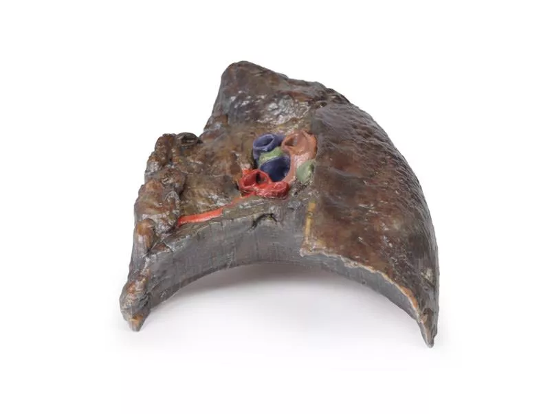
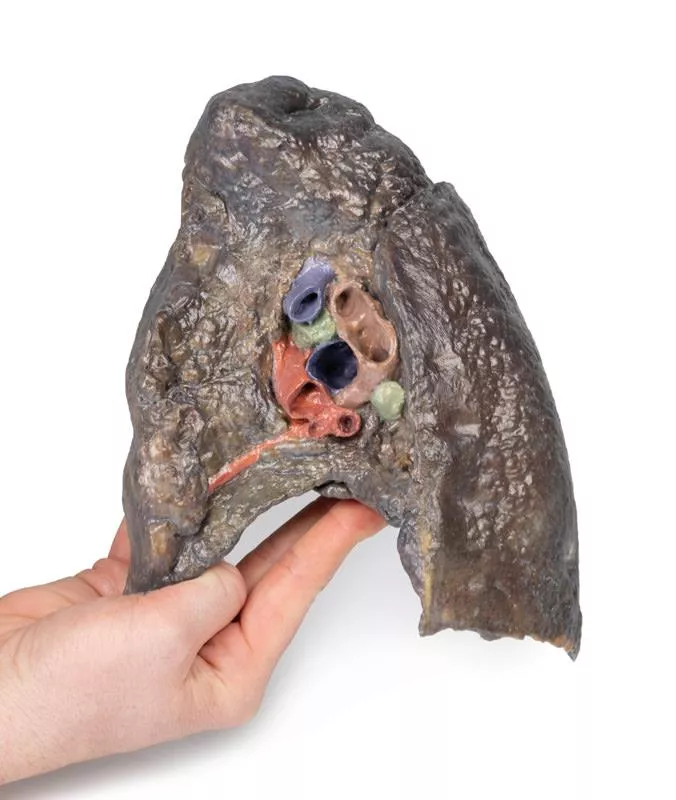
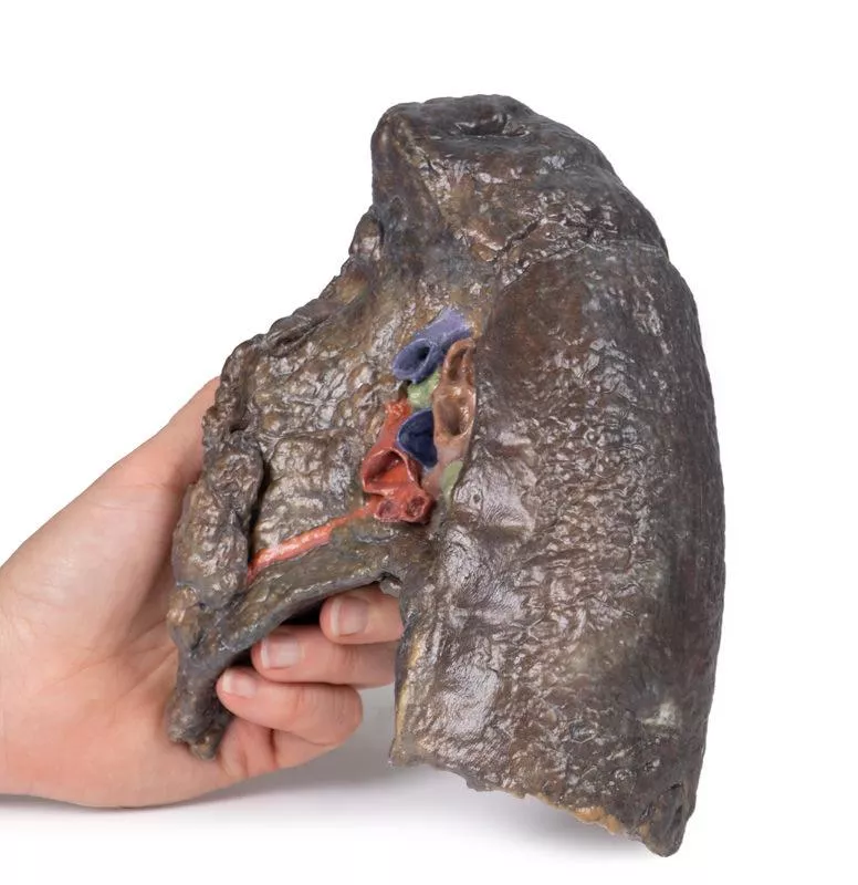
This high-quality 3D model presents a sagittal section of the right lung, focused on the hilum, where key anatomical structures enter and exit the lung.
It serves as an essential tool for understanding pulmonary vascular and bronchial anatomy, with clear orientation from apex to base and medial to lateral surfaces.
Key Features:
Hilum Structure:
- The hilum marks the transition between visceral and parietal pleura and serves as the lung’s only anatomical connection to the body via the pulmonary ligament.
- Major structures entering the lung at this point include:
Visible Anatomical Landmarks:
- Cardiac impression (formed by the right atrium) is visible just anterior to the hilum.
- The oesophageal groove is preserved along the posterior surface, tracing the path of the descending oesophagus.
- Oblique and horizontal fissures are well-defined on the lateral surface, demarcating the lung's three lobes.
- Hilar lymph nodes are observed around the medial hilum.
Lung Surfaces:
- Diaphragmatic surface (inferior), showing the concave interface with the diaphragm.
- Costal visceral surface (posterior), where the lung contacts the thoracic wall.
It serves as an essential tool for understanding pulmonary vascular and bronchial anatomy, with clear orientation from apex to base and medial to lateral surfaces.
Key Features:
Hilum Structure:
- The hilum marks the transition between visceral and parietal pleura and serves as the lung’s only anatomical connection to the body via the pulmonary ligament.
- Major structures entering the lung at this point include:
- Pulmonary artery (superior in the hilum) – carrying deoxygenated blood from the heart.
- Superior and inferior pulmonary veins (anterior and inferior) – returning oxygenated blood to the heart.
- Right main bronchus and its lobar branches – located posteriorly in the hilum.
- Associated nerves and lymphatics.
- Superior and inferior pulmonary veins (anterior and inferior) – returning oxygenated blood to the heart.
- Right main bronchus and its lobar branches – located posteriorly in the hilum.
- Associated nerves and lymphatics.
Visible Anatomical Landmarks:
- Cardiac impression (formed by the right atrium) is visible just anterior to the hilum.
- The oesophageal groove is preserved along the posterior surface, tracing the path of the descending oesophagus.
- Oblique and horizontal fissures are well-defined on the lateral surface, demarcating the lung's three lobes.
- Hilar lymph nodes are observed around the medial hilum.
Lung Surfaces:
- Diaphragmatic surface (inferior), showing the concave interface with the diaphragm.
- Costal visceral surface (posterior), where the lung contacts the thoracic wall.
Login
Erler-Zimmer
Erler-Zimmer Medical GmbH
Hauptstrasse 27
77886 Lauf
Germany
info@erler-zimmer.de
Achtung! Medizinisches Ausbildungsmaterial, kein Spielzeug. Nicht geeignet für Personen unter 14 Jahren.
Attention! Medical training material, not a toy. Not suitable for persons under 14 years of age.
Other customers also bought

Parotid Gland and Facial Nerve dissection
This 3D model presents a detailed superficial dissection of the lateral face, focusing on the parotid gland and its anatomical relationship to key neurovascular structures and surface landmarks. It serves as a valuable reference for clinicians involved in Mohs surgery, skin cancer treatment, and plastic or reconstructive facial procedures.Key Features:Dissection WindowThe exposed region extends from just anterior to the external ear, bounded superiorly by the zygomatic arch and inferiorly by the angle of the mandible, covering the area between the masseter and the sternocleidomastoid muscle. Parotid Gland & Facial Nerve- The parotid gland is fully exposed, with superior portions removed to reveal:- The superficial temporal artery- The facial nerve, dividing into its temporal, zygomatic, and buccal branches- The parotid duct is visible as it traverses the dissection field toward its termination in the buccinator muscleAssociated NervesA branch of the great auricular nerve is preserved along the inferior and posterior margins of the gland, running anterior to the sternocleidomastoid

Pericardial space
This detailed 3D anatomical model displays the pericardial cavity and reflections with the heart removed, allowing clear visualization of key structures within the middle mediastinum.Key Features:Pericardium:Shows the full extent of the parietal pericardium, continuous with the visceral layer (epicardium), and false-colored to indicate the positions of the atria, ventricles, and great vessels. Mediastinal Landmarks:Highlights the base, apex, diaphragmatic, and pulmonary surfaces of the heart by their impressions within the pericardial cavity.Great Vessels:- Aorta (ascending, arch, and descending)- Superior and Inferior Vena Cava- Pulmonary trunk and arteries- Pulmonary veins (4 total) - All shown in their natural positions relative to the pericardium.Pericardial Sinuses:- Transverse sinus: Located between arteries and veins; relevant for surgical access.- Oblique sinus: Posterior recess between pulmonary veins.Educational Use:- Ideal for teaching thoracic and cardiac anatomy, including pericardial reflections, mediastinal relationships, and surgical landmarks.- Useful in medical training, surgical planning, and radiological orientation.

Lung Slab, Hilum removed
This 3D anatomical model presents a left lung dissected in a parasagittal plane, offering a unique internal view of pulmonary structures and anatomical landmarks. The mediastinal surface has been removed, allowing detailed observation of internal lung anatomy and its relationship to the heart and diaphragm.Key Features:Bronchial Tree (Internal View):- The primary bronchi are not visible due to prior branching.- Subdivided bronchi are preserved, though the dissection depth makes it unclear whether they represent secondary (lobar) or tertiary (segmental) bronchi. Vascular Structures:- Pulmonary arteries and veins are typically seen at the hilum, but precise levels of subdivision are undetermined in this section.Cardiac Impression:- A clear concave impression remains on the medial lung surface, formed by the left ventricle of the heart pressing against the lung.- Despite the dissection, this landmark remains distinctly visible.Diaphragmatic Surface:- The lung’s base is concave, resting atop the diaphragm.- While the pleura is not preserved, the model indicates where the diaphragmatic recess would form—between the costal and diaphragmatic pleura.

Heart
This 3D model depicts a life-sized adult heart with light dissection of the epicardium, offering a clear view of the coronary arteries, cardiac veins, and great vessels at the heart's base.Key Anatomical Features:Great Vessels:- Superior vena cava and azygos vein drain into the right atrium.- Aortic arch preserved, with two main branches: - A combined brachiocephalic trunk (giving rise to both right and left common carotid and right subclavian arteries). - Left subclavian artery arising independently.- Pulmonary trunk and pulmonary arteries intact.- Ligamentum arteriosum visible between the aortic arch and left pulmonary artery. Coronary Arteries:- Right coronary artery (RCA) descends from the ascending aorta, wrapping to the posterior interventricular sulcus.- Left coronary artery (LCA) branches: - Anterior interventricular artery (LAD) runs toward the apex.- Diagonal branches dive into the myocardium.- Circumflex artery runs posteriorly, near the great cardiac vein.Venous Drainage:Coronary sinus clearly preserved on the posterior heart surface, draining into the right atrium near the inferior vena cava.

Hilum of the left lung
This high-resolution 3D model offers a detailed view of the left lung hilum and associated structures, sectioned sagittally through the cardiac notch. It clearly demonstrates the relationships between the bronchi, pulmonary vessels, pleura, and supporting structures, making it ideal for advanced anatomical education.Key Features:Hilum Anatomy:- Entry/exit point for the pulmonary artery, superior and inferior pulmonary veins, main bronchus, lymphatics, and nerves.- Shows the meeting point of visceral and parietal pleura, forming the pulmonary ligament—the lung’s sole anatomical connection to the body. Pulmonary Circulation:- Pulmonary artery (superior in position) carries deoxygenated blood from the heart.- Pulmonary veins (anterior and inferior) return oxygenated blood to the heart.Bronchial Structure:Left main bronchus and its lobar branches are visible, located posteriorly within the hilum.Additional Views:- Oblique fissure along the lateral lung surface.- Diaphragmatic surface at the base; costal visceral surface posteriorly.- Pulmonary lymph nodes surrounding the hilum, both medially and laterally.

Abdomen with bilateral Hernias
This 3D model represents one of the largest and most complex in the series, consisting of a partial torso from the diaphragm to the proximal thigh with a complete abdominal cavity preserving varying levels of dissection. This 3D model also records the rare, simultaneous occurrence of indirect and direct inguinal hernias allowing for a consideration of the anatomical underpinnings for both conditions. Given the scale of the dissection this 3D model description is divided into discrete parts based on views and regions.The diaphragmThe diaphragm is preserved on the model’s superior aspect, with both domes and costodiaphragmatic recesses visible despite some distortion from rib removal. The fibrous pericardium rests on the central tendon, with the terminal inferior vena cava seen in the caval foramen. Lateral to this lies the oesophagus in the oesophageal hiatus, and the descending thoracic aorta approaching the aortic hiatus near the vertebrae. The epigastric and hypochondriac regionsIn the abdomen, removal of the anterior wall, greater omentum, and much of the GI tract reveals retroperitoneal structures. The terminal oesophagus enters just left of the liver. With the stomach removed, the pancreas is fully exposed from head to tail, reaching the spleen in the left hypochondrium. Above it, the splenic and common hepatic arteries span the narrow space between pancreas, diaphragm, and liver. The tortuous splenic artery divides near the splenic vein; the common hepatic gives rise to the gastroduodenal and right gastric arteries, superficial to the portal vein. The superior mesenteric vessels pass near the pancreatic head, and the ileocolic artery leads to the caecum. The inferior mesenteric vein arises from the superior rectal vein and crosses the descending aorta. Below the liver, the gallbladder lies between the lobes. On the left, renal vessels pass deep to the pancreas, with ureters descending across the psoas muscles. The umbilical and lumbar regionsMost abdominal organs in the umbilical and lumbar regions have been removed to reveal the posterior abdominal wall. Centrally, the descending aorta and inferior vena cava are prominent, with testicular vessels traceable toward the inguinal region. Two right lumbar arteries branch from the aorta, and the inferior mesenteric artery gives rise to the left colic, sigmoid, and superior rectal arteries. On the right, subcostal, iliohypogastric, and ilioinguinal nerves are visible, along with the circumflex iliac artery.The hypogastrium and iliac regionsThe abdominal aorta bifurcates into the common, internal, and external iliac arteries, with matching iliac veins merging into the inferior vena cava. The obturator artery, ureters, and testicular vessels are visible. In the true pelvis, the peritoneum covers the bladder, while the rectum remains obscured. The right iliac fossa contains the terminal ileum, caecum, and appendix, with nearby vessels and nerves. On the left, the sigmoid colon crosses the iliac fossa, where an epiploic appendage extends into an indirect hernia near the inferior epigastric artery. The inguinal region and perineumThis model uniquely preserves both direct (right) and indirect (left) inguinal hernias, with the inferior epigastric vessels retained for anatomical orientation. The right hernia lies medial to these vessels; the left hernia sac extends laterally into the spermatic cord, containing an epiploic appendage. The perineum reveals the penis, testes, and spermatic cords. On the right, the cord remains intact; on the left, it’s opened, showing a varicose testicular vein linked to the indirect hernia. The thighThe femoral triangle has been dissected on both thighs. On the right, the femoral sheath was removed to reveal the femoral artery, vein, deep inguinal lymph nodes, and femoral nerve. On the left, a broader view exposes anterior and medial thigh muscles, with the femoral artery, profunda femoris, and circumflex iliac artery visible. The model ends mid-thigh, showing cross-sectional anatomy including the femoral shaft, vessels, and muscles in the subsartorial canal.

Thorax with heart and vessels
This highly detailed 3D model depicts the key anatomy of the superior thoracic aperture, mediastinum, and adjacent neck and thoracic structures, with both clavicles and select muscular and venous elements removed to enhance visibility and educational impact.Anatomical Highlights:Superior Thoracic Aperture:- Trachea visible at the top, with a robust ring of cartilage.- Rib 1 exposed from lateral to medial, including insertion of the anterior scalene muscle.- Removal of clavicles allows unobstructed views into the upper thoracic corridor. Vascular Anatomy:- Right subclavian artery, situated above rib 1, gives rise to the thyrocervical trunk.- Left subclavian artery, also above rib 1, branches into the suprascapular artery.- Both common carotid arteries are visible; the left carotid sheath includes the left vagus nerve.Nervous System:- Left vagus nerve follows the left carotid artery within the carotid sheath; left recurrent laryngeal nerve loops under the aorta.- Right vagus nerve and right phrenic nerve retracted during dissection; left phrenic nerve remains anterior to the heart, tracing to the diaphragm.- Elements of the left brachial plexus are visible—from roots to trunks—including the dorsal scapular nerve.Mediastinal & Cardiac Orientation:- Arch of the aorta, with brachiocephalic trunk, left common carotid, and left subclavian artery, sits just above the heart.- Pulmonary trunk emerges immediately superior to the heart.- Left anterior descending artery (LAD) courses along the anterior heart surface.- Superior vena cava lies to the right and posterior to the ascending aorta.- Right phrenic nerve positioned posteriorly to the heart; left phrenic nerve runs in its connective tissue anteriorly.Inferior Thorax & Diaphragm:- Ribs 8–12 and associated external intercostal musculature visible; muscle fibers run inferomedially into fascial layers.- Right hemidiaphragm sits higher than the left, reflecting the presence of the liver beneath. Applications & BenefitsClinical Relevance:Ideal for visualizing neck and thoracic anatomy relevant to cardiovascular, respiratory, and nerve-related procedures, including laryngeal, thoracic, and brachial plexus surgery.Educational Utility: Offers a clean, accessible view of mediastinal compartments, vascular pathways, and nerve trajectories without obstruction by bone or superficial structures. Enhanced Learning: The model’s clarity supports instruction in radiographic interpretation, thoracic surgical approaches, and anatomy exams.

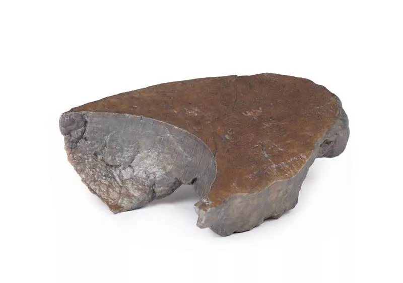
Right lung, hilum removed
This 3D model provides a detailed sectional view of the right lung, serving as a complementary piece to the TW 63 Right Lung Hilum and contrasting with the TW 61 Left Lung Section.It highlights the macrostructure of the lung from apex to base, offering a valuable perspective for anatomical study and comparison between lung sides.Key Features:Lobar Organization:- Clearly defined oblique and horizontal fissures segment the lung into the superior, middle, and inferior lobes.- The depth of these fissures is visible, showing their extension into the internal lung structure. Surface Impressions:- Prominent rib impressions run longitudinally from the apex to the base on the lateral aspect, indicating close contact with the thoracic cage.Diaphragmatic Surface:- The deeply concave base reflects the domed shape of the right diaphragm, which is elevated in life due to the position of the underlying liver.

Thoracic cross section at T6
This detailed 3D model presents a transverse cross-section of the thorax at the level of the T6 vertebra, offering a clear view of thoracic anatomy in relation to skeletal, vascular, respiratory, and cardiac structures.Key Features:Posterior Structures- Begins medially with the spinal cord within the vertebral canal- Costovertebral joints of the 6th ribs are visible, along with surrounding ribs forming the thoracic wall Anterior Thoracic Wall- Costosternal joints show the connection between ribs and sternumMajor Thoracic Organs- Oesophagus located anterior to the vertebral body- Descending aorta situated lateral to the vertebral bodyLungs and Pleural Cavities- Within the pleural spaces (lined by parietal pleura):- Right lung: middle and inferior lobes- Left lung: inferior lobeHeart and Mediastinum- In the middle mediastinum, the heart is shown within the pericardium, sectioned to display internal anatomy:- Left atrium (posterior)- Aortic valve, right ventricle, and right atrium in clockwise order

Continuous innovation

Social responsibility

Active customer orientation

Understanding quality

Sustainable actions

ISO 9001 certification