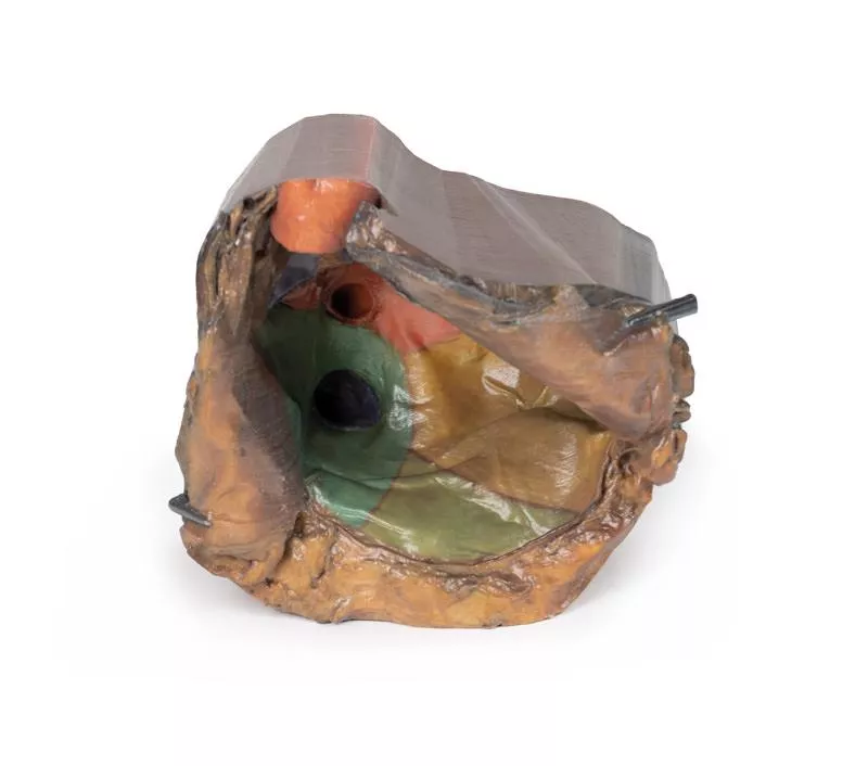
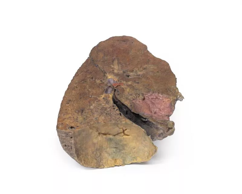
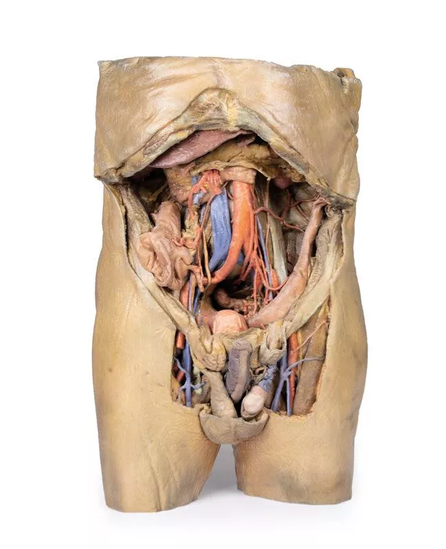
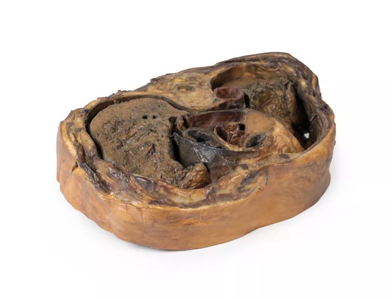
Product information "Right lung, hilum removed"
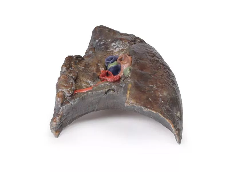
This 3D model provides a detailed sectional view of the right lung, serving as a complementary piece to the TW 63 Right Lung Hilum and contrasting with the TW 61 Left Lung Section.
It highlights the macrostructure of the lung from apex to base, offering a valuable perspective for anatomical study and comparison between lung sides.
Key Features:
Lobar Organization:
- Clearly defined oblique and horizontal fissures segment the lung into the superior, middle, and inferior lobes.
- The depth of these fissures is visible, showing their extension into the internal lung structure.
Surface Impressions:
- Prominent rib impressions run longitudinally from the apex to the base on the lateral aspect, indicating close contact with the thoracic cage.
Diaphragmatic Surface:
- The deeply concave base reflects the domed shape of the right diaphragm, which is elevated in life due to the position of the underlying liver.
Erler-Zimmer
Erler-Zimmer GmbH & Co.KG
Hauptstrasse 27
77886 Lauf
Germany
info@erler-zimmer.de
Achtung! Medizinisches Ausbildungsmaterial, kein Spielzeug. Nicht geeignet für Personen unter 14 Jahren.
Attention! Medical training material, not a toy. Not suitable for persons under 14 years of age.


