


Product information "Pericardial space"
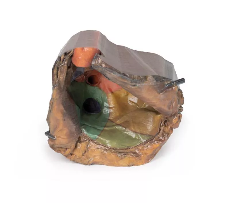
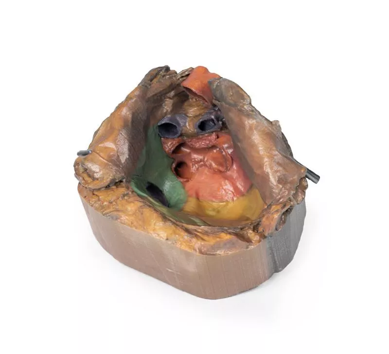
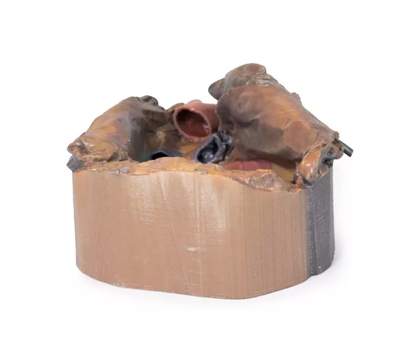
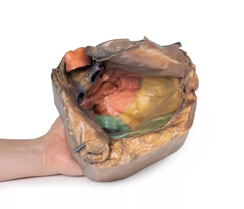
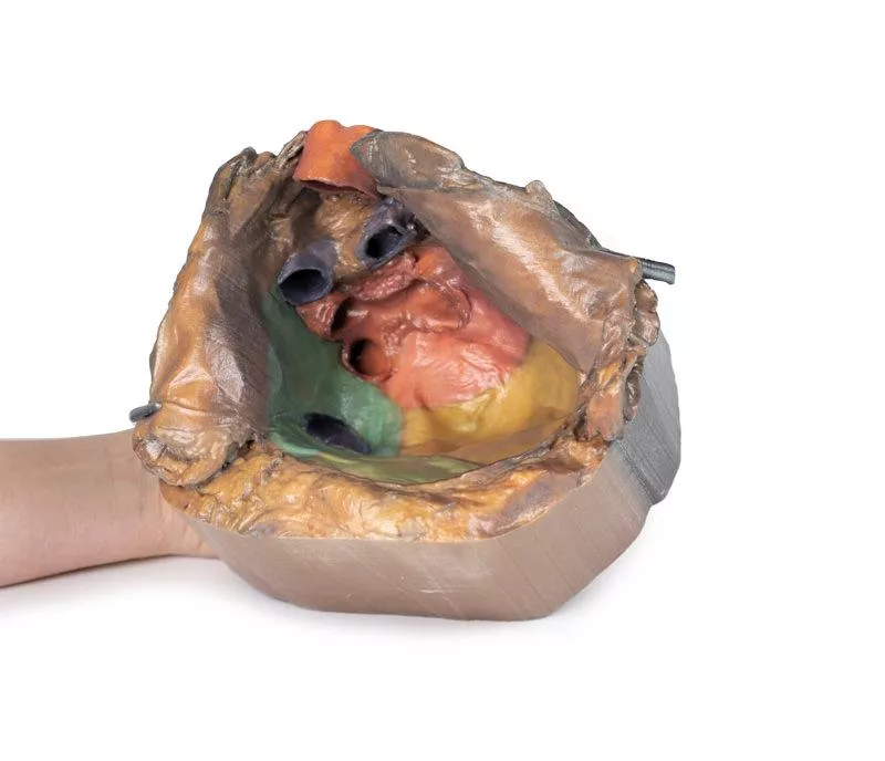
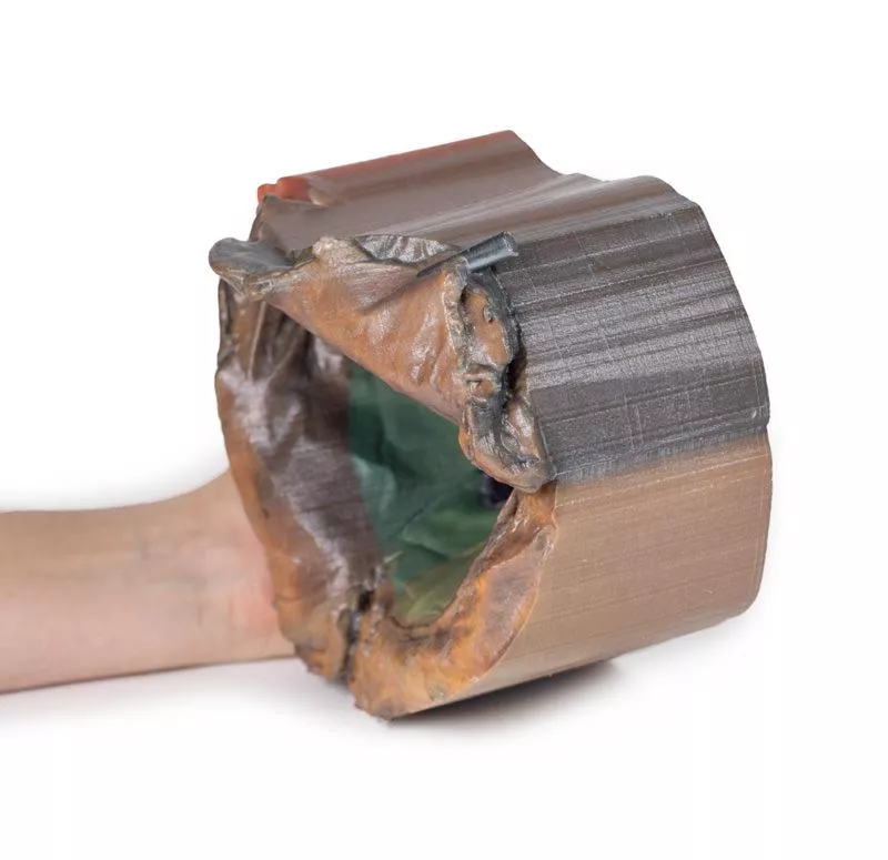
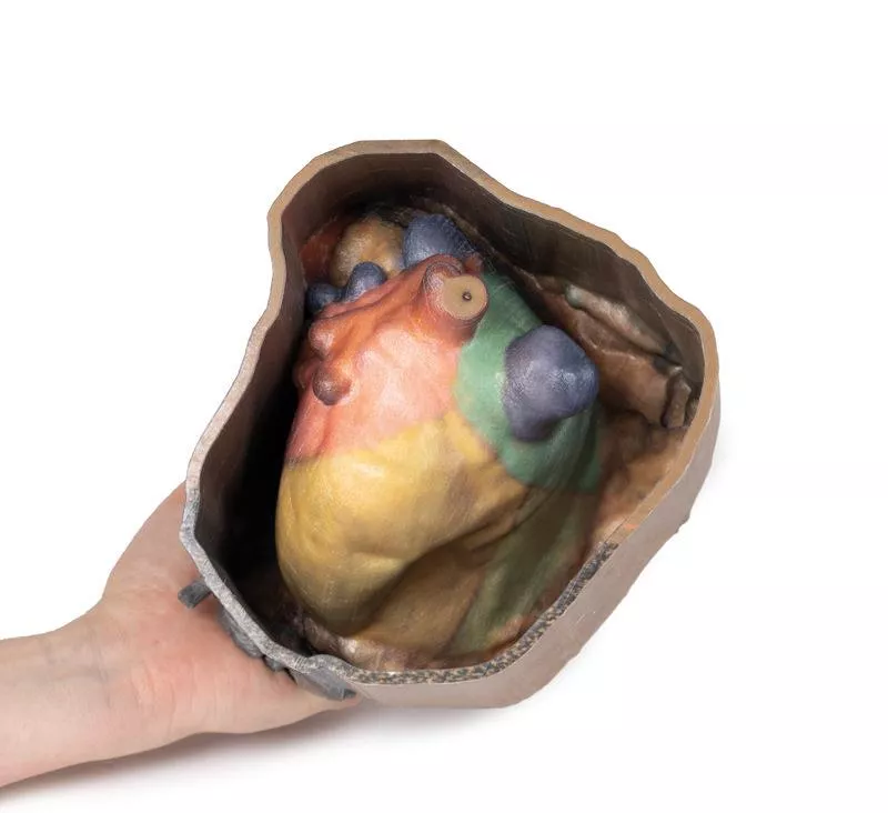
This detailed 3D anatomical model displays the pericardial cavity and reflections with the heart removed, allowing clear visualization of key structures within the middle mediastinum.
Key Features:
Pericardium:
Shows the full extent of the parietal pericardium, continuous with the visceral layer (epicardium), and false-colored to indicate the positions of the atria, ventricles, and great vessels.
Mediastinal Landmarks:
Highlights the base, apex, diaphragmatic, and pulmonary surfaces of the heart by their impressions within the pericardial cavity.
Great Vessels:
- Aorta (ascending, arch, and descending)
- Superior and Inferior Vena Cava
- Pulmonary trunk and arteries
- Pulmonary veins (4 total) - All shown in their natural positions relative to the pericardium.
Pericardial Sinuses:
- Transverse sinus: Located between arteries and veins; relevant for surgical access.
- Oblique sinus: Posterior recess between pulmonary veins.
Educational Use:
- Ideal for teaching thoracic and cardiac anatomy, including pericardial reflections, mediastinal relationships, and surgical landmarks.
- Useful in medical training, surgical planning, and radiological orientation.
Login
Erler-Zimmer
Erler-Zimmer Medical GmbH
Hauptstrasse 27
77886 Lauf
Germany
info@erler-zimmer.de
Achtung! Medizinisches Ausbildungsmaterial, kein Spielzeug. Nicht geeignet für Personen unter 14 Jahren.
Attention! Medical training material, not a toy. Not suitable for persons under 14 years of age.
Other customers also bought

Hilum of the left lung
This high-resolution 3D model offers a detailed view of the left lung hilum and associated structures, sectioned sagittally through the cardiac notch. It clearly demonstrates the relationships between the bronchi, pulmonary vessels, pleura, and supporting structures, making it ideal for advanced anatomical education.Key Features:Hilum Anatomy:- Entry/exit point for the pulmonary artery, superior and inferior pulmonary veins, main bronchus, lymphatics, and nerves.- Shows the meeting point of visceral and parietal pleura, forming the pulmonary ligament—the lung’s sole anatomical connection to the body. Pulmonary Circulation:- Pulmonary artery (superior in position) carries deoxygenated blood from the heart.- Pulmonary veins (anterior and inferior) return oxygenated blood to the heart.Bronchial Structure:Left main bronchus and its lobar branches are visible, located posteriorly within the hilum.Additional Views:- Oblique fissure along the lateral lung surface.- Diaphragmatic surface at the base; costal visceral surface posteriorly.- Pulmonary lymph nodes surrounding the hilum, both medially and laterally.

Hilum of the right lung
This high-quality 3D model presents a sagittal section of the right lung, focused on the hilum, where key anatomical structures enter and exit the lung. It serves as an essential tool for understanding pulmonary vascular and bronchial anatomy, with clear orientation from apex to base and medial to lateral surfaces.Key Features:Hilum Structure:- The hilum marks the transition between visceral and parietal pleura and serves as the lung’s only anatomical connection to the body via the pulmonary ligament.- Major structures entering the lung at this point include:- Pulmonary artery (superior in the hilum) – carrying deoxygenated blood from the heart.- Superior and inferior pulmonary veins (anterior and inferior) – returning oxygenated blood to the heart.- Right main bronchus and its lobar branches – located posteriorly in the hilum. - Associated nerves and lymphatics. Visible Anatomical Landmarks:- Cardiac impression (formed by the right atrium) is visible just anterior to the hilum.- The oesophageal groove is preserved along the posterior surface, tracing the path of the descending oesophagus.- Oblique and horizontal fissures are well-defined on the lateral surface, demarcating the lung's three lobes.- Hilar lymph nodes are observed around the medial hilum.Lung Surfaces:- Diaphragmatic surface (inferior), showing the concave interface with the diaphragm.- Costal visceral surface (posterior), where the lung contacts the thoracic wall.

Parotid Gland and Facial Nerve dissection
This 3D model presents a detailed superficial dissection of the lateral face, focusing on the parotid gland and its anatomical relationship to key neurovascular structures and surface landmarks. It serves as a valuable reference for clinicians involved in Mohs surgery, skin cancer treatment, and plastic or reconstructive facial procedures.Key Features:Dissection WindowThe exposed region extends from just anterior to the external ear, bounded superiorly by the zygomatic arch and inferiorly by the angle of the mandible, covering the area between the masseter and the sternocleidomastoid muscle. Parotid Gland & Facial Nerve- The parotid gland is fully exposed, with superior portions removed to reveal:- The superficial temporal artery- The facial nerve, dividing into its temporal, zygomatic, and buccal branches- The parotid duct is visible as it traverses the dissection field toward its termination in the buccinator muscleAssociated NervesA branch of the great auricular nerve is preserved along the inferior and posterior margins of the gland, running anterior to the sternocleidomastoid

Superficial Facial nerves & Parotid Gland
This 3D model provides a detailed view of the superficial anatomy of the face and head, expanding upon our HW 44 model with a broader dissection of the scalp, occipital region, and areas below the external ear.Key Features:Extended Facial AnatomyIncludes the terminal branches of the facial nerve (CN VII) traced from the parotid gland, with the platysma muscle preserved and extending from the mandible to the neck. Enhanced Posterior Dissection- Broader exposure across the posterior scalp and occipital region- Includes the retromandibular vein, great auricular nerve, and lesser occipital nerve- Shows the course of the occipital artery and vein near the trapeziusNeurovascular HighlightsImproved visualization of the supraorbital, supratrochlear, and superficial temporal arteries and nervesMusculaturePreserves fibers of the auricularis and occipitalis muscles, integrated into the epicranius (occipitofrontalis)

Superficial Face
This detailed 3D model features a superficial dissection of the left face just anterior to the ear, with false colouring highlighting key neurovascular structures and muscles of facial expression. It serves as a focused complement to the broader dissection in our HW 45 model. Undissected areas have been digitally removed for clarity.Key Features:Parotid Region & Facial Nerve Branches:Exposes the parotid gland and duct, along with terminal branches of the facial nerve (CN VII): cervical, mandibular, buccal, zygomatic, and temporal. Facial Vessels & Nerve-Vessel Relationships:Shows the facial artery and vein in relation to CN VII branches. Vessels are traced from the mandible to the orbit, offering anatomical landmarks.Muscles of Facial Expression (Highlighted):Includes masseter, depressor anguli oris, zygomaticus major & minor, orbicularis oris, nasalis, levator labii superioris alaeque nasi, procerus, and orbicularis oculi.Temporal & Forehead Structures:Displays the auriculotemporal nerve and superficial temporal artery over the temporal fascia, with part of the temporalis muscle visible.Superiorly, the supraorbital nerve and vessels ascend on the epicranial aponeurosis, overlying the frontalis muscle.This model offers a concise yet richly detailed view of facial anatomy, ideal for teaching nerve-muscle-vascular relationships in the superficial face.

Thorax with heart and vessels
This highly detailed 3D model depicts the key anatomy of the superior thoracic aperture, mediastinum, and adjacent neck and thoracic structures, with both clavicles and select muscular and venous elements removed to enhance visibility and educational impact.Anatomical Highlights:Superior Thoracic Aperture:- Trachea visible at the top, with a robust ring of cartilage.- Rib 1 exposed from lateral to medial, including insertion of the anterior scalene muscle.- Removal of clavicles allows unobstructed views into the upper thoracic corridor. Vascular Anatomy:- Right subclavian artery, situated above rib 1, gives rise to the thyrocervical trunk.- Left subclavian artery, also above rib 1, branches into the suprascapular artery.- Both common carotid arteries are visible; the left carotid sheath includes the left vagus nerve.Nervous System:- Left vagus nerve follows the left carotid artery within the carotid sheath; left recurrent laryngeal nerve loops under the aorta.- Right vagus nerve and right phrenic nerve retracted during dissection; left phrenic nerve remains anterior to the heart, tracing to the diaphragm.- Elements of the left brachial plexus are visible—from roots to trunks—including the dorsal scapular nerve.Mediastinal & Cardiac Orientation:- Arch of the aorta, with brachiocephalic trunk, left common carotid, and left subclavian artery, sits just above the heart.- Pulmonary trunk emerges immediately superior to the heart.- Left anterior descending artery (LAD) courses along the anterior heart surface.- Superior vena cava lies to the right and posterior to the ascending aorta.- Right phrenic nerve positioned posteriorly to the heart; left phrenic nerve runs in its connective tissue anteriorly.Inferior Thorax & Diaphragm:- Ribs 8–12 and associated external intercostal musculature visible; muscle fibers run inferomedially into fascial layers.- Right hemidiaphragm sits higher than the left, reflecting the presence of the liver beneath. Applications & BenefitsClinical Relevance:Ideal for visualizing neck and thoracic anatomy relevant to cardiovascular, respiratory, and nerve-related procedures, including laryngeal, thoracic, and brachial plexus surgery.Educational Utility: Offers a clean, accessible view of mediastinal compartments, vascular pathways, and nerve trajectories without obstruction by bone or superficial structures. Enhanced Learning: The model’s clarity supports instruction in radiographic interpretation, thoracic surgical approaches, and anatomy exams.

Continuous innovation

Social responsibility

Active customer orientation

Understanding quality

Sustainable actions

ISO 9001 certification