


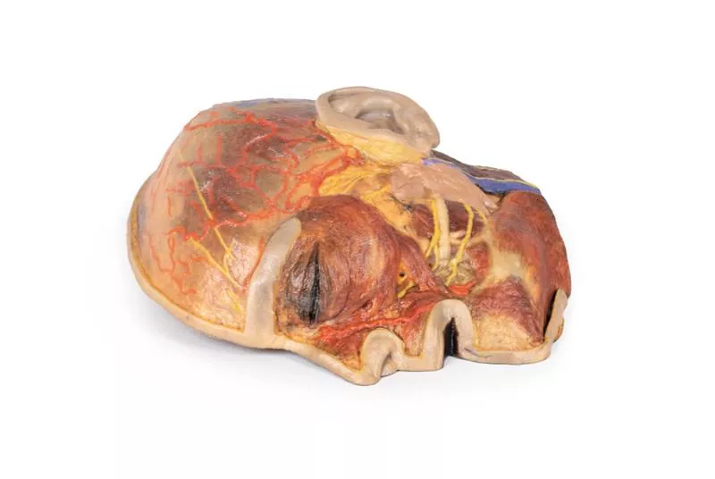


Product information "Superficial Facial nerves & Parotid Gland"
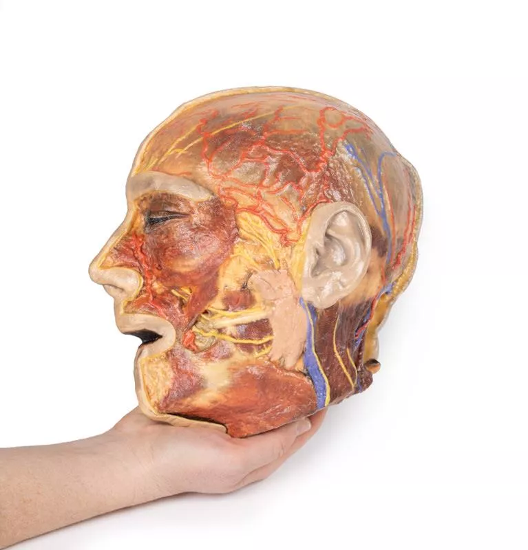
This 3D model provides a detailed view of the superficial anatomy of the face and head, expanding upon our HW 44 model with a broader dissection of the scalp, occipital region, and areas below the external ear.
Key Features:
Extended Facial Anatomy
Includes the terminal branches of the facial nerve (CN VII) traced from the parotid gland, with the platysma muscle preserved and extending from the mandible to the neck.
Enhanced Posterior Dissection
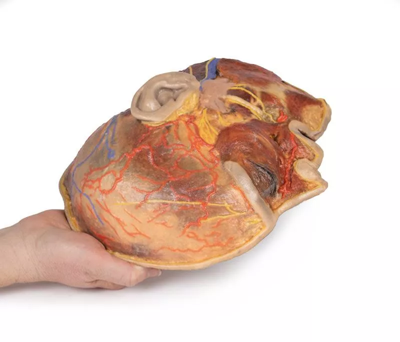
- Broader exposure across the posterior scalp and occipital region
- Includes the retromandibular vein, great auricular nerve, and lesser occipital nerve
- Shows the course of the occipital artery and vein near the trapezius
Neurovascular Highlights
Improved visualization of the supraorbital, supratrochlear, and superficial temporal arteries and nerves
Musculature
Preserves fibers of the auricularis and occipitalis muscles, integrated into the epicranius (occipitofrontalis)
Key Features:
Extended Facial Anatomy
Includes the terminal branches of the facial nerve (CN VII) traced from the parotid gland, with the platysma muscle preserved and extending from the mandible to the neck.
Enhanced Posterior Dissection
- Broader exposure across the posterior scalp and occipital region
- Includes the retromandibular vein, great auricular nerve, and lesser occipital nerve
- Shows the course of the occipital artery and vein near the trapezius
Neurovascular Highlights
Improved visualization of the supraorbital, supratrochlear, and superficial temporal arteries and nerves
Musculature
Preserves fibers of the auricularis and occipitalis muscles, integrated into the epicranius (occipitofrontalis)
Login
Erler-Zimmer
Erler-Zimmer Medical GmbH
Hauptstrasse 27
77886 Lauf
Germany
info@erler-zimmer.de
Achtung! Medizinisches Ausbildungsmaterial, kein Spielzeug. Nicht geeignet für Personen unter 14 Jahren.
Attention! Medical training material, not a toy. Not suitable for persons under 14 years of age.
Other customers also bought

Sinus Pathways
This 3D model shows a midsagittal to parasagittal section of the right head, highlighting the drainage pathways of the paranasal sinuses into the nasal cavity using colored markers. Anteriorly, the nasolacrimal duct (white) opens beneath the inferior nasal concha. The middle concha has been sectioned to reveal the maxillary sinus opening via the semilunar hiatus (green), and the drainage of the frontal sinus (blue), anterior (orange) and middle (yellow) ethmoidal cells. Posterior ethmoidal cells open into the superior meatus (purple), while the sphenoid sinus drains above the nasopharynx (red).The model also preserves key anatomical structures:The nasal cavity from nostril to auditory tube, the soft palate, uvula, and pharynx down to the epiglottis. The oral cavity is sectioned to show genioglossus and geniohyoid muscles. Within the cranial cavity, parts of the frontal lobe, optic structures, and pituitary gland are visible, along with the brainstem, cerebellum, and tentorium cerebelli. The transverse and sigmoid sinuses are seen bilaterally, with a portion of the medial temporal lobe and lateral ventricle also present.

Sagittal Section of Head and Neck with Infratemporal Fossa and Carotid Sheath Dissection
This 3D model complements the H11 and H12 head and neck specimens, offering a clear view of the endocranial cavity without the brain, alongside a lateral dissection of the face, infratemporal region, and neck.Key Features:Endocranial Cavity- Brain removed; dura mater, tentorium cerebelli, and superior sagittal sinus fully visible- Several cranial nerves (CN II, III, V, VI, VII, VIII) seen piercing the dura- Pituitary gland preserved in sella turcica, left vertebral artery visible in posterior fossa Lateral Facial & Infratemporal Region- Facial artery and vein retained, dissected free of superficial tissues- Partial removal of mandible and zygomatic arch reveals:- Inferior alveolar and lingual nerves, posterior deep temporal artery, and TMJ articulation- Visible branches of the external carotid artery, including maxillary and superficial temporal arteriesNeck Anatomy- Facial nerve (CN VII) near posterior belly of digastric- Dissected carotid sheath showing internal/external carotid arteries, internal jugular vein, and vagus nerve (CN X)- Hypoglossal nerve (CN XII) and facial artery near submandibular gland- Hyoid bone, thyroid gland, and larynx visible- Cervical plexus branches on scalene muscles; brachial plexus roots preserved near internal jugular vein

Upper Limb Ligaments
This 3D printed model presents the entire upper limb skeleton, from the pectoral girdle to the hand, with detailed ligamentous anatomy and select tendon and muscle insertions.Shoulder and Pectoral GirdleVisible ligaments include the acromioclavicular, coracoclavicular, coracoacromial, and superior transverse scapular ligament. A small portion of the supraspinatus muscle and tendon demonstrates its path beneath the coracoacromial ligament. Key preserved structures:- Subscapularis tendon (reflected to expose the glenohumeral capsule)- Tendons of teres major, latissimus dorsi, and long head of triceps- Long head of biceps tendon within the intertubercular groove and capsule Elbow JointThe elbow capsule is opened to show the articulation between the humerus, radius, and ulna.Preserved:- Ulnar and radial collateral ligaments- Anular ligament of radius- Biceps tendon insertion on the radial tuberosity Wrist and HandDistally, the model includes key wrist ligaments:- Palmar/dorsal radiocarpal and ulnocarpal ligaments- Radial/ulnar collateral ligaments, pisohamate, carpometacarpal, and othersIn the hand:- MCP and IP joint capsules with collateral ligaments and volar plates- Flexor digitorum superficialis/profundus and flexor pollicis longus tendons are preserved and shown at their insertions

Liver with vessels and gallbladder
This liver specimen displays notable differences compared to a typical liver. It is less wedge-shaped and elongated in the superoinferior dimension, resulting in a greater vertical height when viewed from the posterior. Size- Measures approximately 18 cm along the midclavicular line.- Typical livers measure under 16 cm in this dimension.- The increased length suggests mild hepatomegaly (enlargement). Important Notes- Size estimates may be affected by specimen preservation and fixing, which can cause some distortion.- Diagnosing hepatomegaly based on a single measurement is limited and varies with individual anatomy, measurement technique, sex, and body mass index (BMI). Anatomical VariationsThis specimen does not match common anatomical variations often confused with hepatomegaly such as:- Riedel’s lobe: a downward projection of the right lobe- Beaver tail liver: elongated left lobe- Papillary process from the caudate lobe

Abdomen with inguinal hernia
Diaphragm and Xyphoid ProcessThe diaphragm has been secured to the superior border of the dissected specimen with sutures to ensure an unobstructed view of the abdomen. The xyphoid process is in the middle of this sutured border. Liver and GallbladderThe liver in the right hypochondrium is pushed laterally to reveal the kidney behind it. The falciform ligament divides the liver’s right and left lobes and contains the ligamentum teres, a remnant of the fetal umbilical vein. Below the ligamentum teres, the gallbladder sits between the liver lobes. Stomach and Splenic VasculatureThe deflated stomach is pushed up to reveal the twisted splenic artery and vein, which branch into the spleen’s hilum.Spleen and PancreasThe spleen sits in the left hypochondrium, with its gastric impression marking the stomach’s greater curvature. The pancreas tail, intraperitoneal and fused to the spleen’s hilum, lies near its inferior pole. KidneysThe kidneys are mostly retroperitoneal, but the covering peritoneum is removed in this specimen. Normally, the right kidney lies lower due to the liver, but here it is smaller and higher than the left. The left kidney is enlarged, with two accessory renal arteries from the aorta supplying its hilum and lower pole.Adrenal GlandsThe left adrenal gland is detached from its usual position on the superior pole of the kidney. The middle adrenal artery originates directly from the aorta, left of the coeliac trunk, whilst the inferior adrenal artery is derived from the left renal artery: both supply the adrenal gland. The superior adrenal artery has been obscured by connective tissue.Rectum and BladderAlthough the majority of the peritoneum in the abdomen has been removed below the level of the sacral prominence (S1), a layer of peritoneum remains intact, which overlays the rectum and bladder. Notably this the first part of the rectum, which is intraperitoneal. Gastrointestinal TractThe final part of the ascending duodenum and the descending colon at the left colic flexure has been ligated with twine, with the intestine in between being removed in order to provide a better view of the abdomen. Pelvic RegionIn this specimen, the sigmoid colon has herniated indirectly through the inguinal canal. On the right, the vas deferens exits the superficial inguinal ring toward the right testicle, while other spermatic cord contents are removed. Sutures below the vas deferens mark the embalming entry via the right femoral artery.Abdominal VasculatureThe coeliac trunk, located just below the stomach, typically branches into the left gastric, splenic, and common hepatic arteries to supply the foregut.In this 3D model, the coeliac trunk gives off right and left gastric arteries, the splenic artery, and a gastroduodenal branch that splits into two superior pancreaticoduodenal arteries. The proper hepatic artery arises independently from the abdominal aorta and gives off the right inferior phrenic artery. The iliolumbar artery emerges deep to the right psoas, connecting with branches of the right deep circumflex iliac artery along the iliac crest.

Head, Neck and Shoulder with angiosomes
This large, multipart 3D printed specimen showcases detailed anatomy of the head, neck, thorax, axillae, and upper limbs.Head and Neck:The calotte has been removed ~2?cm above the orbits to expose the brain and endocranial cavity. A transverse cerebral section shows grey and white matter, lateral ventricles, and choroid plexus. The right side retains skin and fascia, false-coloured to highlight facial and neck angiosomes. The left side reveals facial expression and mastication muscles, and infratemporal structures including the lingual nerve and terminal branches of the external carotid artery. The carotid sheaths are opened bilaterally, exposing the common, internal, and external carotid arteries, and vagus nerves. The sternocleidomastoid and internal jugular veins are mostly removed. On the right, the great auricular and hypoglossal nerves are visible, along with the stylohyoid ligament and supra-/infrahyoid muscles. The thyroid gland is prominent, with preserved superior and inferior thyroid vessels.Root of the Neck and Axilla:On the left, partial clavicle removal reveals the first rib, anterior scalene, and brachial plexus roots (C5–T1) forming trunks between scalene muscles. The subclavian artery passes posterior to scalenus anterior, transitioning to the axillary artery, closely related to the brachial plexus cords.The left axilla displays brachial plexus divisions and cords. The formation of the median nerve around the axillary artery is distinct. The ulnar, musculocutaneous, axillary, thoracodorsal, and long thoracic nerves are clearly identified with their courses and muscular targets.On the right, the clavicle and subclavius muscle are intact, showing the cervico-axillary canal. Pectoralis major and minor have been reflected, exposing deeper structures.Thorax:A window in the left thoracic wall reveals the mediastinum. The left lung has been removed. Intercostal spaces are visible beneath the parietal pleura; neurovascular bundles are identifiable posteriorly. The heart is exposed without pericardium, showing the left atrium and ventricle, pulmonary vessels, aorta, and both left vagus and recurrent laryngeal nerves. The right thoracic wall remains intact, displaying intercostal and upper limb muscles. From below, the right lung, pleural cavities, and diaphragmatic heart surface are visible. Posterior thoracic skin and fascia are intact, showing cutaneous nerve distribution.Size: 50 x 20 x 41 cm

Parasagittal Section of the head and neck
This high-resolution 3D model features a head and neck specimen sectioned just off the midsagittal plane, preserving critical midline structures often absent in similar models. Ideal for anatomical education, this model offers enhanced visibility due to fixative-induced brain shrinkage, exaggerating spaces between brain and skull.Key Features:Midline Anatomy Preserved:Includes the falx cerebri (anterior portion), septum pellucidum, interventricular foramen (of Monro), and nasal septum.Ventricular & Endocranial Structures:Clear views of the lateral and third ventricles, cerebral aqueduct, fourth ventricle, infundibulum, pituitary gland, and sphenoid sinus.Vascular Highlights:Displays the left vertebral artery, posterior cerebral artery (cross-section), and anterior cerebral artery branches around the corpus callosum.Detailed Nasal & Pharyngeal Regions:Shows relationships between the nasal septum, palate, auditory tube opening, and naso-/oropharynx.Laryngeal and Tracheal Anatomy:Includes epiglottis, arytenoid, thyroid cartilages, hyoid bone, and cross-sectional views of the vestibule, vestibular and vocal folds.This model provides a unique perspective of internal head and neck anatomy, combining anatomical depth with high educational value.

Superficial Face
This detailed 3D model features a superficial dissection of the left face just anterior to the ear, with false colouring highlighting key neurovascular structures and muscles of facial expression. It serves as a focused complement to the broader dissection in our HW 45 model. Undissected areas have been digitally removed for clarity.Key Features:Parotid Region & Facial Nerve Branches:Exposes the parotid gland and duct, along with terminal branches of the facial nerve (CN VII): cervical, mandibular, buccal, zygomatic, and temporal. Facial Vessels & Nerve-Vessel Relationships:Shows the facial artery and vein in relation to CN VII branches. Vessels are traced from the mandible to the orbit, offering anatomical landmarks.Muscles of Facial Expression (Highlighted):Includes masseter, depressor anguli oris, zygomaticus major & minor, orbicularis oris, nasalis, levator labii superioris alaeque nasi, procerus, and orbicularis oculi.Temporal & Forehead Structures:Displays the auriculotemporal nerve and superficial temporal artery over the temporal fascia, with part of the temporalis muscle visible.Superiorly, the supraorbital nerve and vessels ascend on the epicranial aponeurosis, overlying the frontalis muscle.This model offers a concise yet richly detailed view of facial anatomy, ideal for teaching nerve-muscle-vascular relationships in the superficial face.

Circle of Willis
This highly detailed 3D printed model offers a precise representation of the intracranial arteries supplying the human brain, created from segmented angiographic imaging data. The model displays vascular structures that cannot be physically dissected and viewed in situ, making it a unique and valuable teaching tool.Vertebral and Basilar ArteriesThe paired vertebral arteries are shown entering the cranial cavity through the foramen magnum, where they unite to form the basilar artery. The basilar artery travels along the brainstem and terminates by branching into the posterior cerebral arteries. Just before this bifurcation, the superior cerebellar arteries arise, supplying parts of the cerebellum and midbrain. Internal Carotid Arteries and Carotid SiphonThe internal carotid arteries (ICAs) are traced from their entry at the carotid canal within the temporal bone. From there, they course medially and anteriorly through the petrous portion of the bone and emerge via the upper opening of the foramen lacerum. While the cavernous sinus is not shown, the model beautifully captures the characteristic S-shaped carotid siphon on both sides, clearly positioned lateral to the sella turcica and passing medial to the anterior clinoid processes. Major Cerebral Arteries and the Circle of WillisEach internal carotid artery divides into the anterior cerebral artery (ACA) and middle cerebral artery (MCA). The posterior communicating arteries, connecting the posterior cerebral arteries (PCAs) to the MCAs, are distinctly visible, allowing for an excellent demonstration of the posterior portion of the Circle of Willis.The anterior communicating artery, completing the circle between the paired anterior cerebral arteries, is also present—though its visibility is limited due to the close proximity of the ACAs.

Continuous innovation

Social responsibility

Active customer orientation

Understanding quality

Sustainable actions

ISO 9001 certification