


Product information "Superficial Face"
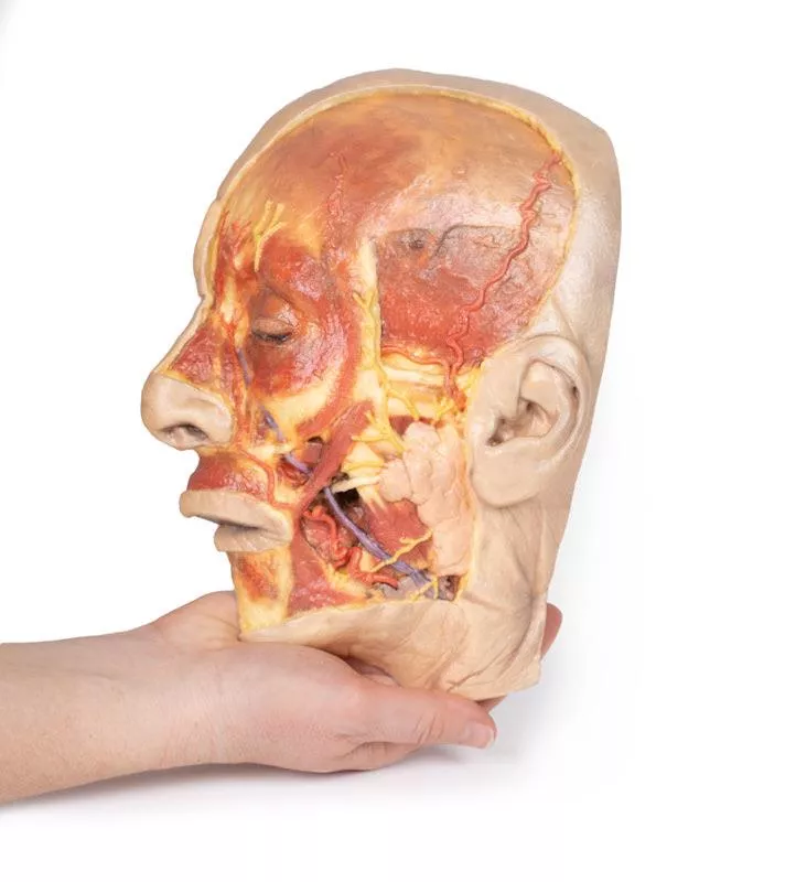
This detailed 3D model features a superficial dissection of the left face just anterior to the ear, with false colouring highlighting key neurovascular structures and muscles of facial expression.
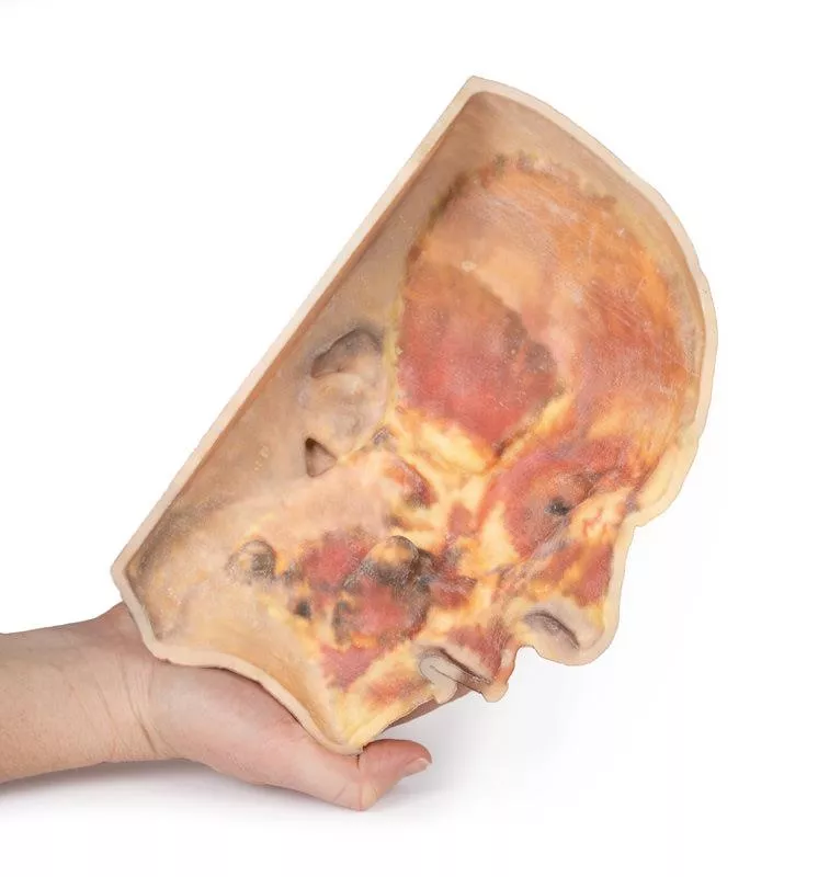
It serves as a focused complement to the broader dissection in our HW 45 model. Undissected areas have been digitally removed for clarity.
Key Features:
Parotid Region & Facial Nerve Branches:
Exposes the parotid gland and duct, along with terminal branches of the facial nerve (CN VII): cervical, mandibular, buccal, zygomatic, and temporal.
Facial Vessels & Nerve-Vessel Relationships:
Shows the facial artery and vein in relation to CN VII branches. Vessels are traced from the mandible to the orbit, offering anatomical landmarks.
Muscles of Facial Expression (Highlighted):
Includes masseter, depressor anguli oris, zygomaticus major & minor, orbicularis oris, nasalis, levator labii superioris alaeque nasi, procerus, and orbicularis oculi.
Temporal & Forehead Structures:
Displays the auriculotemporal nerve and superficial temporal artery over the temporal fascia, with part of the temporalis muscle visible.
Superiorly, the supraorbital nerve and vessels ascend on the epicranial aponeurosis, overlying the frontalis muscle.
This model offers a concise yet richly detailed view of facial anatomy, ideal for teaching nerve-muscle-vascular relationships in the superficial face.
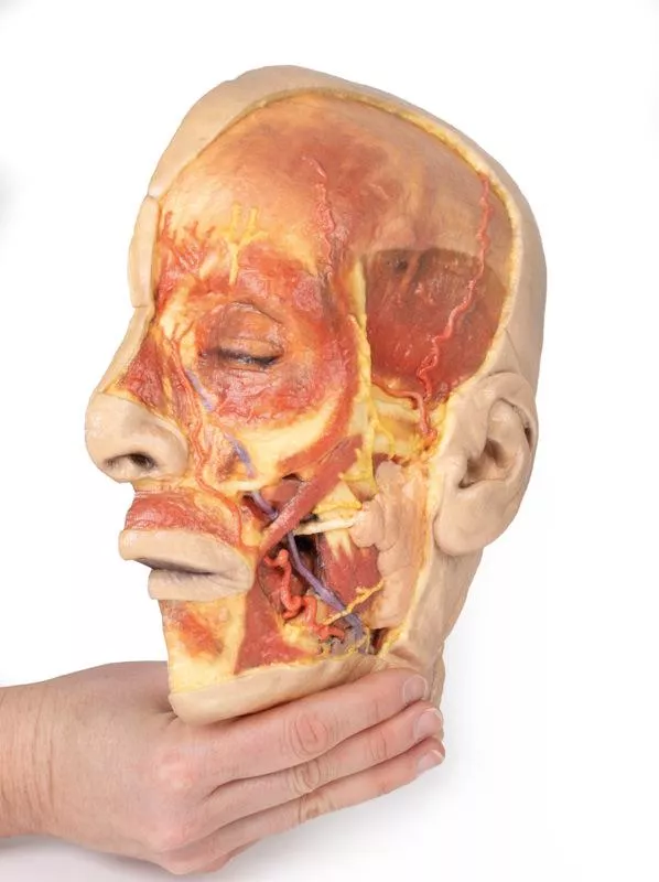
Login
Erler-Zimmer
Erler-Zimmer Medical GmbH
Hauptstrasse 27
77886 Lauf
Germany
info@erler-zimmer.de
Achtung! Medizinisches Ausbildungsmaterial, kein Spielzeug. Nicht geeignet für Personen unter 14 Jahren.
Attention! Medical training material, not a toy. Not suitable for persons under 14 years of age.
Other customers also bought

Liver with vessels and gallbladder
This liver specimen displays notable differences compared to a typical liver. It is less wedge-shaped and elongated in the superoinferior dimension, resulting in a greater vertical height when viewed from the posterior. Size- Measures approximately 18 cm along the midclavicular line.- Typical livers measure under 16 cm in this dimension.- The increased length suggests mild hepatomegaly (enlargement). Important Notes- Size estimates may be affected by specimen preservation and fixing, which can cause some distortion.- Diagnosing hepatomegaly based on a single measurement is limited and varies with individual anatomy, measurement technique, sex, and body mass index (BMI). Anatomical VariationsThis specimen does not match common anatomical variations often confused with hepatomegaly such as:- Riedel’s lobe: a downward projection of the right lobe- Beaver tail liver: elongated left lobe- Papillary process from the caudate lobe

Sinus Pathways
This 3D model shows a midsagittal to parasagittal section of the right head, highlighting the drainage pathways of the paranasal sinuses into the nasal cavity using colored markers. Anteriorly, the nasolacrimal duct (white) opens beneath the inferior nasal concha. The middle concha has been sectioned to reveal the maxillary sinus opening via the semilunar hiatus (green), and the drainage of the frontal sinus (blue), anterior (orange) and middle (yellow) ethmoidal cells. Posterior ethmoidal cells open into the superior meatus (purple), while the sphenoid sinus drains above the nasopharynx (red).The model also preserves key anatomical structures:The nasal cavity from nostril to auditory tube, the soft palate, uvula, and pharynx down to the epiglottis. The oral cavity is sectioned to show genioglossus and geniohyoid muscles. Within the cranial cavity, parts of the frontal lobe, optic structures, and pituitary gland are visible, along with the brainstem, cerebellum, and tentorium cerebelli. The transverse and sigmoid sinuses are seen bilaterally, with a portion of the medial temporal lobe and lateral ventricle also present.

Sagittal Section of Head and Neck with Infratemporal Fossa and Carotid Sheath Dissection
This 3D model complements the H11 and H12 head and neck specimens, offering a clear view of the endocranial cavity without the brain, alongside a lateral dissection of the face, infratemporal region, and neck.Key Features:Endocranial Cavity- Brain removed; dura mater, tentorium cerebelli, and superior sagittal sinus fully visible- Several cranial nerves (CN II, III, V, VI, VII, VIII) seen piercing the dura- Pituitary gland preserved in sella turcica, left vertebral artery visible in posterior fossa Lateral Facial & Infratemporal Region- Facial artery and vein retained, dissected free of superficial tissues- Partial removal of mandible and zygomatic arch reveals:- Inferior alveolar and lingual nerves, posterior deep temporal artery, and TMJ articulation- Visible branches of the external carotid artery, including maxillary and superficial temporal arteriesNeck Anatomy- Facial nerve (CN VII) near posterior belly of digastric- Dissected carotid sheath showing internal/external carotid arteries, internal jugular vein, and vagus nerve (CN X)- Hypoglossal nerve (CN XII) and facial artery near submandibular gland- Hyoid bone, thyroid gland, and larynx visible- Cervical plexus branches on scalene muscles; brachial plexus roots preserved near internal jugular vein

Superficial Facial nerves & Parotid Gland
This 3D model provides a detailed view of the superficial anatomy of the face and head, expanding upon our HW 44 model with a broader dissection of the scalp, occipital region, and areas below the external ear.Key Features:Extended Facial AnatomyIncludes the terminal branches of the facial nerve (CN VII) traced from the parotid gland, with the platysma muscle preserved and extending from the mandible to the neck. Enhanced Posterior Dissection- Broader exposure across the posterior scalp and occipital region- Includes the retromandibular vein, great auricular nerve, and lesser occipital nerve- Shows the course of the occipital artery and vein near the trapeziusNeurovascular HighlightsImproved visualization of the supraorbital, supratrochlear, and superficial temporal arteries and nervesMusculaturePreserves fibers of the auricularis and occipitalis muscles, integrated into the epicranius (occipitofrontalis)

Thoracic cross section at T6
This detailed 3D model presents a transverse cross-section of the thorax at the level of the T6 vertebra, offering a clear view of thoracic anatomy in relation to skeletal, vascular, respiratory, and cardiac structures.Key Features:Posterior Structures- Begins medially with the spinal cord within the vertebral canal- Costovertebral joints of the 6th ribs are visible, along with surrounding ribs forming the thoracic wall Anterior Thoracic Wall- Costosternal joints show the connection between ribs and sternumMajor Thoracic Organs- Oesophagus located anterior to the vertebral body- Descending aorta situated lateral to the vertebral bodyLungs and Pleural Cavities- Within the pleural spaces (lined by parietal pleura):- Right lung: middle and inferior lobes- Left lung: inferior lobeHeart and Mediastinum- In the middle mediastinum, the heart is shown within the pericardium, sectioned to display internal anatomy:- Left atrium (posterior)- Aortic valve, right ventricle, and right atrium in clockwise order

Upper Limb Ligaments
This 3D printed model presents the entire upper limb skeleton, from the pectoral girdle to the hand, with detailed ligamentous anatomy and select tendon and muscle insertions.Shoulder and Pectoral GirdleVisible ligaments include the acromioclavicular, coracoclavicular, coracoacromial, and superior transverse scapular ligament. A small portion of the supraspinatus muscle and tendon demonstrates its path beneath the coracoacromial ligament. Key preserved structures:- Subscapularis tendon (reflected to expose the glenohumeral capsule)- Tendons of teres major, latissimus dorsi, and long head of triceps- Long head of biceps tendon within the intertubercular groove and capsule Elbow JointThe elbow capsule is opened to show the articulation between the humerus, radius, and ulna.Preserved:- Ulnar and radial collateral ligaments- Anular ligament of radius- Biceps tendon insertion on the radial tuberosity Wrist and HandDistally, the model includes key wrist ligaments:- Palmar/dorsal radiocarpal and ulnocarpal ligaments- Radial/ulnar collateral ligaments, pisohamate, carpometacarpal, and othersIn the hand:- MCP and IP joint capsules with collateral ligaments and volar plates- Flexor digitorum superficialis/profundus and flexor pollicis longus tendons are preserved and shown at their insertions

Deep upper limb and hand
This 3D printed model presents a superficial dissection of the right distal arm, forearm, and hand, showcasing key vascular, nervous, and muscular anatomy.Distal Arm & Cubital FossaThe arrangement of the biceps tendon, brachial artery, and median nerve is visible from lateral to medial. The bicipital aponeurosis has been removed to expose deeper structures. The ulnar nerve is reflected from the cubital tunnel, and the radial nerve with its branches is visible near the supinator muscle. Forearm AnatomyOn the anterior forearm, superficial flexors (pronator teres, FCR, FDS, FCU) are preserved; palmaris longus is absent. The radial artery is exposed; the ulnar artery is not visible. Posteriorly, extensors from the common origin are visible, including ECRB, ED, EDM, and ECU. The APL, EPB, and EPL are also shown wrapping around the radius. Hand & SnuffboxThe anatomical snuffbox reveals the radial artery in its floor and cutaneous branches of the radial nerve. The palmar side displays thenar/hypothenar muscles, lumbricals, flexor tendons, and the median nerve beneath the flexor retinaculum. A superficial branch of the radial artery crosses the retinaculum.Ideal for anatomical education, this print offers a clear view of key structures in the distal upper limb.

Posterior Abdominal wall
This detailed 3D printed model presents the male posterior abdominal wall from the diaphragm down to the pelvic brim, including the pelvis and proximal thigh.A focused pelvic and thigh version is also available (MP1770).Muscular Anatomy & DiaphragmThe parietal peritoneum is removed to expose the key muscles of the posterior abdominal wall: psoas major, quadratus lumborum, transversus abdominis, and iliacus below the iliac crest. The diaphragm shows distinct muscular fibers originating from the thoracic cage and lumbar vertebrae (right crus L1–L3, left crus L1–L2), connected by the median arcuate ligament. Major diaphragmatic openings for the esophagus, aorta, and inferior vena cava are visible, although the aorta is removed. NervesSomatic nerves are clearly identifiable, including the subcostal, iliohypogastric, and ilioinguinal nerves (which arise together in this specimen), the lateral cutaneous nerve of the thigh, the genitofemoral nerve (on psoas), and the femoral nerve situated between psoas and iliacus. The sympathetic trunks run alongside the lumbar vertebrae. Vessels & KidneysThe aorta and inferior vena cava are transected at L3, with the aortic bifurcation positioned slightly higher than usual. The renal arteries and veins are preserved, though their full origin is partly obscured by the absence of the great vessels. Both kidneys are dissected free from surrounding fat, showing the typical lower position of the right kidney. The ureters are visible descending from the renal hilum, passing medial to the psoas before crossing the pelvic brim into the true pelvis. This model offers an exceptional view of the complex anatomy of the posterior abdominal wall, pelvis, and proximal thigh, making it ideal for advanced anatomical study, surgical planning, and clinical reference.

Continuous innovation

Social responsibility

Active customer orientation

Understanding quality

Sustainable actions

ISO 9001 certification