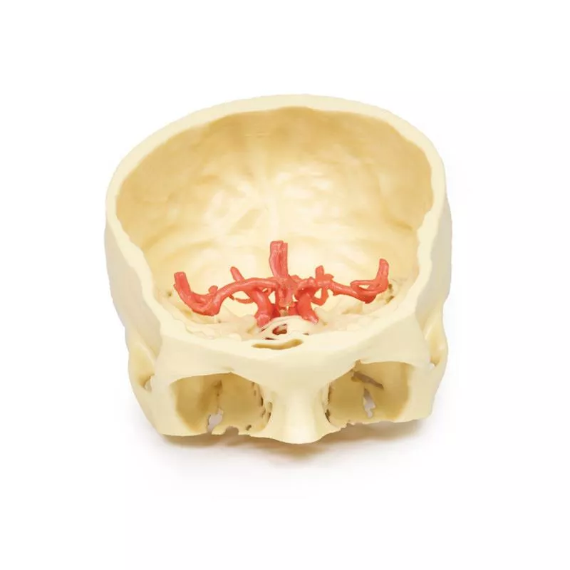


Sagittal Section of Head and Neck with Infratemporal Fossa and Carotid Sheath Dissection
Order for more €200.00 and you will receive your order free of shipping costs (valid for deliveries within Germany).
Product information "Sagittal Section of Head and Neck with Infratemporal Fossa and Carotid Sheath Dissection"
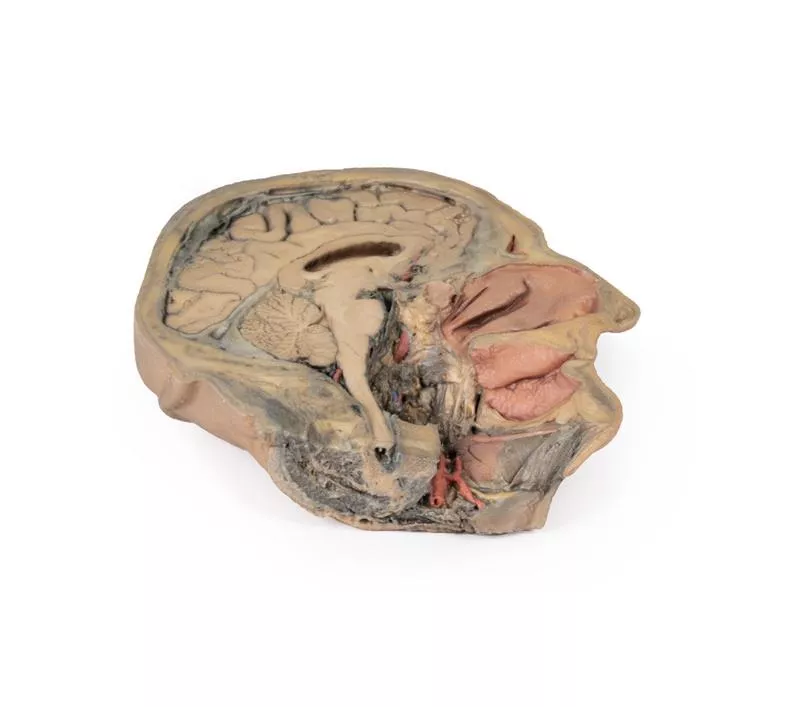
This 3D model complements the H11 and H12 head and neck specimens, offering a clear view of the endocranial cavity without the brain, alongside a lateral dissection of the face, infratemporal region, and neck.
Key Features:
Endocranial Cavity
- Brain removed; dura mater, tentorium cerebelli, and superior sagittal sinus fully visible
- Several cranial nerves (CN II, III, V, VI, VII, VIII) seen piercing the dura
- Pituitary gland preserved in sella turcica, left vertebral artery visible in posterior fossa
Lateral Facial & Infratemporal Region
- Facial artery and vein retained, dissected free of superficial tissues
- Partial removal of mandible and zygomatic arch reveals:
Neck Anatomy
- Facial nerve (CN VII) near posterior belly of digastric
- Dissected carotid sheath showing internal/external carotid arteries, internal jugular vein, and vagus nerve (CN X)
- Hypoglossal nerve (CN XII) and facial artery near submandibular gland
- Hyoid bone, thyroid gland, and larynx visible
- Cervical plexus branches on scalene muscles; brachial plexus roots preserved near internal jugular vein
Key Features:
Endocranial Cavity
- Brain removed; dura mater, tentorium cerebelli, and superior sagittal sinus fully visible
- Several cranial nerves (CN II, III, V, VI, VII, VIII) seen piercing the dura
- Pituitary gland preserved in sella turcica, left vertebral artery visible in posterior fossa
Lateral Facial & Infratemporal Region
- Facial artery and vein retained, dissected free of superficial tissues
- Partial removal of mandible and zygomatic arch reveals:
- Inferior alveolar and lingual nerves, posterior deep temporal artery, and TMJ articulation
- Visible branches of the external carotid artery, including maxillary and superficial temporal arteries
- Visible branches of the external carotid artery, including maxillary and superficial temporal arteries
Neck Anatomy
- Facial nerve (CN VII) near posterior belly of digastric
- Dissected carotid sheath showing internal/external carotid arteries, internal jugular vein, and vagus nerve (CN X)
- Hypoglossal nerve (CN XII) and facial artery near submandibular gland
- Hyoid bone, thyroid gland, and larynx visible
- Cervical plexus branches on scalene muscles; brachial plexus roots preserved near internal jugular vein
Documents
| Datasheet MP1111 | Download |
Erler-Zimmer
Erler-Zimmer GmbH & Co.KG
Hauptstrasse 27
77886 Lauf
Germany
info@erler-zimmer.de
Achtung! Medizinisches Ausbildungsmaterial, kein Spielzeug. Nicht geeignet für Personen unter 14 Jahren.
Attention! Medical training material, not a toy. Not suitable for persons under 14 years of age.
Documents
| Datasheet MP1111 | Download |
Other customers also bought
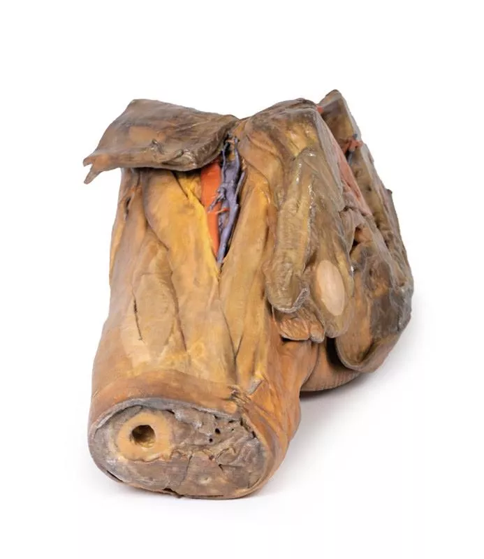
This detailed 3D print displays the left upper limb from the distal arm to the hand, highlighting key muscular, vascular, and neural structures through deep dissection. Elbow Region & Distal ArmThe cubital fossa reveals the classic lateral-to-medial layout: biceps tendon, brachial artery, and median nerve, with the bicipital aponeurosis removed. The ulnar nerve passes behind the medial epicondyle, and the superficial radial nerve is visible between brachioradialis and brachialis. Anterior & Posterior ForearmAnteriorly, the superficial flexors—pronator teres, FCR, FDS, and FCU—are clearly shown (no palmaris longus present). The radial and ulnar arteries, as well as the superficial radial nerve, are identifiable.Posteriorly, extensor muscles from the common extensor origin are preserved, including ECU, extensor digitorum, and ECRB/ECRL. The anatomical snuffbox is visible with the radial artery and cutaneous radial nerve. Palm & Carpal TunnelThe thenar/hypothenar muscles, flexor retinaculum, long tendons, and lumbricals are exposed. The median nerve runs beneath the retinaculum, and the superficial palmar arch (from the ulnar artery) is intact. Digital nerves and vessels are clearly seen, along with the recurrent median nerve branch and the extensor expansion on the middle finger.This model is ideal for studying upper limb anatomy, with special focus on the elbow, forearm, and hand structures.
This 3D print presents an expanded version of the dataset used for our Circle of Willis model, derived from careful segmentation of angiographic data.Intracranial ArteriesLike the original Circle of Willis print, this model demonstrates the internal carotid and vertebral arteries entering the skull and branching into the intracranial arteries that supply the brain. Expanded Arterial NetworkThis expanded model includes the full branching pattern of the cerebral and cerebellar arteries, showing the internal carotid and vertebral artery anastomoses along with the complete Circle of Willis. Detailed Cerebral Artery BranchesThe model highlights the pericallosal arteries (from the anterior cerebral arteries) with their named branches, the superior and inferior divisions of the middle cerebral artery (including sulcal, temporal, and parietal arteries), and the posterior cerebral artery branches, providing a detailed view of the brain’s arterial network.
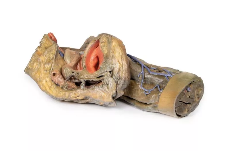
This detailed 3D model displays the left half of a female pelvis, sectioned midsagittally, and extending to the proximal mid-thigh.Pelvic Organs & Peritoneum- Visible structures: Urinary bladder, uterus, vagina, and rectum (from anterior to posterior).- The peritoneum is preserved, showing the vesicouterine and rectouterine pouches.- The broad ligament, uterine tube, fimbriae, and left ovary are identifiable near the pelvic brim. Vessels & Nerves- Common and external iliac arteries pass toward the subinguinal space, alongside the common iliac vein and psoas major.- The ureter crosses over these vessels. - The femoral nerve is visible between psoas major and iliacus muscles. Anterior Thigh & Inguinal Region- Superficial fascia removed, exposing thigh structures up to the perineal edge.- Femoral triangle dissected to show:- Femoral artery and vein, with the vein receiving tributaries from the great saphenous, superficial circumflex iliac, external pudendal, and deep pudendal veins.- Femoral nerve lateral to the artery.- Anterior cutaneous nerves and part of the lateral cutaneous nerve over the sartorius muscle. - Inguinal lymph nodes beneath the inguinal ligament. Posterior Gluteal Region- Gluteus maximus removed to reveal deeper gluteal muscles.- Piriformis reflected, exposing:- Sciatic nerve, formed by tibial and common peroneal nerves.- Superior and inferior gluteal arteries. - Posterior cutaneous nerve of the thigh running parallel to the sciatic nerve.- Obturator internus, gemelli, and quadratus femoris muscles exposed.- Internal pudendal artery and pudendal nerve track toward the ischioanal fossa.- Their branches, including the inferior rectal nerve, are visible near the pelvic diaphragm and external anal sphincter.
Coeliac TrunkThe celiac trunk, arising at the T12 level, supplies the embryological foregut. Visible branches include the left gastric artery from the left side, the splenic artery passing to the left hypochondrium, and the common hepatic artery on the right. The common hepatic gives rise to the gastroduodenal artery, which connects to the superior mesenteric artery, and the proper hepatic artery, which branches into the left and right hepatic arteries. The right hepatic artery eventually forms the cystic artery supplying the gallbladder. Superior Mesenteric Artery and Inferior Mesenteric ArteryThe superior and inferior mesenteric arteries arise at L1 and L3, supplying the midgut and hindgut. While not fully preserved, the superior mesenteric artery is visible exiting below the pancreas and branching out, and the inferior mesenteric artery descends along the left side of the aorta. The left colic artery branches from the inferior mesenteric artery, supplying the colon via the marginal arteries. Venous System of the AbdomenThe superior mesenteric vein lies posterior to the artery and appears less tubular. Removal of the liver’s left lobe exposes portal vein branches, which carry nutrients from the gut to hepatocytes before draining via hepatic veins into the inferior vena cava.Hilum of the KidneyThe right kidney shows typical anatomy with the renal vein superior, artery inferior, and ureter descending. The left kidney displays variation: the renal vein is inferior and subdivided, the artery is superior, and the ureter descends medially. Muscles, Nerves and Other VasculatureThe psoas major and iliacus muscles are visible on both sides, with key lumbar plexus nerves around them—especially on the left—including the iliohypogastric, ilioinguinal, femoral, and genitofemoral nerves. Medial to the psoas major, the left testicular artery (from the aorta) and vein (draining to the left renal vein) are seen. The right testicular vein drains directly into the IVC. A branch of the iliolumbar artery passes beneath the testicular vessels and ureter. GallbladderJust inferior to the liver, the gallbladder can be observed with the cystic artery moving inferiorly to meet it. The cystic duct can also be seen moving from the gallbladder, meeting the common hepatic duct moving from the liver to form the common bile duct.
This 3D model provides a combined midsagittal section through the head and superior neck coupled with a deep dissection into the infratemporal fossa region and superficial dissection of the scalp. In the preserved midsagittal section there is preservation of the endocranial contents, the nasal and oral cavities, and the pharynx to the level of the laryngeal cartilages. The nasal cavity is preserved nearly intact, except for a small window excised into the middle nasal concha to expose the ethmoid air cells. A very large sphenoid sinus exists in the individual just superior to the torus of the auditory tube in the nasopharynx. The oral cavity and laryngopharynx are undissected, with the larynx only preserve just distal to the level of the arytenoid cartilages and not including a clear set of vocal folds.Within the endocranial cavity, the sectioned brain is slightly off the midagittal plane, such that neither the superior sagittal sinus nor the third ventricle are clearly defined - but the lateral ventricle is open and part of the fourth ventricle is preserved between the pons and cerebellum. The gyri and sulci of the cerebrum are not well separated, but the cingulate gyrus and corpus callosum can be separated. Cross-sectioned views of the optic tract, pituitary gland, superior and inferior colliculi, superior cerebellar peduncle, and transition between the medulla oblongata and spinal cord are all visible. The tentorium cerebelli and confluence/transverse sinus is positioned between the cerebellar hemisphere and occipital lobe. Small portions of the posterior inferior cerebellar artery, vertebral arteries, basilar artery, and posterior cerebral and anterior cerebral arteries are visible in section.On the opposing side of the model, a superficial and deep dissection has opened a large window into the anatomy of the lateral scalp and infratemporal fossa. Across the scalp there is a well preserved posterior auricular nerve and superficial temporal artery highlighted on the superficial surface of the temporalis muscle. Anteriorly, the temporalis has been dissected to expose the deep temporal arteries arising from across the maxillary artery.The deep level of dissection has exposed parts of the infratemporal fossa (through partial removal of the mandibular ramus and corpus) and dissection of retromandibular tissues. At the inferior margin of the dissection window, the cut edge of the retromandibular vein lies adjacent to the submandibular gland and the ascending path of the facial artery as it cross towards to angle of the mouth. Just superior to the cut retromandibular vein is the posterior belly of the digastric muscle, overlying a small exposure of the deeper internal jugular vein.Just posterior to the retained ascending ramus of the mandible are the external carotid artery and the occipital artery (running in parallel prior to passing posteriorly). Tracing the external carotid artery superiorly, the posterior auricular artery, superficial temporal artery, and maxillary artery are all visible. The maxillary artery passes deep to the lateral pterygoid muscle and into the infratemporal fossa, reappearing superior to the lateral pterygoid as it passes into the pterygomaxillary fissure. Along its course, it gives rise to the posterior deep temporal artery, the inferior alveolar artery (which is exposed in the dissected mandibular corpus), the anterior deep temporal artery, and the posterior superior alveolar artery. Finally, the inferior alveolar nerve can be seen coursing within the opened mandibular corpus, and the lingual nerve resting on the medial pterygoid. The buccinator muscle is also retained, with the distal part of the parotid duct preserved as it enters the muscle towards the oral mucosa.
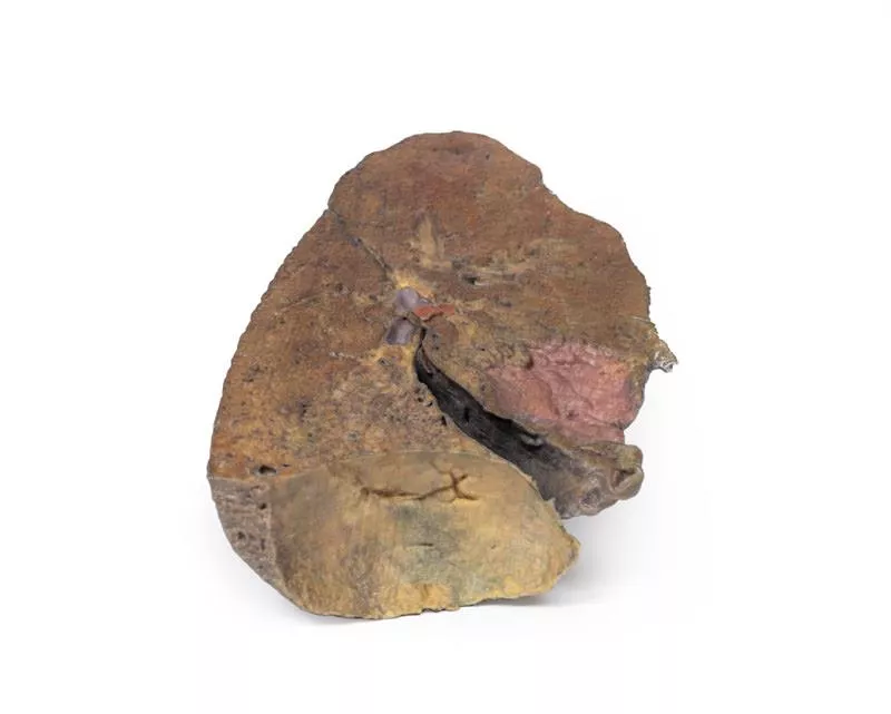
This 3D anatomical model presents a left lung dissected in a parasagittal plane, offering a unique internal view of pulmonary structures and anatomical landmarks. The mediastinal surface has been removed, allowing detailed observation of internal lung anatomy and its relationship to the heart and diaphragm.Key Features:Bronchial Tree (Internal View):- The primary bronchi are not visible due to prior branching.- Subdivided bronchi are preserved, though the dissection depth makes it unclear whether they represent secondary (lobar) or tertiary (segmental) bronchi. Vascular Structures:- Pulmonary arteries and veins are typically seen at the hilum, but precise levels of subdivision are undetermined in this section.Cardiac Impression:- A clear concave impression remains on the medial lung surface, formed by the left ventricle of the heart pressing against the lung.- Despite the dissection, this landmark remains distinctly visible.Diaphragmatic Surface:- The lung’s base is concave, resting atop the diaphragm.- While the pleura is not preserved, the model indicates where the diaphragmatic recess would form—between the costal and diaphragmatic pleura.
This 3D model preserves a right male pelvis sectioned just superior to the L5 vertebra and sectioned at the midsagittal plane, with the thigh preserved to near the midshaft of the femur. This specimen compliments our LW 91 female hemipelvic specimen and thigh. The common iliac artery is preserved with several key branches visible, particularly the distribution of the internal iliac within the true pelvis. Several major vessels including the obturator artery and the partially obliterated umbilical artery passes towards the anterior abdominal wall (to form the medial umbilical ligament) and gives off the superior vesicle artery; while the roots of the iliolumbar, superior gluteal, inferior gluteal and internal pudendal artery are visible lateral to the urinary bladder. The ureter descends superficial to these vessels to approach the urinary bladder which is covered with peritoneum in this model. The ductus deferens is exposed from the entry into the space via the deep inguinal ring and passing posteriorly (though sectioned from its normal insertion pathway and resting on the internal iliac artery). Adjacent to the ureter and on the superficial surface of the psoas major muscle is an enlarged iliac lymph node and part of the lymphatic vasculature ascending along the external iliac artery. The majority of the pelvis has been left undissected, allowing for an appreciation of the rectovesicular pouch and the exposed superior rectal artery and vein approaching the preserved portion of rectum. In cross section, the rectum, seminal vesicle and prostate are visible (the section plane preserves parts of both the prostatic urethra and ejaculatory duct).In the anterior thigh the borders and contents of the femoral triangle are well-preserved, with partial coverage by the flap of the anterior abdominal wall. Posteriorly the skin over the gluteal region and the gluteus maximus muscle have been removed as sequential windows to expose the gluteus medius and minimum muscles, the piriformis, the obturator internus with gemelli muscles, and the quadratus femoris muscle. The superior and inferior gluteal arteries are maintained superior and inferior to the piriformis, respectively; with the sciatic nerve exiting inferior to piriformis before passing deep to the retained portion of the gluteus maximus.
This 3D model combines a midsagittal section of the head with preservation of brain and cranial cavity anatomy, with a unique deep dissection of the pharyngeal region via removal of basicranial bone and the anterior parts of the atlas and axis. As the opposing side is undissected it has been digitally eliminated from the model. Within the endocranial cavity the preservation of dura mater retains the superior sagittal sinus across much of its course from anterior to posterior, reaching the confluence of sinuses visible in cross-section. Both the tentorium cerebelli and the falx cerebelli are preserved. The cerebrum is well-reserved with retention of the cingulate gyrus and sulcus, and removal of the septum pellucidum inferior to the corpus callosum providing a view into the lateral ventricle (with retention of the interventricular foramen at the inferior margin of the septum). The diencephalon and midbrain structures (epithalamus, colliculi, mamillary body, infundibulum) are all appreciable in cross-section as is the cerebellar hemisphere and fourth ventricle. Small views of the anterior cerebral and posterior inferior cerebellar arteries are visible (and false coloured).Outside the endocranium, removal of parts of the occipital, temporal and sphenoid bones (alongside the atlas and axis) has been coupled with removal of the pharyngeal constrictors, carotid sheath and oral mucosa to demonstrate a unique view of several key neurovascular and glandular structures. Within the zone of removed tissue there is partial exposure of the right common carotid artery within the dissected petrous portion of the temporal, as well as partial exposure of the left vertebral artery through disruption of the occipital and dural covering.The medial and lateral pterygoids are exposed near the posterior margin of the largely intact nasal cavity. Between the exposed dura and medulla and the pterygoids (and trapped deep to the sectioned and reflected stylohyoid muscle) the dissected carotid sheath has exposed the internal jugular vein, the vagus nerve, the internal carotid artery (with overriding ascending pharyngeal artery from the external carotid artery), and the sympathetic trunk (with superior cervical ganglion and internal carotid nerve). Immediately anterior to this bundle of neurovascular structures is the external carotid artery, giving rise to the ascending pharyngeal artery, a common trunk for the lingual and facial arteries, and then continuing superiorly out of the plane of dissection. The submandibular gland can be seen resting on the mylohyoid muscle near the lingual artery (which passes deep relative to the gland), with the duct passing towards the genu of the mandible and the origin of the reflected genioglossus muscle. At the inferior border of the specimen, the reflected margin of the dissected tongue the hypoglossal nerve can be seen deep to the lingual artery.
This highly detailed 3D printed model offers a precise representation of the intracranial arteries supplying the human brain, created from segmented angiographic imaging data. The model displays vascular structures that cannot be physically dissected and viewed in situ, making it a unique and valuable teaching tool.Vertebral and Basilar ArteriesThe paired vertebral arteries are shown entering the cranial cavity through the foramen magnum, where they unite to form the basilar artery. The basilar artery travels along the brainstem and terminates by branching into the posterior cerebral arteries. Just before this bifurcation, the superior cerebellar arteries arise, supplying parts of the cerebellum and midbrain. Internal Carotid Arteries and Carotid SiphonThe internal carotid arteries (ICAs) are traced from their entry at the carotid canal within the temporal bone. From there, they course medially and anteriorly through the petrous portion of the bone and emerge via the upper opening of the foramen lacerum. While the cavernous sinus is not shown, the model beautifully captures the characteristic S-shaped carotid siphon on both sides, clearly positioned lateral to the sella turcica and passing medial to the anterior clinoid processes. Major Cerebral Arteries and the Circle of WillisEach internal carotid artery divides into the anterior cerebral artery (ACA) and middle cerebral artery (MCA). The posterior communicating arteries, connecting the posterior cerebral arteries (PCAs) to the MCAs, are distinctly visible, allowing for an excellent demonstration of the posterior portion of the Circle of Willis.The anterior communicating artery, completing the circle between the paired anterior cerebral arteries, is also present—though its visibility is limited due to the close proximity of the ACAs.

Continuous innovation

Social responsibility

Active customer orientation

Understanding quality

Sustainable actions

ISO 9001 certification
Your last viewed products


