


Median Section through head sagittal section of head with deep dissection
Question regarding article:
Product information "Median Section through head sagittal section of head with deep dissection"
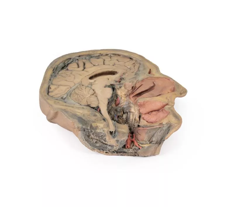
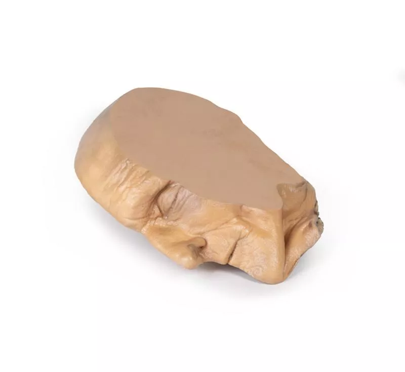
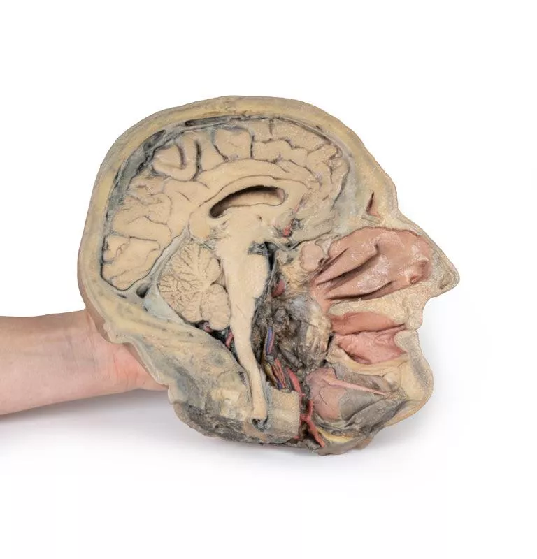
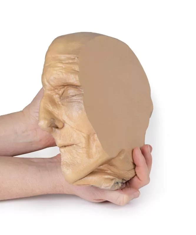
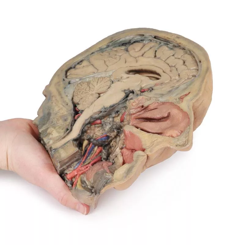
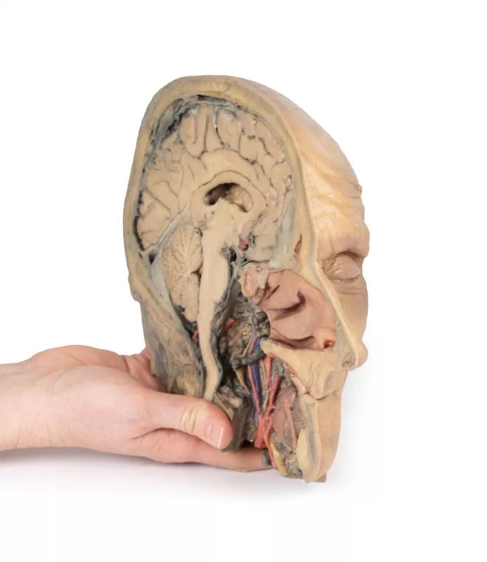
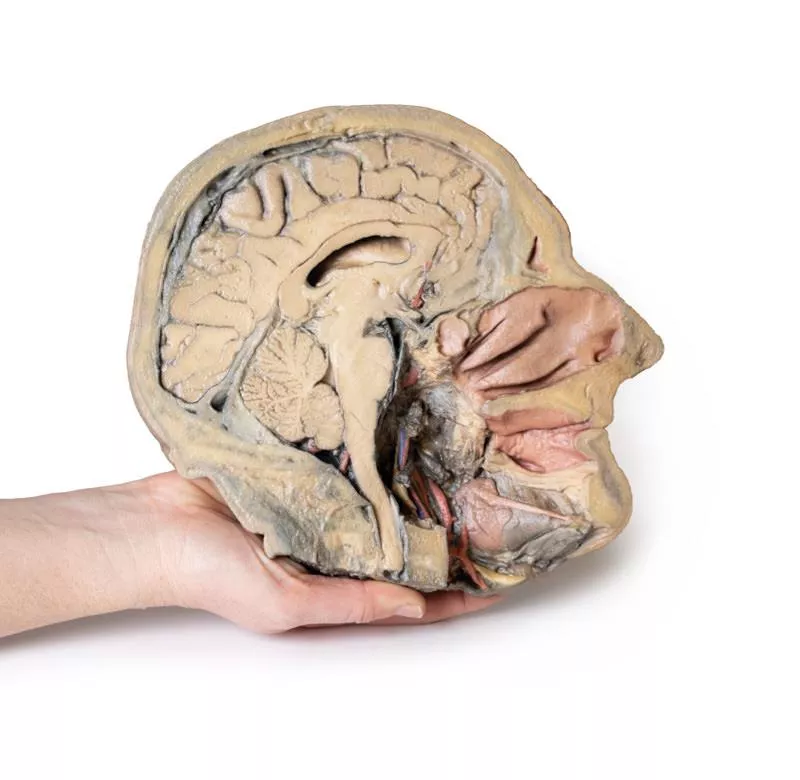
This 3D model combines a midsagittal section of the head with preservation of brain and cranial cavity anatomy, with a unique deep dissection of the pharyngeal region via removal of basicranial bone and the anterior parts of the atlas and axis. As the opposing side is undissected it has been digitally eliminated from the model.
Within the endocranial cavity the preservation of dura mater retains the superior sagittal sinus across much of its course from anterior to posterior, reaching the confluence of sinuses visible in cross-section. Both the tentorium cerebelli and the falx cerebelli are preserved. The cerebrum is well-reserved with retention of the cingulate gyrus and sulcus, and removal of the septum pellucidum inferior to the corpus callosum providing a view into the lateral ventricle (with retention of the interventricular foramen at the inferior margin of the septum). The diencephalon and midbrain structures (epithalamus, colliculi, mamillary body, infundibulum) are all appreciable in cross-section as is the cerebellar hemisphere and fourth ventricle. Small views of the anterior cerebral and posterior inferior cerebellar arteries are visible (and false coloured).
Outside the endocranium, removal of parts of the occipital, temporal and sphenoid bones (alongside the atlas and axis) has been coupled with removal of the pharyngeal constrictors, carotid sheath and oral mucosa to demonstrate a unique view of several key neurovascular and glandular structures. Within the zone of removed tissue there is partial exposure of the right common carotid artery within the dissected petrous portion of the temporal, as well as partial exposure of the left vertebral artery through disruption of the occipital and dural covering.
The medial and lateral pterygoids are exposed near the posterior margin of the largely intact nasal cavity. Between the exposed dura and medulla and the pterygoids (and trapped deep to the sectioned and reflected stylohyoid muscle) the dissected carotid sheath has exposed the internal jugular vein, the vagus nerve, the internal carotid artery (with overriding ascending pharyngeal artery from the external carotid artery), and the sympathetic trunk (with superior cervical ganglion and internal carotid nerve). Immediately anterior to this bundle of neurovascular structures is the external carotid artery, giving rise to the ascending pharyngeal artery, a common trunk for the lingual and facial arteries, and then continuing superiorly out of the plane of dissection. The submandibular gland can be seen resting on the mylohyoid muscle near the lingual artery (which passes deep relative to the gland), with the duct passing towards the genu of the mandible and the origin of the reflected genioglossus muscle. At the inferior border of the specimen, the reflected margin of the dissected tongue the hypoglossal nerve can be seen deep to the lingual artery.
Login
Erler-Zimmer
Erler-Zimmer Medical GmbH
Hauptstrasse 27
77886 Lauf
Germany
info@erler-zimmer.de
Achtung! Medizinisches Ausbildungsmaterial, kein Spielzeug. Nicht geeignet für Personen unter 14 Jahren.
Attention! Medical training material, not a toy. Not suitable for persons under 14 years of age.
Other customers also bought

Parasagittal Section of the head and neck
This high-resolution 3D model features a head and neck specimen sectioned just off the midsagittal plane, preserving critical midline structures often absent in similar models. Ideal for anatomical education, this model offers enhanced visibility due to fixative-induced brain shrinkage, exaggerating spaces between brain and skull.Key Features:Midline Anatomy Preserved:Includes the falx cerebri (anterior portion), septum pellucidum, interventricular foramen (of Monro), and nasal septum.Ventricular & Endocranial Structures:Clear views of the lateral and third ventricles, cerebral aqueduct, fourth ventricle, infundibulum, pituitary gland, and sphenoid sinus.Vascular Highlights:Displays the left vertebral artery, posterior cerebral artery (cross-section), and anterior cerebral artery branches around the corpus callosum.Detailed Nasal & Pharyngeal Regions:Shows relationships between the nasal septum, palate, auditory tube opening, and naso-/oropharynx.Laryngeal and Tracheal Anatomy:Includes epiglottis, arytenoid, thyroid cartilages, hyoid bone, and cross-sectional views of the vestibule, vestibular and vocal folds.This model provides a unique perspective of internal head and neck anatomy, combining anatomical depth with high educational value.

Superficial Face
This detailed 3D model features a superficial dissection of the left face just anterior to the ear, with false colouring highlighting key neurovascular structures and muscles of facial expression. It serves as a focused complement to the broader dissection in our HW 45 model. Undissected areas have been digitally removed for clarity.Key Features:Parotid Region & Facial Nerve Branches:Exposes the parotid gland and duct, along with terminal branches of the facial nerve (CN VII): cervical, mandibular, buccal, zygomatic, and temporal. Facial Vessels & Nerve-Vessel Relationships:Shows the facial artery and vein in relation to CN VII branches. Vessels are traced from the mandible to the orbit, offering anatomical landmarks.Muscles of Facial Expression (Highlighted):Includes masseter, depressor anguli oris, zygomaticus major & minor, orbicularis oris, nasalis, levator labii superioris alaeque nasi, procerus, and orbicularis oculi.Temporal & Forehead Structures:Displays the auriculotemporal nerve and superficial temporal artery over the temporal fascia, with part of the temporalis muscle visible.Superiorly, the supraorbital nerve and vessels ascend on the epicranial aponeurosis, overlying the frontalis muscle.This model offers a concise yet richly detailed view of facial anatomy, ideal for teaching nerve-muscle-vascular relationships in the superficial face.

Superficial Facial nerves & Parotid Gland
This 3D model provides a detailed view of the superficial anatomy of the face and head, expanding upon our HW 44 model with a broader dissection of the scalp, occipital region, and areas below the external ear.Key Features:Extended Facial AnatomyIncludes the terminal branches of the facial nerve (CN VII) traced from the parotid gland, with the platysma muscle preserved and extending from the mandible to the neck. Enhanced Posterior Dissection- Broader exposure across the posterior scalp and occipital region- Includes the retromandibular vein, great auricular nerve, and lesser occipital nerve- Shows the course of the occipital artery and vein near the trapeziusNeurovascular HighlightsImproved visualization of the supraorbital, supratrochlear, and superficial temporal arteries and nervesMusculaturePreserves fibers of the auricularis and occipitalis muscles, integrated into the epicranius (occipitofrontalis)

Sagittal Section of head with infratemporal Fossa Dissection
This 3D model provides a combined midsagittal section through the head and superior neck coupled with a deep dissection into the infratemporal fossa region and superficial dissection of the scalp. In the preserved midsagittal section there is preservation of the endocranial contents, the nasal and oral cavities, and the pharynx to the level of the laryngeal cartilages. The nasal cavity is preserved nearly intact, except for a small window excised into the middle nasal concha to expose the ethmoid air cells. A very large sphenoid sinus exists in the individual just superior to the torus of the auditory tube in the nasopharynx. The oral cavity and laryngopharynx are undissected, with the larynx only preserve just distal to the level of the arytenoid cartilages and not including a clear set of vocal folds.Within the endocranial cavity, the sectioned brain is slightly off the midagittal plane, such that neither the superior sagittal sinus nor the third ventricle are clearly defined - but the lateral ventricle is open and part of the fourth ventricle is preserved between the pons and cerebellum. The gyri and sulci of the cerebrum are not well separated, but the cingulate gyrus and corpus callosum can be separated. Cross-sectioned views of the optic tract, pituitary gland, superior and inferior colliculi, superior cerebellar peduncle, and transition between the medulla oblongata and spinal cord are all visible. The tentorium cerebelli and confluence/transverse sinus is positioned between the cerebellar hemisphere and occipital lobe. Small portions of the posterior inferior cerebellar artery, vertebral arteries, basilar artery, and posterior cerebral and anterior cerebral arteries are visible in section.On the opposing side of the model, a superficial and deep dissection has opened a large window into the anatomy of the lateral scalp and infratemporal fossa. Across the scalp there is a well preserved posterior auricular nerve and superficial temporal artery highlighted on the superficial surface of the temporalis muscle. Anteriorly, the temporalis has been dissected to expose the deep temporal arteries arising from across the maxillary artery.The deep level of dissection has exposed parts of the infratemporal fossa (through partial removal of the mandibular ramus and corpus) and dissection of retromandibular tissues. At the inferior margin of the dissection window, the cut edge of the retromandibular vein lies adjacent to the submandibular gland and the ascending path of the facial artery as it cross towards to angle of the mouth. Just superior to the cut retromandibular vein is the posterior belly of the digastric muscle, overlying a small exposure of the deeper internal jugular vein.Just posterior to the retained ascending ramus of the mandible are the external carotid artery and the occipital artery (running in parallel prior to passing posteriorly). Tracing the external carotid artery superiorly, the posterior auricular artery, superficial temporal artery, and maxillary artery are all visible. The maxillary artery passes deep to the lateral pterygoid muscle and into the infratemporal fossa, reappearing superior to the lateral pterygoid as it passes into the pterygomaxillary fissure. Along its course, it gives rise to the posterior deep temporal artery, the inferior alveolar artery (which is exposed in the dissected mandibular corpus), the anterior deep temporal artery, and the posterior superior alveolar artery. Finally, the inferior alveolar nerve can be seen coursing within the opened mandibular corpus, and the lingual nerve resting on the medial pterygoid. The buccinator muscle is also retained, with the distal part of the parotid duct preserved as it enters the muscle towards the oral mucosa.

Internal abdominal wall
This detailed 3D model captures the internal surface of the anterior abdominal wall—a region often removed or damaged during dissections. It complements our MP1130 abdominal specimen, where the anterior wall has been removed, providing a clear view of key muscle and connective tissue structures. Key Features:Muscle Fibers & Aponeurosis:The horizontally oriented transversus abdominus muscle fibers converge toward their aponeurosis (tendon sheet), visible especially along the specimen’s superior margins. Arcuate Line:Located in the lower third of the model, this landmark marks where the aponeurosis shifts relative to the rectus abdominus muscle.- Above the arcuate line: Aponeurosis fibers split evenly around the rectus abdominus.- Below the arcuate line: All aponeurotic fibers pass anteriorly to the rectus abdominus, reflecting a change in abdominal wall structure. Vascular Structures:Inferior Epigastric Arteries & Veins:These vessels originate from the external iliac arteries and veins, ascending superiorly through the anterior abdominal wall. Hesselbach’s Triangle:On the right side of the model, the orientation of the inferior epigastric artery relative to the rectus abdominus muscle defines the apex of the inguinal (Hesselbach’s) triangle—a critical anatomical region often associated with direct inguinal hernias. (Note: The inguinal ligament forming the base of this triangle is not present in this specimen.) Embryological Remnant: Median Abdominal Ligament:Positioned midline between the two rectus abdominus muscles, this fold of parietal peritoneum covers the urachus, a fibrous remnant from embryological development extending from the bladder to the umbilicus.

Transverse Section of the head
This 3D model features a transverse section through the cranial cavity with a deep dissection of the face, orbit, and temporomandibular joint (TMJ) region. It offers a comprehensive view of both intracranial and facial structures.Key Features:Cranial Cavity & Brain- Partial dura mater removal; dissection reveals lateral and third ventricles, falx cerebri, choroid plexus, and optic pathways- Middle cerebral artery visible in the lateral fissure- Key vascular structures: internal carotid, anterior and middle cerebral arteries Left OrbitRoof removed; exposure of: Frontal nerve, lacrimal gland, superior oblique, medial rectus, nasociliary nerveRight OrbitSuperficial tissues removed; shows: All extraocular muscles, including inferior oblique, and levator palpebrae superiorisFace & TMJ (Right Side)- Exposed: infraorbital nerve/artery, masseter (both heads), and temporalis muscle near pterion- Parotid gland dissected to show mandibular condyle in glenoid fossa and external ear alignment

Abdomen with bilateral Hernias
This 3D model represents one of the largest and most complex in the series, consisting of a partial torso from the diaphragm to the proximal thigh with a complete abdominal cavity preserving varying levels of dissection. This 3D model also records the rare, simultaneous occurrence of indirect and direct inguinal hernias allowing for a consideration of the anatomical underpinnings for both conditions. Given the scale of the dissection this 3D model description is divided into discrete parts based on views and regions.The diaphragmThe diaphragm is preserved on the model’s superior aspect, with both domes and costodiaphragmatic recesses visible despite some distortion from rib removal. The fibrous pericardium rests on the central tendon, with the terminal inferior vena cava seen in the caval foramen. Lateral to this lies the oesophagus in the oesophageal hiatus, and the descending thoracic aorta approaching the aortic hiatus near the vertebrae. The epigastric and hypochondriac regionsIn the abdomen, removal of the anterior wall, greater omentum, and much of the GI tract reveals retroperitoneal structures. The terminal oesophagus enters just left of the liver. With the stomach removed, the pancreas is fully exposed from head to tail, reaching the spleen in the left hypochondrium. Above it, the splenic and common hepatic arteries span the narrow space between pancreas, diaphragm, and liver. The tortuous splenic artery divides near the splenic vein; the common hepatic gives rise to the gastroduodenal and right gastric arteries, superficial to the portal vein. The superior mesenteric vessels pass near the pancreatic head, and the ileocolic artery leads to the caecum. The inferior mesenteric vein arises from the superior rectal vein and crosses the descending aorta. Below the liver, the gallbladder lies between the lobes. On the left, renal vessels pass deep to the pancreas, with ureters descending across the psoas muscles. The umbilical and lumbar regionsMost abdominal organs in the umbilical and lumbar regions have been removed to reveal the posterior abdominal wall. Centrally, the descending aorta and inferior vena cava are prominent, with testicular vessels traceable toward the inguinal region. Two right lumbar arteries branch from the aorta, and the inferior mesenteric artery gives rise to the left colic, sigmoid, and superior rectal arteries. On the right, subcostal, iliohypogastric, and ilioinguinal nerves are visible, along with the circumflex iliac artery.The hypogastrium and iliac regionsThe abdominal aorta bifurcates into the common, internal, and external iliac arteries, with matching iliac veins merging into the inferior vena cava. The obturator artery, ureters, and testicular vessels are visible. In the true pelvis, the peritoneum covers the bladder, while the rectum remains obscured. The right iliac fossa contains the terminal ileum, caecum, and appendix, with nearby vessels and nerves. On the left, the sigmoid colon crosses the iliac fossa, where an epiploic appendage extends into an indirect hernia near the inferior epigastric artery. The inguinal region and perineumThis model uniquely preserves both direct (right) and indirect (left) inguinal hernias, with the inferior epigastric vessels retained for anatomical orientation. The right hernia lies medial to these vessels; the left hernia sac extends laterally into the spermatic cord, containing an epiploic appendage. The perineum reveals the penis, testes, and spermatic cords. On the right, the cord remains intact; on the left, it’s opened, showing a varicose testicular vein linked to the indirect hernia. The thighThe femoral triangle has been dissected on both thighs. On the right, the femoral sheath was removed to reveal the femoral artery, vein, deep inguinal lymph nodes, and femoral nerve. On the left, a broader view exposes anterior and medial thigh muscles, with the femoral artery, profunda femoris, and circumflex iliac artery visible. The model ends mid-thigh, showing cross-sectional anatomy including the femoral shaft, vessels, and muscles in the subsartorial canal.

Spleen and pancreas
This detailed 3D anatomical model preserves the deep foregut organs, including the descending, horizontal, and ascending parts of the duodenum, the pancreas, and the spleen. It offers a unique and insightful view into the complex anatomy of this region. DuodenumA small window is opened in the duodenum to reveal the plicae circularis, the characteristic circular folds of the proximal small intestine. This contrasts with the prominent rugae of the stomach, providing an educational comparison of mucosal patterns in the upper gastrointestinal tract. PancreasThe pancreas is preserved in its natural anatomical position, nestled within the curvature of the duodenum. The head of the pancreas is clearly visible, including the distinct uncus located at its distal margin, adjacent to the origin of the superior mesenteric artery (SMA). In this model, the SMA is already divided into its major named branches, highlighting its vascular complexity.The body of the pancreas features the superior margin where the celiac trunk, sectioned from the descending abdominal aorta, is located. The complete splenic artery is shown following its tortuous path from the celiac trunk to the spleen. The model also displays the origins of the left gastric artery and the common hepatic artery branching from the celiac trunk.Adjacent to the celiac trunk, a segment of the splenic vein is visible emerging from the pancreas’ capsule. This vein runs alongside the splenic artery en route to the spleen. Additionally, a portion of the superior mesenteric vein is seen adhering to the posterior pancreas, representing its path before converging with the splenic vein to form the hepatic portal vein.The tail of the pancreas is embedded within the splenic capsule, partially obscuring the splenic artery branches before entering the spleen. For more detailed views of this region, refer to our other spleen models (MP1130 and MP1134), which illustrate further anatomical and spatial relationships.

Continuous innovation

Social responsibility

Active customer orientation

Understanding quality

Sustainable actions

ISO 9001 certification