


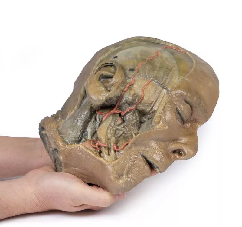
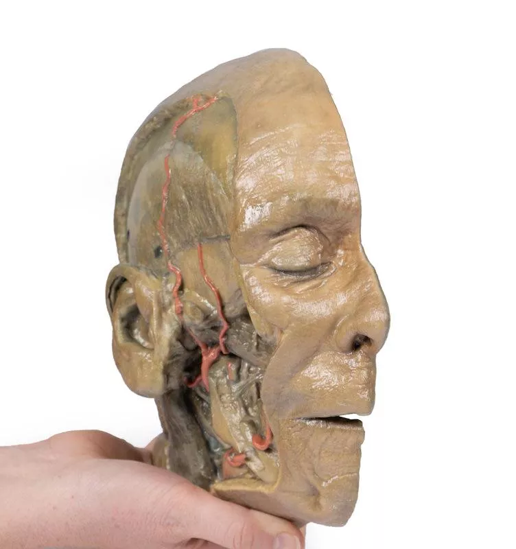

Product information "Sagittal Section of head with infratemporal Fossa Dissection"
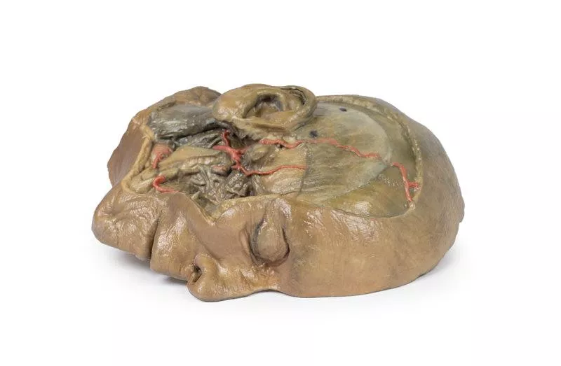
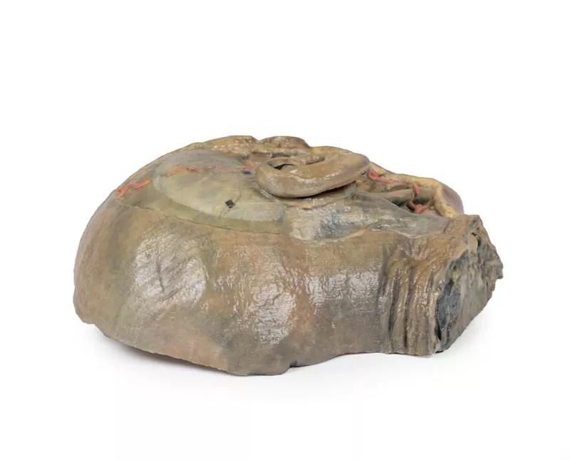
This 3D model provides a combined midsagittal section through the head and superior neck coupled with a deep dissection into the infratemporal fossa region and superficial dissection of the scalp.
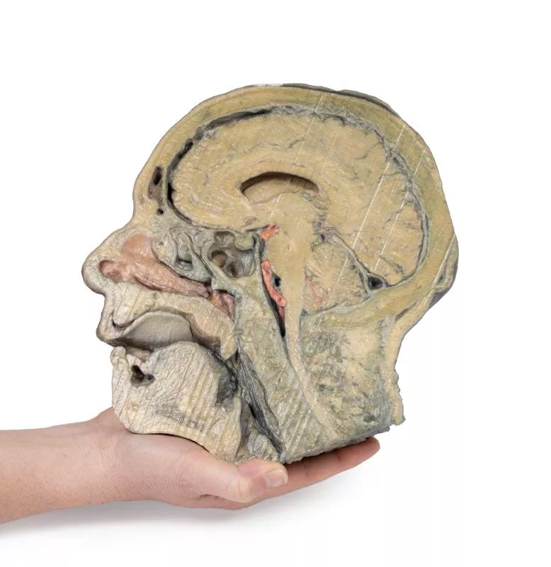
In the preserved midsagittal section there is preservation of the endocranial contents, the nasal and oral cavities, and the pharynx to the level of the laryngeal cartilages. The nasal cavity is preserved nearly intact, except for a small window excised into the middle nasal concha to expose the ethmoid air cells. A very large sphenoid sinus exists in the individual just superior to the torus of the auditory tube in the nasopharynx. The oral cavity and laryngopharynx are undissected, with the larynx only preserve just distal to the level of the arytenoid cartilages and not including a clear set of vocal folds.
Within the endocranial cavity, the sectioned brain is slightly off the midagittal plane, such that neither the superior sagittal sinus nor the third ventricle are clearly defined - but the lateral ventricle is open and part of the fourth ventricle is preserved between the pons and cerebellum. The gyri and sulci of the cerebrum are not well separated, but the cingulate gyrus and corpus callosum can be separated. Cross-sectioned views of the optic tract, pituitary gland, superior and inferior colliculi, superior cerebellar peduncle, and transition between the medulla oblongata and spinal cord are all visible. The tentorium cerebelli and confluence/transverse sinus is positioned between the cerebellar hemisphere and occipital lobe. Small portions of the posterior inferior cerebellar artery, vertebral arteries, basilar artery, and posterior cerebral and anterior cerebral arteries are visible in section.
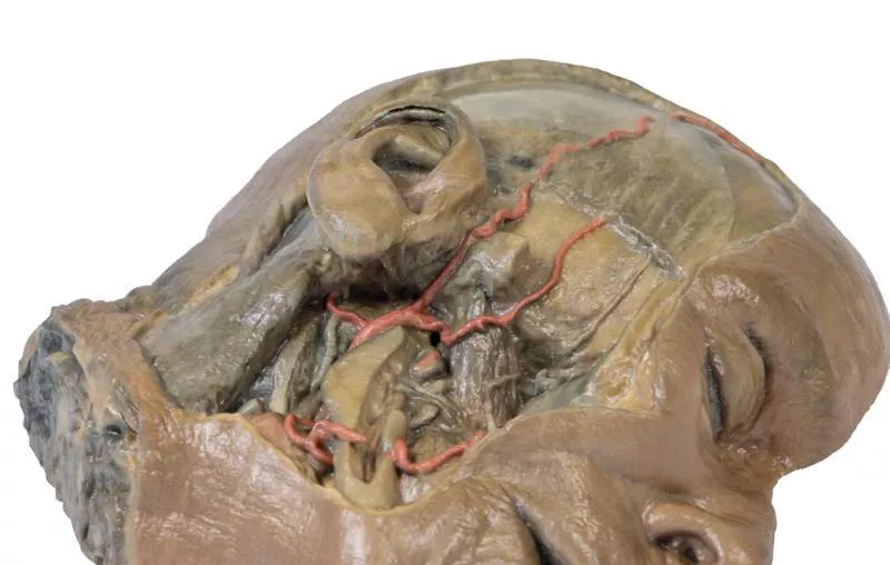
On the opposing side of the model, a superficial and deep dissection has opened a large window into the anatomy of the lateral scalp and infratemporal fossa. Across the scalp there is a well preserved posterior auricular nerve and superficial temporal artery highlighted on the superficial surface of the temporalis muscle. Anteriorly, the temporalis has been dissected to expose the deep temporal arteries arising from across the maxillary artery.
The deep level of dissection has exposed parts of the infratemporal fossa (through partial removal of the mandibular ramus and corpus) and dissection of retromandibular tissues. At the inferior margin of the dissection window, the cut edge of the retromandibular vein lies adjacent to the submandibular gland and the ascending path of the facial artery as it cross towards to angle of the mouth. Just superior to the cut retromandibular vein is the posterior belly of the digastric muscle, overlying a small exposure of the deeper internal jugular vein.
Just posterior to the retained ascending ramus of the mandible are the external carotid artery and the occipital artery (running in parallel prior to passing posteriorly). Tracing the external carotid artery superiorly, the posterior auricular artery, superficial temporal artery, and maxillary artery are all visible. The maxillary artery passes deep to the lateral pterygoid muscle and into the infratemporal fossa, reappearing superior to the lateral pterygoid as it passes into the pterygomaxillary fissure. Along its course, it gives rise to the posterior deep temporal artery, the inferior alveolar artery (which is exposed in the dissected mandibular corpus), the anterior deep temporal artery, and the posterior superior alveolar artery. Finally, the inferior alveolar nerve can be seen coursing within the opened mandibular corpus, and the lingual nerve resting on the medial pterygoid. The buccinator muscle is also retained, with the distal part of the parotid duct preserved as it enters the muscle towards the oral mucosa.
In the preserved midsagittal section there is preservation of the endocranial contents, the nasal and oral cavities, and the pharynx to the level of the laryngeal cartilages. The nasal cavity is preserved nearly intact, except for a small window excised into the middle nasal concha to expose the ethmoid air cells. A very large sphenoid sinus exists in the individual just superior to the torus of the auditory tube in the nasopharynx. The oral cavity and laryngopharynx are undissected, with the larynx only preserve just distal to the level of the arytenoid cartilages and not including a clear set of vocal folds.
Within the endocranial cavity, the sectioned brain is slightly off the midagittal plane, such that neither the superior sagittal sinus nor the third ventricle are clearly defined - but the lateral ventricle is open and part of the fourth ventricle is preserved between the pons and cerebellum. The gyri and sulci of the cerebrum are not well separated, but the cingulate gyrus and corpus callosum can be separated. Cross-sectioned views of the optic tract, pituitary gland, superior and inferior colliculi, superior cerebellar peduncle, and transition between the medulla oblongata and spinal cord are all visible. The tentorium cerebelli and confluence/transverse sinus is positioned between the cerebellar hemisphere and occipital lobe. Small portions of the posterior inferior cerebellar artery, vertebral arteries, basilar artery, and posterior cerebral and anterior cerebral arteries are visible in section.
On the opposing side of the model, a superficial and deep dissection has opened a large window into the anatomy of the lateral scalp and infratemporal fossa. Across the scalp there is a well preserved posterior auricular nerve and superficial temporal artery highlighted on the superficial surface of the temporalis muscle. Anteriorly, the temporalis has been dissected to expose the deep temporal arteries arising from across the maxillary artery.
The deep level of dissection has exposed parts of the infratemporal fossa (through partial removal of the mandibular ramus and corpus) and dissection of retromandibular tissues. At the inferior margin of the dissection window, the cut edge of the retromandibular vein lies adjacent to the submandibular gland and the ascending path of the facial artery as it cross towards to angle of the mouth. Just superior to the cut retromandibular vein is the posterior belly of the digastric muscle, overlying a small exposure of the deeper internal jugular vein.
Just posterior to the retained ascending ramus of the mandible are the external carotid artery and the occipital artery (running in parallel prior to passing posteriorly). Tracing the external carotid artery superiorly, the posterior auricular artery, superficial temporal artery, and maxillary artery are all visible. The maxillary artery passes deep to the lateral pterygoid muscle and into the infratemporal fossa, reappearing superior to the lateral pterygoid as it passes into the pterygomaxillary fissure. Along its course, it gives rise to the posterior deep temporal artery, the inferior alveolar artery (which is exposed in the dissected mandibular corpus), the anterior deep temporal artery, and the posterior superior alveolar artery. Finally, the inferior alveolar nerve can be seen coursing within the opened mandibular corpus, and the lingual nerve resting on the medial pterygoid. The buccinator muscle is also retained, with the distal part of the parotid duct preserved as it enters the muscle towards the oral mucosa.
Login
Erler-Zimmer
Erler-Zimmer Medical GmbH
Hauptstrasse 27
77886 Lauf
Germany
info@erler-zimmer.de
Achtung! Medizinisches Ausbildungsmaterial, kein Spielzeug. Nicht geeignet für Personen unter 14 Jahren.
Attention! Medical training material, not a toy. Not suitable for persons under 14 years of age.
Other customers also bought

Transverse Section of the head
This 3D model features a transverse section through the cranial cavity with a deep dissection of the face, orbit, and temporomandibular joint (TMJ) region. It offers a comprehensive view of both intracranial and facial structures.Key Features:Cranial Cavity & Brain- Partial dura mater removal; dissection reveals lateral and third ventricles, falx cerebri, choroid plexus, and optic pathways- Middle cerebral artery visible in the lateral fissure- Key vascular structures: internal carotid, anterior and middle cerebral arteries Left OrbitRoof removed; exposure of: Frontal nerve, lacrimal gland, superior oblique, medial rectus, nasociliary nerveRight OrbitSuperficial tissues removed; shows: All extraocular muscles, including inferior oblique, and levator palpebrae superiorisFace & TMJ (Right Side)- Exposed: infraorbital nerve/artery, masseter (both heads), and temporalis muscle near pterion- Parotid gland dissected to show mandibular condyle in glenoid fossa and external ear alignment

Hilum of the right lung
This high-quality 3D model presents a sagittal section of the right lung, focused on the hilum, where key anatomical structures enter and exit the lung. It serves as an essential tool for understanding pulmonary vascular and bronchial anatomy, with clear orientation from apex to base and medial to lateral surfaces.Key Features:Hilum Structure:- The hilum marks the transition between visceral and parietal pleura and serves as the lung’s only anatomical connection to the body via the pulmonary ligament.- Major structures entering the lung at this point include:- Pulmonary artery (superior in the hilum) – carrying deoxygenated blood from the heart.- Superior and inferior pulmonary veins (anterior and inferior) – returning oxygenated blood to the heart.- Right main bronchus and its lobar branches – located posteriorly in the hilum. - Associated nerves and lymphatics. Visible Anatomical Landmarks:- Cardiac impression (formed by the right atrium) is visible just anterior to the hilum.- The oesophageal groove is preserved along the posterior surface, tracing the path of the descending oesophagus.- Oblique and horizontal fissures are well-defined on the lateral surface, demarcating the lung's three lobes.- Hilar lymph nodes are observed around the medial hilum.Lung Surfaces:- Diaphragmatic surface (inferior), showing the concave interface with the diaphragm.- Costal visceral surface (posterior), where the lung contacts the thoracic wall.

Internal abdominal wall
This detailed 3D model captures the internal surface of the anterior abdominal wall—a region often removed or damaged during dissections. It complements our MP1130 abdominal specimen, where the anterior wall has been removed, providing a clear view of key muscle and connective tissue structures. Key Features:Muscle Fibers & Aponeurosis:The horizontally oriented transversus abdominus muscle fibers converge toward their aponeurosis (tendon sheet), visible especially along the specimen’s superior margins. Arcuate Line:Located in the lower third of the model, this landmark marks where the aponeurosis shifts relative to the rectus abdominus muscle.- Above the arcuate line: Aponeurosis fibers split evenly around the rectus abdominus.- Below the arcuate line: All aponeurotic fibers pass anteriorly to the rectus abdominus, reflecting a change in abdominal wall structure. Vascular Structures:Inferior Epigastric Arteries & Veins:These vessels originate from the external iliac arteries and veins, ascending superiorly through the anterior abdominal wall. Hesselbach’s Triangle:On the right side of the model, the orientation of the inferior epigastric artery relative to the rectus abdominus muscle defines the apex of the inguinal (Hesselbach’s) triangle—a critical anatomical region often associated with direct inguinal hernias. (Note: The inguinal ligament forming the base of this triangle is not present in this specimen.) Embryological Remnant: Median Abdominal Ligament:Positioned midline between the two rectus abdominus muscles, this fold of parietal peritoneum covers the urachus, a fibrous remnant from embryological development extending from the bladder to the umbilicus.

Thoracic cross section at T6
This detailed 3D model presents a transverse cross-section of the thorax at the level of the T6 vertebra, offering a clear view of thoracic anatomy in relation to skeletal, vascular, respiratory, and cardiac structures.Key Features:Posterior Structures- Begins medially with the spinal cord within the vertebral canal- Costovertebral joints of the 6th ribs are visible, along with surrounding ribs forming the thoracic wall Anterior Thoracic Wall- Costosternal joints show the connection between ribs and sternumMajor Thoracic Organs- Oesophagus located anterior to the vertebral body- Descending aorta situated lateral to the vertebral bodyLungs and Pleural Cavities- Within the pleural spaces (lined by parietal pleura):- Right lung: middle and inferior lobes- Left lung: inferior lobeHeart and Mediastinum- In the middle mediastinum, the heart is shown within the pericardium, sectioned to display internal anatomy:- Left atrium (posterior)- Aortic valve, right ventricle, and right atrium in clockwise order

Median Section through head sagittal section of head with deep dissection
This 3D model combines a midsagittal section of the head with preservation of brain and cranial cavity anatomy, with a unique deep dissection of the pharyngeal region via removal of basicranial bone and the anterior parts of the atlas and axis. As the opposing side is undissected it has been digitally eliminated from the model. Within the endocranial cavity the preservation of dura mater retains the superior sagittal sinus across much of its course from anterior to posterior, reaching the confluence of sinuses visible in cross-section. Both the tentorium cerebelli and the falx cerebelli are preserved. The cerebrum is well-reserved with retention of the cingulate gyrus and sulcus, and removal of the septum pellucidum inferior to the corpus callosum providing a view into the lateral ventricle (with retention of the interventricular foramen at the inferior margin of the septum). The diencephalon and midbrain structures (epithalamus, colliculi, mamillary body, infundibulum) are all appreciable in cross-section as is the cerebellar hemisphere and fourth ventricle. Small views of the anterior cerebral and posterior inferior cerebellar arteries are visible (and false coloured).Outside the endocranium, removal of parts of the occipital, temporal and sphenoid bones (alongside the atlas and axis) has been coupled with removal of the pharyngeal constrictors, carotid sheath and oral mucosa to demonstrate a unique view of several key neurovascular and glandular structures. Within the zone of removed tissue there is partial exposure of the right common carotid artery within the dissected petrous portion of the temporal, as well as partial exposure of the left vertebral artery through disruption of the occipital and dural covering.The medial and lateral pterygoids are exposed near the posterior margin of the largely intact nasal cavity. Between the exposed dura and medulla and the pterygoids (and trapped deep to the sectioned and reflected stylohyoid muscle) the dissected carotid sheath has exposed the internal jugular vein, the vagus nerve, the internal carotid artery (with overriding ascending pharyngeal artery from the external carotid artery), and the sympathetic trunk (with superior cervical ganglion and internal carotid nerve). Immediately anterior to this bundle of neurovascular structures is the external carotid artery, giving rise to the ascending pharyngeal artery, a common trunk for the lingual and facial arteries, and then continuing superiorly out of the plane of dissection. The submandibular gland can be seen resting on the mylohyoid muscle near the lingual artery (which passes deep relative to the gland), with the duct passing towards the genu of the mandible and the origin of the reflected genioglossus muscle. At the inferior border of the specimen, the reflected margin of the dissected tongue the hypoglossal nerve can be seen deep to the lingual artery.

Vasculature of the spleen
This anatomical model vividly displays the splenic hilum, focusing on the critical vascular structures supplying and draining the spleen.Key Features:Splenic Artery and Vein:Both vessels enter the spleen at the hilum. The splenic vein’s opening is kept patent using inserted silicon tubing, allowing clear visualization of venous drainage. The model shows the most superior branch of the splenic vein carefully sectioned to reveal its course. Tortuous Splenic Artery:The model highlights the distinctive twisted and curled shape of the splenic artery as it branches at the hilum, reflecting its natural, winding path from the coeliac trunk to the spleen. Branching Vessels:The splenic artery and vein give rise to the short gastric arteries and the left gastro-omental artery. In this specimen, these branches are cut beyond their origin, so they are not fully visible, providing a focused view of the main vessels at the hilum.Ligament Attachments (Not Present):- Splenorenal Ligament: Connects the spleen to the left kidney and contains the splenic artery, vein, and tail of the pancreas. Formed embryologically from the dorsal mesentery’s peritoneum, this ligament is removed in the model to expose the splenic vessels clearly.- Gastrosplenic Ligament: Connects the stomach to the spleen, containing the short gastric arteries and part of the left gastro-omental artery. This ligament is also absent in the model due to dissection beyond the splenic artery branch. Spleen Capsule:The outer surface of the spleen is covered by a thin fibrous capsule. This delicate layer is prone to rupture because of the spleen’s high blood content, an important clinical consideration highlighted by the model.

Hilum of the left lung
This high-resolution 3D model offers a detailed view of the left lung hilum and associated structures, sectioned sagittally through the cardiac notch. It clearly demonstrates the relationships between the bronchi, pulmonary vessels, pleura, and supporting structures, making it ideal for advanced anatomical education.Key Features:Hilum Anatomy:- Entry/exit point for the pulmonary artery, superior and inferior pulmonary veins, main bronchus, lymphatics, and nerves.- Shows the meeting point of visceral and parietal pleura, forming the pulmonary ligament—the lung’s sole anatomical connection to the body. Pulmonary Circulation:- Pulmonary artery (superior in position) carries deoxygenated blood from the heart.- Pulmonary veins (anterior and inferior) return oxygenated blood to the heart.Bronchial Structure:Left main bronchus and its lobar branches are visible, located posteriorly within the hilum.Additional Views:- Oblique fissure along the lateral lung surface.- Diaphragmatic surface at the base; costal visceral surface posteriorly.- Pulmonary lymph nodes surrounding the hilum, both medially and laterally.

Continuous innovation

Social responsibility

Active customer orientation

Understanding quality

Sustainable actions

ISO 9001 certification