


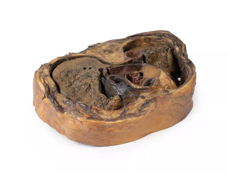
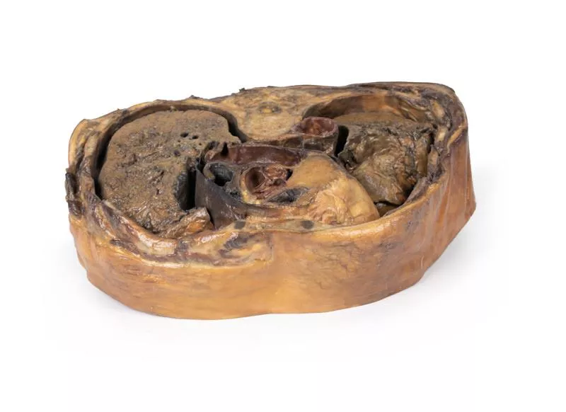
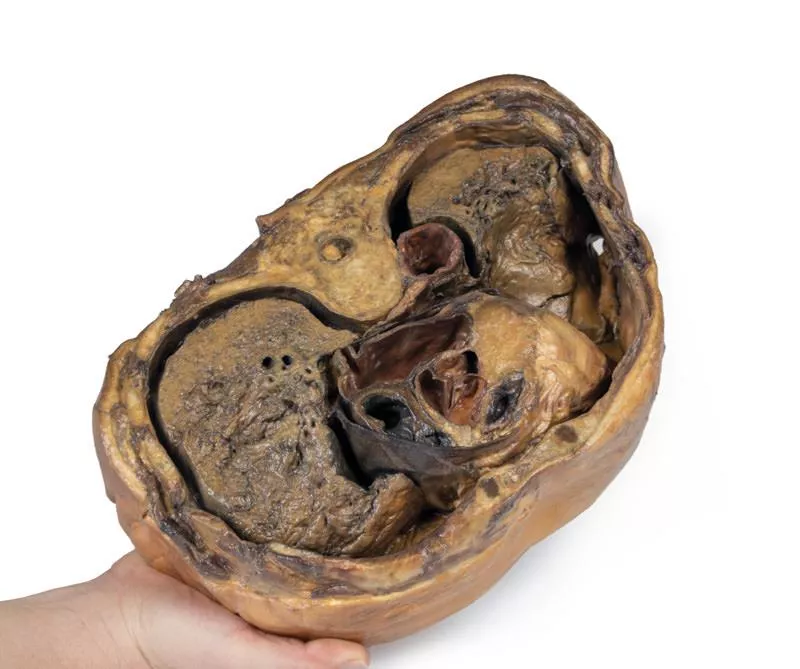
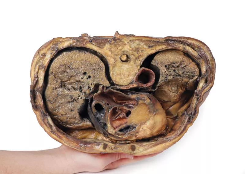
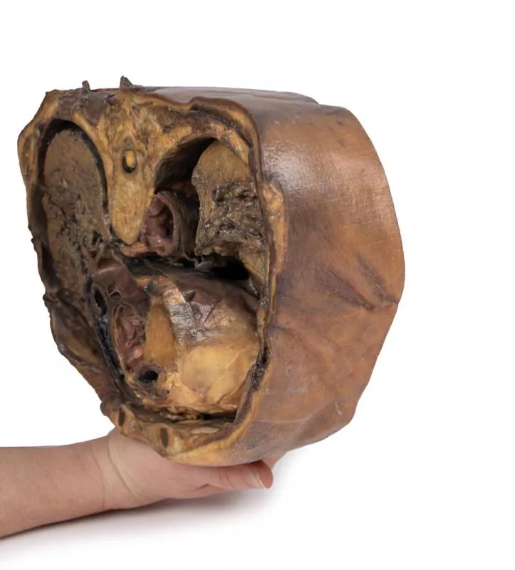
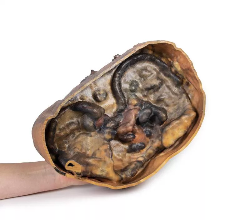

Product information "Thoracic cross section at T6"
This detailed 3D model presents a transverse cross-section of the thorax at the level of the T6 vertebra, offering a clear view of thoracic anatomy in relation to skeletal, vascular, respiratory, and cardiac structures.
Key Features:
Posterior Structures
- Begins medially with the spinal cord within the vertebral canal
- Costovertebral joints of the 6th ribs are visible, along with surrounding ribs forming the thoracic wall
Anterior Thoracic Wall
- Costosternal joints show the connection between ribs and sternum
Major Thoracic Organs
- Oesophagus located anterior to the vertebral body
- Descending aorta situated lateral to the vertebral body
Lungs and Pleural Cavities
- Within the pleural spaces (lined by parietal pleura):
Heart and Mediastinum
- In the middle mediastinum, the heart is shown within the pericardium, sectioned to display internal anatomy:
Key Features:
Posterior Structures
- Begins medially with the spinal cord within the vertebral canal
- Costovertebral joints of the 6th ribs are visible, along with surrounding ribs forming the thoracic wall
Anterior Thoracic Wall
- Costosternal joints show the connection between ribs and sternum
Major Thoracic Organs
- Oesophagus located anterior to the vertebral body
- Descending aorta situated lateral to the vertebral body
Lungs and Pleural Cavities
- Within the pleural spaces (lined by parietal pleura):
- Right lung: middle and inferior lobes
- Left lung: inferior lobe
- Left lung: inferior lobe
Heart and Mediastinum
- In the middle mediastinum, the heart is shown within the pericardium, sectioned to display internal anatomy:
- Left atrium (posterior)
- Aortic valve, right ventricle, and right atrium in clockwise order
- Aortic valve, right ventricle, and right atrium in clockwise order
Login
Erler-Zimmer
Erler-Zimmer Medical GmbH
Hauptstrasse 27
77886 Lauf
Germany
info@erler-zimmer.de
Achtung! Medizinisches Ausbildungsmaterial, kein Spielzeug. Nicht geeignet für Personen unter 14 Jahren.
Attention! Medical training material, not a toy. Not suitable for persons under 14 years of age.
Other customers also bought
")
Brain (Cerebrum)
This 3D model provides a unique perspective on the anatomy of the cerebrum relative to the meninges. The cerebrum has been separated from the brainstem and cerebellum, with only parts of the midbrain and cerebral peduncles visible on the inferior surface. Adjacent to the cut section the olfactory tracts and bulbs can be seen extending along the inferior margin of the frontal lobes of the cerebrum. Varying dissection between the left and right cerebral hemispheres allows an appreciation for the organisation of the brain and meninges as it would normally appear within the cranial cavity. In the midline, the dura mater has been preserved from anterior (rostral) to posterior. The central portion of the true (endosteal) dura opened to expose the superior sagittal sinus (between endosteal and meningeal layers of dura mater). Numerous arachnoid granulations (clusters of arachnoid villi) are visible within the opened superior sagittal sinus – as well as across the margins of the preserved dura. On the right cerebral hemisphere, the dura mater has been completely removed to expose the underlying arachnoid mater, which obscures the appearance of the underlying cerebral gyri and sulci as well as the terminal branches of cerebral arteries. In contrast, the arachnoid mater has dissected across most of the hemisphere (excepting a margin for reference) to expose the gyri and sulci covered in pia mater. This allows a clear view of the lateral sulcus and the central sulcus, with the latter defining the boundaries of the frontal and parietal lobes - and separating the primary sensory and motor cortical areas on the gyri on either side of the sulcus.

Superficial Face
This detailed 3D model features a superficial dissection of the left face just anterior to the ear, with false colouring highlighting key neurovascular structures and muscles of facial expression. It serves as a focused complement to the broader dissection in our HW 45 model. Undissected areas have been digitally removed for clarity.Key Features:Parotid Region & Facial Nerve Branches:Exposes the parotid gland and duct, along with terminal branches of the facial nerve (CN VII): cervical, mandibular, buccal, zygomatic, and temporal. Facial Vessels & Nerve-Vessel Relationships:Shows the facial artery and vein in relation to CN VII branches. Vessels are traced from the mandible to the orbit, offering anatomical landmarks.Muscles of Facial Expression (Highlighted):Includes masseter, depressor anguli oris, zygomaticus major & minor, orbicularis oris, nasalis, levator labii superioris alaeque nasi, procerus, and orbicularis oculi.Temporal & Forehead Structures:Displays the auriculotemporal nerve and superficial temporal artery over the temporal fascia, with part of the temporalis muscle visible.Superiorly, the supraorbital nerve and vessels ascend on the epicranial aponeurosis, overlying the frontalis muscle.This model offers a concise yet richly detailed view of facial anatomy, ideal for teaching nerve-muscle-vascular relationships in the superficial face.

Liver with vessels and gallbladder
This liver specimen displays notable differences compared to a typical liver. It is less wedge-shaped and elongated in the superoinferior dimension, resulting in a greater vertical height when viewed from the posterior. Size- Measures approximately 18 cm along the midclavicular line.- Typical livers measure under 16 cm in this dimension.- The increased length suggests mild hepatomegaly (enlargement). Important Notes- Size estimates may be affected by specimen preservation and fixing, which can cause some distortion.- Diagnosing hepatomegaly based on a single measurement is limited and varies with individual anatomy, measurement technique, sex, and body mass index (BMI). Anatomical VariationsThis specimen does not match common anatomical variations often confused with hepatomegaly such as:- Riedel’s lobe: a downward projection of the right lobe- Beaver tail liver: elongated left lobe- Papillary process from the caudate lobe

Internal abdominal wall
This detailed 3D model captures the internal surface of the anterior abdominal wall—a region often removed or damaged during dissections. It complements our MP1130 abdominal specimen, where the anterior wall has been removed, providing a clear view of key muscle and connective tissue structures. Key Features:Muscle Fibers & Aponeurosis:The horizontally oriented transversus abdominus muscle fibers converge toward their aponeurosis (tendon sheet), visible especially along the specimen’s superior margins. Arcuate Line:Located in the lower third of the model, this landmark marks where the aponeurosis shifts relative to the rectus abdominus muscle.- Above the arcuate line: Aponeurosis fibers split evenly around the rectus abdominus.- Below the arcuate line: All aponeurotic fibers pass anteriorly to the rectus abdominus, reflecting a change in abdominal wall structure. Vascular Structures:Inferior Epigastric Arteries & Veins:These vessels originate from the external iliac arteries and veins, ascending superiorly through the anterior abdominal wall. Hesselbach’s Triangle:On the right side of the model, the orientation of the inferior epigastric artery relative to the rectus abdominus muscle defines the apex of the inguinal (Hesselbach’s) triangle—a critical anatomical region often associated with direct inguinal hernias. (Note: The inguinal ligament forming the base of this triangle is not present in this specimen.) Embryological Remnant: Median Abdominal Ligament:Positioned midline between the two rectus abdominus muscles, this fold of parietal peritoneum covers the urachus, a fibrous remnant from embryological development extending from the bladder to the umbilicus.

Transverse Section of the head
This 3D model features a transverse section through the cranial cavity with a deep dissection of the face, orbit, and temporomandibular joint (TMJ) region. It offers a comprehensive view of both intracranial and facial structures.Key Features:Cranial Cavity & Brain- Partial dura mater removal; dissection reveals lateral and third ventricles, falx cerebri, choroid plexus, and optic pathways- Middle cerebral artery visible in the lateral fissure- Key vascular structures: internal carotid, anterior and middle cerebral arteries Left OrbitRoof removed; exposure of: Frontal nerve, lacrimal gland, superior oblique, medial rectus, nasociliary nerveRight OrbitSuperficial tissues removed; shows: All extraocular muscles, including inferior oblique, and levator palpebrae superiorisFace & TMJ (Right Side)- Exposed: infraorbital nerve/artery, masseter (both heads), and temporalis muscle near pterion- Parotid gland dissected to show mandibular condyle in glenoid fossa and external ear alignment

Thorax with heart and vessels
This highly detailed 3D model depicts the key anatomy of the superior thoracic aperture, mediastinum, and adjacent neck and thoracic structures, with both clavicles and select muscular and venous elements removed to enhance visibility and educational impact.Anatomical Highlights:Superior Thoracic Aperture:- Trachea visible at the top, with a robust ring of cartilage.- Rib 1 exposed from lateral to medial, including insertion of the anterior scalene muscle.- Removal of clavicles allows unobstructed views into the upper thoracic corridor. Vascular Anatomy:- Right subclavian artery, situated above rib 1, gives rise to the thyrocervical trunk.- Left subclavian artery, also above rib 1, branches into the suprascapular artery.- Both common carotid arteries are visible; the left carotid sheath includes the left vagus nerve.Nervous System:- Left vagus nerve follows the left carotid artery within the carotid sheath; left recurrent laryngeal nerve loops under the aorta.- Right vagus nerve and right phrenic nerve retracted during dissection; left phrenic nerve remains anterior to the heart, tracing to the diaphragm.- Elements of the left brachial plexus are visible—from roots to trunks—including the dorsal scapular nerve.Mediastinal & Cardiac Orientation:- Arch of the aorta, with brachiocephalic trunk, left common carotid, and left subclavian artery, sits just above the heart.- Pulmonary trunk emerges immediately superior to the heart.- Left anterior descending artery (LAD) courses along the anterior heart surface.- Superior vena cava lies to the right and posterior to the ascending aorta.- Right phrenic nerve positioned posteriorly to the heart; left phrenic nerve runs in its connective tissue anteriorly.Inferior Thorax & Diaphragm:- Ribs 8–12 and associated external intercostal musculature visible; muscle fibers run inferomedially into fascial layers.- Right hemidiaphragm sits higher than the left, reflecting the presence of the liver beneath. Applications & BenefitsClinical Relevance:Ideal for visualizing neck and thoracic anatomy relevant to cardiovascular, respiratory, and nerve-related procedures, including laryngeal, thoracic, and brachial plexus surgery.Educational Utility: Offers a clean, accessible view of mediastinal compartments, vascular pathways, and nerve trajectories without obstruction by bone or superficial structures. Enhanced Learning: The model’s clarity supports instruction in radiographic interpretation, thoracic surgical approaches, and anatomy exams.

Continuous innovation

Social responsibility

Active customer orientation

Understanding quality

Sustainable actions

ISO 9001 certification