


Product information "Brain (Cerebrum)"
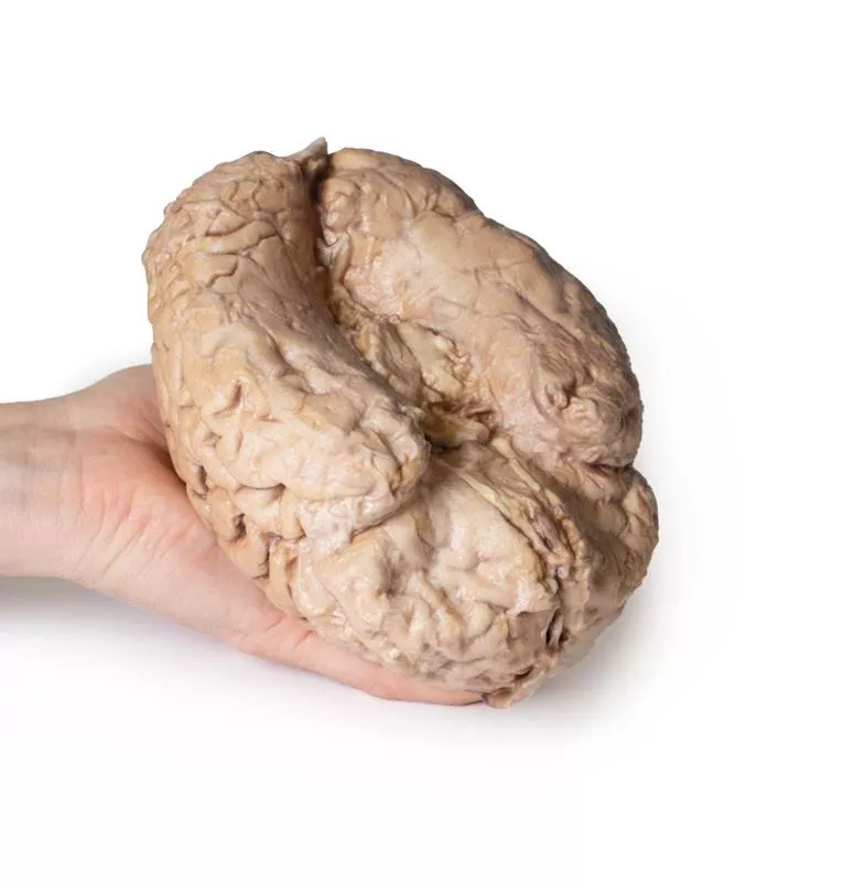
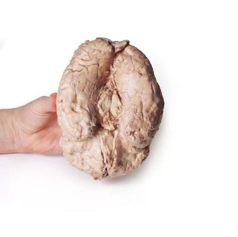
This 3D model provides a unique perspective on the anatomy of the cerebrum relative to the meninges.
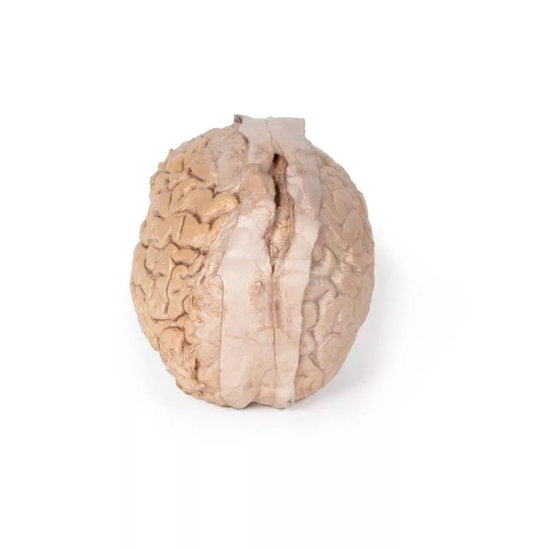
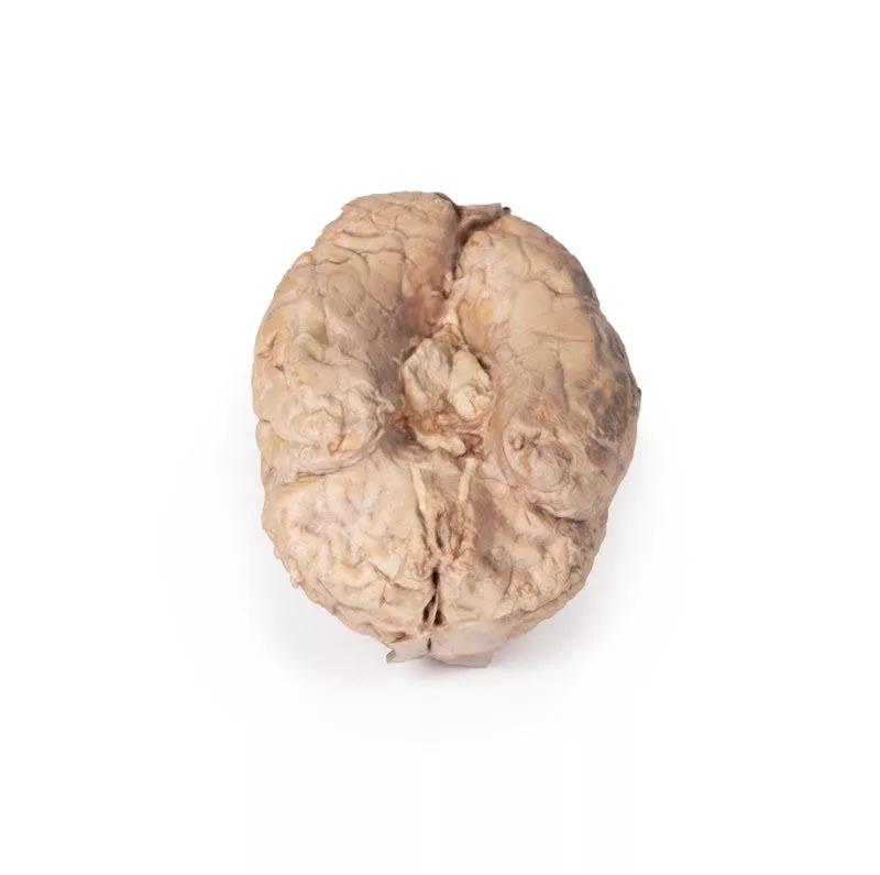
The cerebrum has been separated from the brainstem and cerebellum, with only parts of the midbrain and cerebral peduncles visible on the inferior surface. Adjacent to the cut section the olfactory tracts and bulbs can be seen extending along the inferior margin of the frontal lobes of the cerebrum.
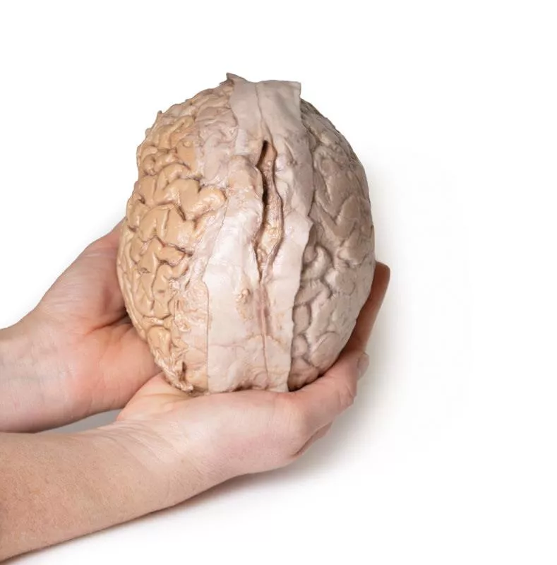
Varying dissection between the left and right cerebral hemispheres allows an appreciation for the organisation of the brain and meninges as it would normally appear within the cranial cavity. In the midline, the dura mater has been preserved from anterior (rostral) to posterior. The central portion of the true (endosteal) dura opened to expose the superior sagittal sinus (between endosteal and meningeal layers of dura mater).
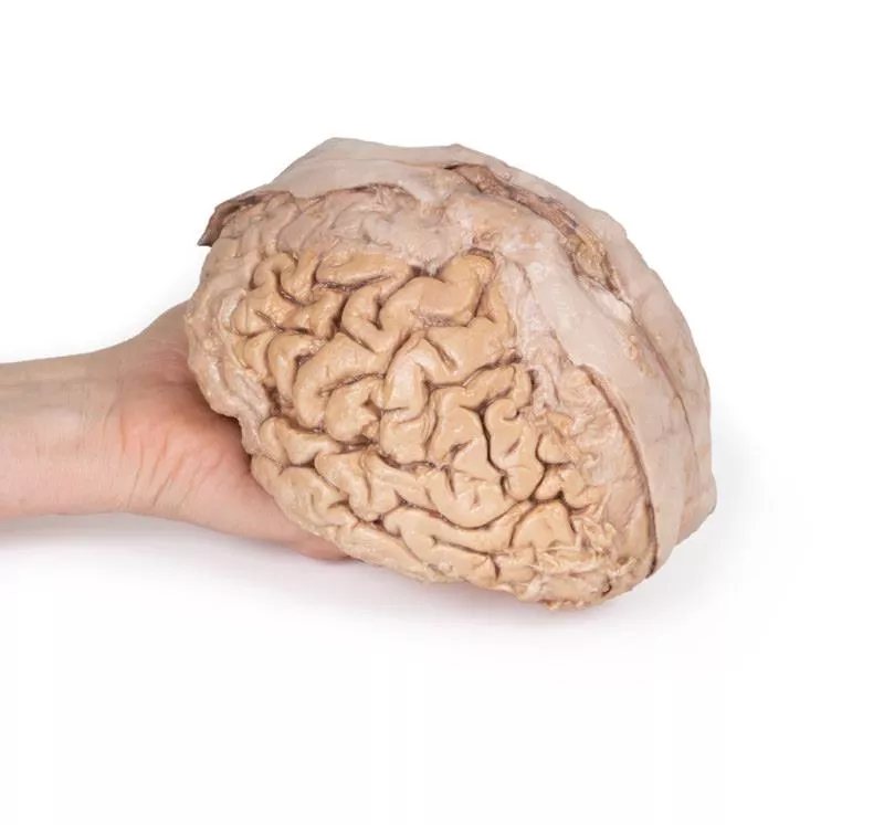
Numerous arachnoid granulations (clusters of arachnoid villi) are visible within the opened superior sagittal sinus – as well as across the margins of the preserved dura. On the right cerebral hemisphere, the dura mater has been completely removed to expose the underlying arachnoid mater, which obscures the appearance of the underlying cerebral gyri and sulci as well as the terminal branches of cerebral arteries. In contrast, the arachnoid mater has dissected across most of the hemisphere (excepting a margin for reference) to expose the gyri and sulci covered in pia mater. This allows a clear view of the lateral sulcus and the central sulcus, with the latter defining the boundaries of the frontal and parietal lobes - and separating the primary sensory and motor cortical areas on the gyri on either side of the sulcus.
The cerebrum has been separated from the brainstem and cerebellum, with only parts of the midbrain and cerebral peduncles visible on the inferior surface. Adjacent to the cut section the olfactory tracts and bulbs can be seen extending along the inferior margin of the frontal lobes of the cerebrum.
Varying dissection between the left and right cerebral hemispheres allows an appreciation for the organisation of the brain and meninges as it would normally appear within the cranial cavity. In the midline, the dura mater has been preserved from anterior (rostral) to posterior. The central portion of the true (endosteal) dura opened to expose the superior sagittal sinus (between endosteal and meningeal layers of dura mater).
Numerous arachnoid granulations (clusters of arachnoid villi) are visible within the opened superior sagittal sinus – as well as across the margins of the preserved dura. On the right cerebral hemisphere, the dura mater has been completely removed to expose the underlying arachnoid mater, which obscures the appearance of the underlying cerebral gyri and sulci as well as the terminal branches of cerebral arteries. In contrast, the arachnoid mater has dissected across most of the hemisphere (excepting a margin for reference) to expose the gyri and sulci covered in pia mater. This allows a clear view of the lateral sulcus and the central sulcus, with the latter defining the boundaries of the frontal and parietal lobes - and separating the primary sensory and motor cortical areas on the gyri on either side of the sulcus.
Login
Erler-Zimmer
Erler-Zimmer Medical GmbH
Hauptstrasse 27
77886 Lauf
Germany
info@erler-zimmer.de
Achtung! Medizinisches Ausbildungsmaterial, kein Spielzeug. Nicht geeignet für Personen unter 14 Jahren.
Attention! Medical training material, not a toy. Not suitable for persons under 14 years of age.
Other customers also bought

Brain Hemisection
This 3D model is a midsagittal hemisection through a whole brain, preserving the right side anatomy and deep brain structures and spaces visible in the midline. In lateral view, the right cerebral and cerebellar hemispheres are covered in the arachnoid mater. In the midline view, the brain regions from the cerebrum to the medulla oblongata are preserved. Centrally, the third ventricle is opened, with an intact septum pellucidum superiorly positioned and obscuring the lateral ventricles within the cerebral hemisphere. On the inferior margin of the third ventricle both the right mamillary body and right optic tract can be observed, whereas posteriorly the cerebral aqueduct can be observed extending across the midbrain between the tectum and tegmentum towards the fourth ventricle (between the cerebellum and pons). The cerebellum is separated from the occipital lobe by a preserved portion of the tentorium cerebelli, and in cross-section the cerebellar cortex helps form the prominent arbor vitae. A series of arterial branches have been false coloured to contrast their course across the preserved brain structures. In the midsagittal view the anterior cerebral artery courses from around the corpus callosum to supply the cingulate gyrus and other midline cortical regions. The base of the middle cerebral artery can be seen passing deep between the temporal and frontal lobes, with the posterior communicating artery connecting it to a small remnant of the posterior cerebral artery. Adjacent to the posterior cerebral is the superior cerebellar artery, extending laterally to pass between the temporal lobe and the cerebellum before passing deep into the transverse fissure.

Transverse Section of the head
This 3D model features a transverse section through the cranial cavity with a deep dissection of the face, orbit, and temporomandibular joint (TMJ) region. It offers a comprehensive view of both intracranial and facial structures.Key Features:Cranial Cavity & Brain- Partial dura mater removal; dissection reveals lateral and third ventricles, falx cerebri, choroid plexus, and optic pathways- Middle cerebral artery visible in the lateral fissure- Key vascular structures: internal carotid, anterior and middle cerebral arteries Left OrbitRoof removed; exposure of: Frontal nerve, lacrimal gland, superior oblique, medial rectus, nasociliary nerveRight OrbitSuperficial tissues removed; shows: All extraocular muscles, including inferior oblique, and levator palpebrae superiorisFace & TMJ (Right Side)- Exposed: infraorbital nerve/artery, masseter (both heads), and temporalis muscle near pterion- Parotid gland dissected to show mandibular condyle in glenoid fossa and external ear alignment

Brain stem, deep cerebral and diencephalic structures
This 3D model preserves the several deep cerebral and diencephalic structures through to the proximal medulla oblongata and compliment the other isolated brainstem (MP1101) in our series.Superiorly, on the right side of the 3D model, the lentiform (lenticular) nucleus is in place and the corona radiata of the internal capsule is seen emerging around it. On the left, the lentiform nucleus is absent, but the caudate nucleus head and body are present medially on both sides, wrapping medial to the preserved internal capsule margins and leading to the amygdaloid bodies on each side. The thalami are present bilaterally, and the third ventricle is opened slightly in the midline inferior to the epithalamus (pineal gland).Anteriorly, the cerebral peduncles are present, with the optic nerves extending from the preserved chiasm and tracts. The interpeduncular region is exposed with both the mammillary bodies and the sectioned infundibulum visible. Caudal to the interpeduncular region is the pons preserving the origins of the middle cerebellar peduncles as well as the origins of cranial nerves V, VII, and VIII. The portion of the medulla oblongata preserved possesses prominent pyramids and olives.Posteriorly, the superior and inferior colliculi sit just superior to the sectioned superior cerebellar peduncles, and the fourth ventricle is opened to expose the rhomboid fossa and features of the floor: the medial eminence, facial colliculus, hypoglossal triangle, the vestibular triangle and the vagal triangle.

Pericardial space
This detailed 3D anatomical model displays the pericardial cavity and reflections with the heart removed, allowing clear visualization of key structures within the middle mediastinum.Key Features:Pericardium:Shows the full extent of the parietal pericardium, continuous with the visceral layer (epicardium), and false-colored to indicate the positions of the atria, ventricles, and great vessels. Mediastinal Landmarks:Highlights the base, apex, diaphragmatic, and pulmonary surfaces of the heart by their impressions within the pericardial cavity.Great Vessels:- Aorta (ascending, arch, and descending)- Superior and Inferior Vena Cava- Pulmonary trunk and arteries- Pulmonary veins (4 total) - All shown in their natural positions relative to the pericardium.Pericardial Sinuses:- Transverse sinus: Located between arteries and veins; relevant for surgical access.- Oblique sinus: Posterior recess between pulmonary veins.Educational Use:- Ideal for teaching thoracic and cardiac anatomy, including pericardial reflections, mediastinal relationships, and surgical landmarks.- Useful in medical training, surgical planning, and radiological orientation.

Sagittal Section of Head and Neck with Infratemporal Fossa and Carotid Sheath Dissection
This 3D model complements the H11 and H12 head and neck specimens, offering a clear view of the endocranial cavity without the brain, alongside a lateral dissection of the face, infratemporal region, and neck.Key Features:Endocranial Cavity- Brain removed; dura mater, tentorium cerebelli, and superior sagittal sinus fully visible- Several cranial nerves (CN II, III, V, VI, VII, VIII) seen piercing the dura- Pituitary gland preserved in sella turcica, left vertebral artery visible in posterior fossa Lateral Facial & Infratemporal Region- Facial artery and vein retained, dissected free of superficial tissues- Partial removal of mandible and zygomatic arch reveals:- Inferior alveolar and lingual nerves, posterior deep temporal artery, and TMJ articulation- Visible branches of the external carotid artery, including maxillary and superficial temporal arteriesNeck Anatomy- Facial nerve (CN VII) near posterior belly of digastric- Dissected carotid sheath showing internal/external carotid arteries, internal jugular vein, and vagus nerve (CN X)- Hypoglossal nerve (CN XII) and facial artery near submandibular gland- Hyoid bone, thyroid gland, and larynx visible- Cervical plexus branches on scalene muscles; brachial plexus roots preserved near internal jugular vein

Brain Stem, isolated anatomy from midbrain to medulla oblongata
This 3D model provides a view of the isolated brainstem anatomy from the midbrain to the medulla oblongata, and compliments the other diencephalon/brainstem 3D model (MP1100) in our series. Rostrally, the 3D model has been sectioned at an angle from the overlying diencephalon while retaining the mamillary bodies of the hypothalamus between the cerebral peduncles (anteriorly) and the pineal gland/epithalamus (posteriorly). Posteriorly, the corpora quadrigemina (the collective superior and inferior colliculi) of the midbrain are prominent adjacent to the superior cerebellar peduncles. The cerebellum itself has been removed, leaving the cross-section of the middle and inferior cerebellar peduncles on each side. Inferior to the sectioned peduncles is the partially opened fourth ventricle and remnants of the posterior inferior cerebellar arteries. On the ventral aspect of the 3D model the pons is preserved with the origin of the trigeminal nerve (CN V) preserved (particularly on the left side). Inferior to the pons on the medulla oblongata, both the pyramids and olives are visible on both sides (particularly clear on the right).

Continuous innovation

Social responsibility

Active customer orientation

Understanding quality

Sustainable actions

ISO 9001 certification