


Brain Stem, isolated anatomy from midbrain to medulla oblongata
Question regarding article:
Product information "Brain Stem, isolated anatomy from midbrain to medulla oblongata"
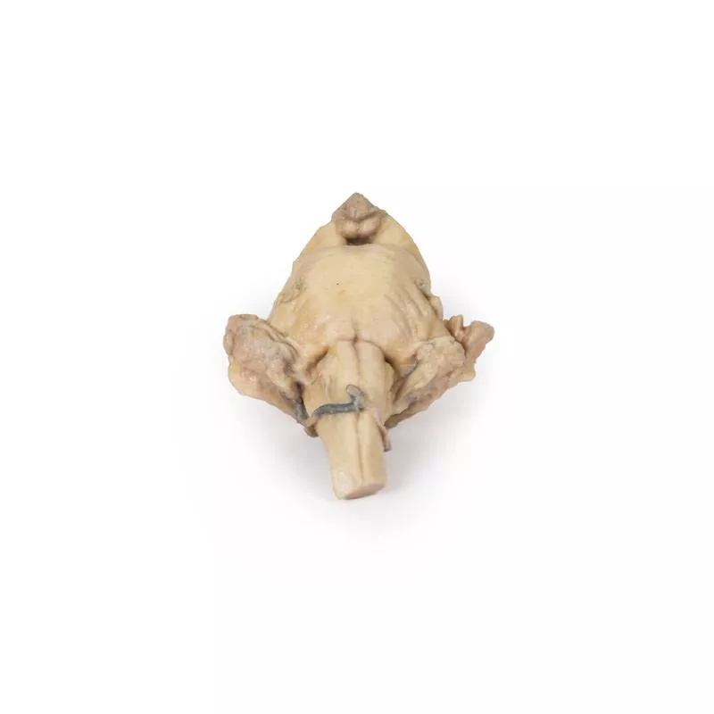
This 3D model provides a view of the isolated brainstem anatomy from the midbrain to the medulla oblongata, and compliments the other diencephalon/brainstem 3D model (MP1100) in our series.
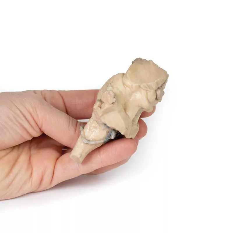
Rostrally, the 3D model has been sectioned at an angle from the overlying diencephalon while retaining the mamillary bodies of the hypothalamus between the cerebral peduncles (anteriorly) and the pineal gland/epithalamus (posteriorly).
Posteriorly, the corpora quadrigemina (the collective superior and inferior colliculi) of the midbrain are prominent adjacent to the superior cerebellar peduncles. The cerebellum itself has been removed, leaving the cross-section of the middle and inferior cerebellar peduncles on each side. Inferior to the sectioned peduncles is the partially opened fourth ventricle and remnants of the posterior inferior cerebellar arteries.
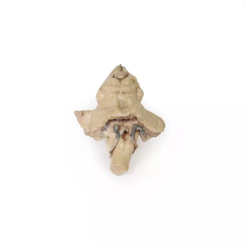
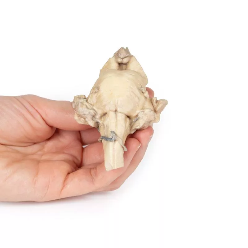
On the ventral aspect of the 3D model the pons is preserved with the origin of the trigeminal nerve (CN V) preserved (particularly on the left side). Inferior to the pons on the medulla oblongata, both the pyramids and olives are visible on both sides (particularly clear on the right).
Rostrally, the 3D model has been sectioned at an angle from the overlying diencephalon while retaining the mamillary bodies of the hypothalamus between the cerebral peduncles (anteriorly) and the pineal gland/epithalamus (posteriorly).
Posteriorly, the corpora quadrigemina (the collective superior and inferior colliculi) of the midbrain are prominent adjacent to the superior cerebellar peduncles. The cerebellum itself has been removed, leaving the cross-section of the middle and inferior cerebellar peduncles on each side. Inferior to the sectioned peduncles is the partially opened fourth ventricle and remnants of the posterior inferior cerebellar arteries.
On the ventral aspect of the 3D model the pons is preserved with the origin of the trigeminal nerve (CN V) preserved (particularly on the left side). Inferior to the pons on the medulla oblongata, both the pyramids and olives are visible on both sides (particularly clear on the right).
Login
Erler-Zimmer
Erler-Zimmer Medical GmbH
Hauptstrasse 27
77886 Lauf
Germany
info@erler-zimmer.de
Achtung! Medizinisches Ausbildungsmaterial, kein Spielzeug. Nicht geeignet für Personen unter 14 Jahren.
Attention! Medical training material, not a toy. Not suitable for persons under 14 years of age.
Other customers also bought

Median Section through head sagittal section of head with deep dissection
This 3D model combines a midsagittal section of the head with preservation of brain and cranial cavity anatomy, with a unique deep dissection of the pharyngeal region via removal of basicranial bone and the anterior parts of the atlas and axis. As the opposing side is undissected it has been digitally eliminated from the model. Within the endocranial cavity the preservation of dura mater retains the superior sagittal sinus across much of its course from anterior to posterior, reaching the confluence of sinuses visible in cross-section. Both the tentorium cerebelli and the falx cerebelli are preserved. The cerebrum is well-reserved with retention of the cingulate gyrus and sulcus, and removal of the septum pellucidum inferior to the corpus callosum providing a view into the lateral ventricle (with retention of the interventricular foramen at the inferior margin of the septum). The diencephalon and midbrain structures (epithalamus, colliculi, mamillary body, infundibulum) are all appreciable in cross-section as is the cerebellar hemisphere and fourth ventricle. Small views of the anterior cerebral and posterior inferior cerebellar arteries are visible (and false coloured).Outside the endocranium, removal of parts of the occipital, temporal and sphenoid bones (alongside the atlas and axis) has been coupled with removal of the pharyngeal constrictors, carotid sheath and oral mucosa to demonstrate a unique view of several key neurovascular and glandular structures. Within the zone of removed tissue there is partial exposure of the right common carotid artery within the dissected petrous portion of the temporal, as well as partial exposure of the left vertebral artery through disruption of the occipital and dural covering.The medial and lateral pterygoids are exposed near the posterior margin of the largely intact nasal cavity. Between the exposed dura and medulla and the pterygoids (and trapped deep to the sectioned and reflected stylohyoid muscle) the dissected carotid sheath has exposed the internal jugular vein, the vagus nerve, the internal carotid artery (with overriding ascending pharyngeal artery from the external carotid artery), and the sympathetic trunk (with superior cervical ganglion and internal carotid nerve). Immediately anterior to this bundle of neurovascular structures is the external carotid artery, giving rise to the ascending pharyngeal artery, a common trunk for the lingual and facial arteries, and then continuing superiorly out of the plane of dissection. The submandibular gland can be seen resting on the mylohyoid muscle near the lingual artery (which passes deep relative to the gland), with the duct passing towards the genu of the mandible and the origin of the reflected genioglossus muscle. At the inferior border of the specimen, the reflected margin of the dissected tongue the hypoglossal nerve can be seen deep to the lingual artery.
")
Brain (Cerebrum)
This 3D model provides a unique perspective on the anatomy of the cerebrum relative to the meninges. The cerebrum has been separated from the brainstem and cerebellum, with only parts of the midbrain and cerebral peduncles visible on the inferior surface. Adjacent to the cut section the olfactory tracts and bulbs can be seen extending along the inferior margin of the frontal lobes of the cerebrum. Varying dissection between the left and right cerebral hemispheres allows an appreciation for the organisation of the brain and meninges as it would normally appear within the cranial cavity. In the midline, the dura mater has been preserved from anterior (rostral) to posterior. The central portion of the true (endosteal) dura opened to expose the superior sagittal sinus (between endosteal and meningeal layers of dura mater). Numerous arachnoid granulations (clusters of arachnoid villi) are visible within the opened superior sagittal sinus – as well as across the margins of the preserved dura. On the right cerebral hemisphere, the dura mater has been completely removed to expose the underlying arachnoid mater, which obscures the appearance of the underlying cerebral gyri and sulci as well as the terminal branches of cerebral arteries. In contrast, the arachnoid mater has dissected across most of the hemisphere (excepting a margin for reference) to expose the gyri and sulci covered in pia mater. This allows a clear view of the lateral sulcus and the central sulcus, with the latter defining the boundaries of the frontal and parietal lobes - and separating the primary sensory and motor cortical areas on the gyri on either side of the sulcus.

Superficial Face
This detailed 3D model features a superficial dissection of the left face just anterior to the ear, with false colouring highlighting key neurovascular structures and muscles of facial expression. It serves as a focused complement to the broader dissection in our HW 45 model. Undissected areas have been digitally removed for clarity.Key Features:Parotid Region & Facial Nerve Branches:Exposes the parotid gland and duct, along with terminal branches of the facial nerve (CN VII): cervical, mandibular, buccal, zygomatic, and temporal. Facial Vessels & Nerve-Vessel Relationships:Shows the facial artery and vein in relation to CN VII branches. Vessels are traced from the mandible to the orbit, offering anatomical landmarks.Muscles of Facial Expression (Highlighted):Includes masseter, depressor anguli oris, zygomaticus major & minor, orbicularis oris, nasalis, levator labii superioris alaeque nasi, procerus, and orbicularis oculi.Temporal & Forehead Structures:Displays the auriculotemporal nerve and superficial temporal artery over the temporal fascia, with part of the temporalis muscle visible.Superiorly, the supraorbital nerve and vessels ascend on the epicranial aponeurosis, overlying the frontalis muscle.This model offers a concise yet richly detailed view of facial anatomy, ideal for teaching nerve-muscle-vascular relationships in the superficial face.

Brain Hemisection
This 3D model is a midsagittal hemisection through a whole brain, preserving the right side anatomy and deep brain structures and spaces visible in the midline. In lateral view, the right cerebral and cerebellar hemispheres are covered in the arachnoid mater. In the midline view, the brain regions from the cerebrum to the medulla oblongata are preserved. Centrally, the third ventricle is opened, with an intact septum pellucidum superiorly positioned and obscuring the lateral ventricles within the cerebral hemisphere. On the inferior margin of the third ventricle both the right mamillary body and right optic tract can be observed, whereas posteriorly the cerebral aqueduct can be observed extending across the midbrain between the tectum and tegmentum towards the fourth ventricle (between the cerebellum and pons). The cerebellum is separated from the occipital lobe by a preserved portion of the tentorium cerebelli, and in cross-section the cerebellar cortex helps form the prominent arbor vitae. A series of arterial branches have been false coloured to contrast their course across the preserved brain structures. In the midsagittal view the anterior cerebral artery courses from around the corpus callosum to supply the cingulate gyrus and other midline cortical regions. The base of the middle cerebral artery can be seen passing deep between the temporal and frontal lobes, with the posterior communicating artery connecting it to a small remnant of the posterior cerebral artery. Adjacent to the posterior cerebral is the superior cerebellar artery, extending laterally to pass between the temporal lobe and the cerebellum before passing deep into the transverse fissure.

Brain stem, deep cerebral and diencephalic structures
This 3D model preserves the several deep cerebral and diencephalic structures through to the proximal medulla oblongata and compliment the other isolated brainstem (MP1101) in our series.Superiorly, on the right side of the 3D model, the lentiform (lenticular) nucleus is in place and the corona radiata of the internal capsule is seen emerging around it. On the left, the lentiform nucleus is absent, but the caudate nucleus head and body are present medially on both sides, wrapping medial to the preserved internal capsule margins and leading to the amygdaloid bodies on each side. The thalami are present bilaterally, and the third ventricle is opened slightly in the midline inferior to the epithalamus (pineal gland).Anteriorly, the cerebral peduncles are present, with the optic nerves extending from the preserved chiasm and tracts. The interpeduncular region is exposed with both the mammillary bodies and the sectioned infundibulum visible. Caudal to the interpeduncular region is the pons preserving the origins of the middle cerebellar peduncles as well as the origins of cranial nerves V, VII, and VIII. The portion of the medulla oblongata preserved possesses prominent pyramids and olives.Posteriorly, the superior and inferior colliculi sit just superior to the sectioned superior cerebellar peduncles, and the fourth ventricle is opened to expose the rhomboid fossa and features of the floor: the medial eminence, facial colliculus, hypoglossal triangle, the vestibular triangle and the vagal triangle.

Sagittal Section of head with infratemporal Fossa Dissection
This 3D model provides a combined midsagittal section through the head and superior neck coupled with a deep dissection into the infratemporal fossa region and superficial dissection of the scalp. In the preserved midsagittal section there is preservation of the endocranial contents, the nasal and oral cavities, and the pharynx to the level of the laryngeal cartilages. The nasal cavity is preserved nearly intact, except for a small window excised into the middle nasal concha to expose the ethmoid air cells. A very large sphenoid sinus exists in the individual just superior to the torus of the auditory tube in the nasopharynx. The oral cavity and laryngopharynx are undissected, with the larynx only preserve just distal to the level of the arytenoid cartilages and not including a clear set of vocal folds.Within the endocranial cavity, the sectioned brain is slightly off the midagittal plane, such that neither the superior sagittal sinus nor the third ventricle are clearly defined - but the lateral ventricle is open and part of the fourth ventricle is preserved between the pons and cerebellum. The gyri and sulci of the cerebrum are not well separated, but the cingulate gyrus and corpus callosum can be separated. Cross-sectioned views of the optic tract, pituitary gland, superior and inferior colliculi, superior cerebellar peduncle, and transition between the medulla oblongata and spinal cord are all visible. The tentorium cerebelli and confluence/transverse sinus is positioned between the cerebellar hemisphere and occipital lobe. Small portions of the posterior inferior cerebellar artery, vertebral arteries, basilar artery, and posterior cerebral and anterior cerebral arteries are visible in section.On the opposing side of the model, a superficial and deep dissection has opened a large window into the anatomy of the lateral scalp and infratemporal fossa. Across the scalp there is a well preserved posterior auricular nerve and superficial temporal artery highlighted on the superficial surface of the temporalis muscle. Anteriorly, the temporalis has been dissected to expose the deep temporal arteries arising from across the maxillary artery.The deep level of dissection has exposed parts of the infratemporal fossa (through partial removal of the mandibular ramus and corpus) and dissection of retromandibular tissues. At the inferior margin of the dissection window, the cut edge of the retromandibular vein lies adjacent to the submandibular gland and the ascending path of the facial artery as it cross towards to angle of the mouth. Just superior to the cut retromandibular vein is the posterior belly of the digastric muscle, overlying a small exposure of the deeper internal jugular vein.Just posterior to the retained ascending ramus of the mandible are the external carotid artery and the occipital artery (running in parallel prior to passing posteriorly). Tracing the external carotid artery superiorly, the posterior auricular artery, superficial temporal artery, and maxillary artery are all visible. The maxillary artery passes deep to the lateral pterygoid muscle and into the infratemporal fossa, reappearing superior to the lateral pterygoid as it passes into the pterygomaxillary fissure. Along its course, it gives rise to the posterior deep temporal artery, the inferior alveolar artery (which is exposed in the dissected mandibular corpus), the anterior deep temporal artery, and the posterior superior alveolar artery. Finally, the inferior alveolar nerve can be seen coursing within the opened mandibular corpus, and the lingual nerve resting on the medial pterygoid. The buccinator muscle is also retained, with the distal part of the parotid duct preserved as it enters the muscle towards the oral mucosa.

Sinus Pathways
This 3D model shows a midsagittal to parasagittal section of the right head, highlighting the drainage pathways of the paranasal sinuses into the nasal cavity using colored markers. Anteriorly, the nasolacrimal duct (white) opens beneath the inferior nasal concha. The middle concha has been sectioned to reveal the maxillary sinus opening via the semilunar hiatus (green), and the drainage of the frontal sinus (blue), anterior (orange) and middle (yellow) ethmoidal cells. Posterior ethmoidal cells open into the superior meatus (purple), while the sphenoid sinus drains above the nasopharynx (red).The model also preserves key anatomical structures:The nasal cavity from nostril to auditory tube, the soft palate, uvula, and pharynx down to the epiglottis. The oral cavity is sectioned to show genioglossus and geniohyoid muscles. Within the cranial cavity, parts of the frontal lobe, optic structures, and pituitary gland are visible, along with the brainstem, cerebellum, and tentorium cerebelli. The transverse and sigmoid sinuses are seen bilaterally, with a portion of the medial temporal lobe and lateral ventricle also present.

Parasagittal Section of the head and neck
This high-resolution 3D model features a head and neck specimen sectioned just off the midsagittal plane, preserving critical midline structures often absent in similar models. Ideal for anatomical education, this model offers enhanced visibility due to fixative-induced brain shrinkage, exaggerating spaces between brain and skull.Key Features:Midline Anatomy Preserved:Includes the falx cerebri (anterior portion), septum pellucidum, interventricular foramen (of Monro), and nasal septum.Ventricular & Endocranial Structures:Clear views of the lateral and third ventricles, cerebral aqueduct, fourth ventricle, infundibulum, pituitary gland, and sphenoid sinus.Vascular Highlights:Displays the left vertebral artery, posterior cerebral artery (cross-section), and anterior cerebral artery branches around the corpus callosum.Detailed Nasal & Pharyngeal Regions:Shows relationships between the nasal septum, palate, auditory tube opening, and naso-/oropharynx.Laryngeal and Tracheal Anatomy:Includes epiglottis, arytenoid, thyroid cartilages, hyoid bone, and cross-sectional views of the vestibule, vestibular and vocal folds.This model provides a unique perspective of internal head and neck anatomy, combining anatomical depth with high educational value.

Continuous innovation

Social responsibility

Active customer orientation

Understanding quality

Sustainable actions

ISO 9001 certification