


Product information "Brain Hemisection"
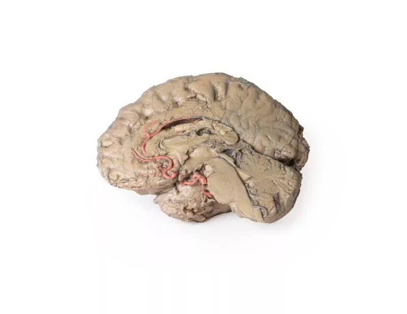
This 3D model is a midsagittal hemisection through a whole brain, preserving the right side anatomy and deep brain structures and spaces visible in the midline.
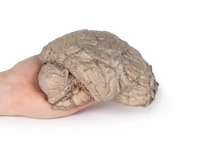
In lateral view, the right cerebral and cerebellar hemispheres are covered in the arachnoid mater. In the midline view, the brain regions from the cerebrum to the medulla oblongata are preserved. Centrally, the third ventricle is opened, with an intact septum pellucidum superiorly positioned and obscuring the lateral ventricles within the cerebral hemisphere. On the inferior margin of the third ventricle both the right mamillary body and right optic tract can be observed, whereas posteriorly the cerebral aqueduct can be observed extending across the midbrain between the tectum and tegmentum towards the fourth ventricle (between the cerebellum and pons). The cerebellum is separated from the occipital lobe by a preserved portion of the tentorium cerebelli, and in cross-section the cerebellar cortex helps form the prominent arbor vitae.
A series of arterial branches have been false coloured to contrast their course across the preserved brain structures. In the midsagittal view the anterior cerebral artery courses from around the corpus callosum to supply the cingulate gyrus and other midline cortical regions. The base of the middle cerebral artery can be seen passing deep between the temporal and frontal lobes, with the posterior communicating artery connecting it to a small remnant of the posterior cerebral artery. Adjacent to the posterior cerebral is the superior cerebellar artery, extending laterally to pass between the temporal lobe and the cerebellum before passing deep into the transverse fissure.
Login
Erler-Zimmer
Erler-Zimmer Medical GmbH
Hauptstrasse 27
77886 Lauf
Germany
info@erler-zimmer.de
Achtung! Medizinisches Ausbildungsmaterial, kein Spielzeug. Nicht geeignet für Personen unter 14 Jahren.
Attention! Medical training material, not a toy. Not suitable for persons under 14 years of age.
Other customers also bought

Pericardial space
This detailed 3D anatomical model displays the pericardial cavity and reflections with the heart removed, allowing clear visualization of key structures within the middle mediastinum.Key Features:Pericardium:Shows the full extent of the parietal pericardium, continuous with the visceral layer (epicardium), and false-colored to indicate the positions of the atria, ventricles, and great vessels. Mediastinal Landmarks:Highlights the base, apex, diaphragmatic, and pulmonary surfaces of the heart by their impressions within the pericardial cavity.Great Vessels:- Aorta (ascending, arch, and descending)- Superior and Inferior Vena Cava- Pulmonary trunk and arteries- Pulmonary veins (4 total) - All shown in their natural positions relative to the pericardium.Pericardial Sinuses:- Transverse sinus: Located between arteries and veins; relevant for surgical access.- Oblique sinus: Posterior recess between pulmonary veins.Educational Use:- Ideal for teaching thoracic and cardiac anatomy, including pericardial reflections, mediastinal relationships, and surgical landmarks.- Useful in medical training, surgical planning, and radiological orientation.

Thoracic cross section at T6
This detailed 3D model presents a transverse cross-section of the thorax at the level of the T6 vertebra, offering a clear view of thoracic anatomy in relation to skeletal, vascular, respiratory, and cardiac structures.Key Features:Posterior Structures- Begins medially with the spinal cord within the vertebral canal- Costovertebral joints of the 6th ribs are visible, along with surrounding ribs forming the thoracic wall Anterior Thoracic Wall- Costosternal joints show the connection between ribs and sternumMajor Thoracic Organs- Oesophagus located anterior to the vertebral body- Descending aorta situated lateral to the vertebral bodyLungs and Pleural Cavities- Within the pleural spaces (lined by parietal pleura):- Right lung: middle and inferior lobes- Left lung: inferior lobeHeart and Mediastinum- In the middle mediastinum, the heart is shown within the pericardium, sectioned to display internal anatomy:- Left atrium (posterior)- Aortic valve, right ventricle, and right atrium in clockwise order

Liver with vessels and gallbladder
This liver specimen displays notable differences compared to a typical liver. It is less wedge-shaped and elongated in the superoinferior dimension, resulting in a greater vertical height when viewed from the posterior. Size- Measures approximately 18 cm along the midclavicular line.- Typical livers measure under 16 cm in this dimension.- The increased length suggests mild hepatomegaly (enlargement). Important Notes- Size estimates may be affected by specimen preservation and fixing, which can cause some distortion.- Diagnosing hepatomegaly based on a single measurement is limited and varies with individual anatomy, measurement technique, sex, and body mass index (BMI). Anatomical VariationsThis specimen does not match common anatomical variations often confused with hepatomegaly such as:- Riedel’s lobe: a downward projection of the right lobe- Beaver tail liver: elongated left lobe- Papillary process from the caudate lobe

Stomach
This 3D model is an isolated stomach with two dissection windows to expose the rugae and pylorus. A small portion of the terminal oesophagus is preserved at the cardiac region, and a small portion of the proximal duodenum beyond the pyloric sphincter. The large window within the body of the stomach allows for a clear view into the fundus and the well-developed rugae on the posterior aspect of the wall of the organ. The smaller window, opened just at the pyloric region, allows for an appreciation of the thickening of the organ wall at the pyloric sphincter just proximal to the start of the duodenum.

Parasagittal Section of the head and neck
This high-resolution 3D model features a head and neck specimen sectioned just off the midsagittal plane, preserving critical midline structures often absent in similar models. Ideal for anatomical education, this model offers enhanced visibility due to fixative-induced brain shrinkage, exaggerating spaces between brain and skull.Key Features:Midline Anatomy Preserved:Includes the falx cerebri (anterior portion), septum pellucidum, interventricular foramen (of Monro), and nasal septum.Ventricular & Endocranial Structures:Clear views of the lateral and third ventricles, cerebral aqueduct, fourth ventricle, infundibulum, pituitary gland, and sphenoid sinus.Vascular Highlights:Displays the left vertebral artery, posterior cerebral artery (cross-section), and anterior cerebral artery branches around the corpus callosum.Detailed Nasal & Pharyngeal Regions:Shows relationships between the nasal septum, palate, auditory tube opening, and naso-/oropharynx.Laryngeal and Tracheal Anatomy:Includes epiglottis, arytenoid, thyroid cartilages, hyoid bone, and cross-sectional views of the vestibule, vestibular and vocal folds.This model provides a unique perspective of internal head and neck anatomy, combining anatomical depth with high educational value.

Female pelvis deep dissection
This high-detail 3D model showcases a deep dissection of the female pelvis, isolated from surrounding regions, with emphasis on visceral, vascular, and ligamentous structures in relation to bony landmarks.Pelvic Organs & Peritoneal Structures- Sigmoid colon descends into the rectum over the pelvic brim, crossing the common and external iliac vessels.- Nearby: Sigmoid and superior rectal arteries, and the descending ureter.- Urinary bladder (collapsed) and uterus are positioned anteriorly in the true pelvis.- The broad ligament is retained, though ovaries, uterine tubes, ovarian and round ligaments are present but indistinct due to age-related atrophy.- Suspensory and round ligaments are detached from the peritoneum to expose surrounding vessels. Arteries & Veins- Internal iliac artery branches are visible bilaterally.- Median sacral artery is seen in the midline between the common iliac arteries.- Left side: Uterine artery only.- Right side: Uterine, superior vesical, and obturator arteries.- Inferior epigastric artery and vein arise from the external iliac vessels, visible near the inferior abdominal wall. Musculoskeletal Features- Right side: Entire femur and thigh muscles removed to expose:- Obturator membrane- Acetabular cartilage- Transverse acetabular ligament- Posterior dissection reveals:- Superior gluteal foramen and artery- Sacrospinous ligament (with sacrotuberous ligament removed)- Inferior rectal artery branches within the ischioanal fossa Nerves & Ligaments- Left sciatic nerve preserved within the greater sciatic foramen- Sacrotuberous ligament retained on the left- Ischioanal fossae on both sides show:- Inferior rectal artery branches- Pelvic diaphragm fibers- External anal sphincter integration with the rectal wall

Sinus Pathways
This 3D model shows a midsagittal to parasagittal section of the right head, highlighting the drainage pathways of the paranasal sinuses into the nasal cavity using colored markers. Anteriorly, the nasolacrimal duct (white) opens beneath the inferior nasal concha. The middle concha has been sectioned to reveal the maxillary sinus opening via the semilunar hiatus (green), and the drainage of the frontal sinus (blue), anterior (orange) and middle (yellow) ethmoidal cells. Posterior ethmoidal cells open into the superior meatus (purple), while the sphenoid sinus drains above the nasopharynx (red).The model also preserves key anatomical structures:The nasal cavity from nostril to auditory tube, the soft palate, uvula, and pharynx down to the epiglottis. The oral cavity is sectioned to show genioglossus and geniohyoid muscles. Within the cranial cavity, parts of the frontal lobe, optic structures, and pituitary gland are visible, along with the brainstem, cerebellum, and tentorium cerebelli. The transverse and sigmoid sinuses are seen bilaterally, with a portion of the medial temporal lobe and lateral ventricle also present.

Continuous innovation

Social responsibility

Active customer orientation

Understanding quality

Sustainable actions

ISO 9001 certification