


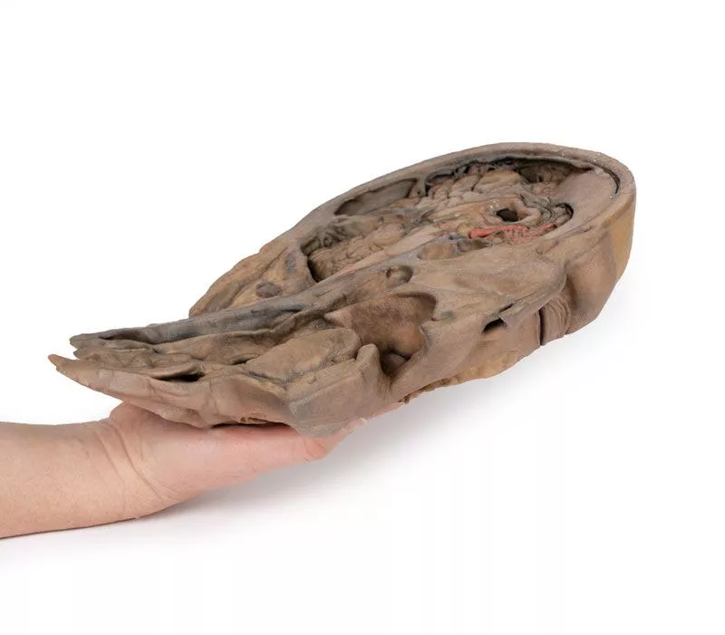


Product information "Parasagittal Section of the head and neck"
This high-resolution 3D model features a head and neck specimen sectioned just off the midsagittal plane, preserving critical midline structures often absent in similar models.
Ideal for anatomical education, this model offers enhanced visibility due to fixative-induced brain shrinkage, exaggerating spaces between brain and skull.
Key Features:
Midline Anatomy Preserved:
Includes the falx cerebri (anterior portion), septum pellucidum, interventricular foramen (of Monro), and nasal septum.
Ventricular & Endocranial Structures:
Clear views of the lateral and third ventricles, cerebral aqueduct, fourth ventricle, infundibulum, pituitary gland, and sphenoid sinus.
Vascular Highlights:
Displays the left vertebral artery, posterior cerebral artery (cross-section), and anterior cerebral artery branches around the corpus callosum.
Detailed Nasal & Pharyngeal Regions:
Shows relationships between the nasal septum, palate, auditory tube opening, and naso-/oropharynx.
Laryngeal and Tracheal Anatomy:
Includes epiglottis, arytenoid, thyroid cartilages, hyoid bone, and cross-sectional views of the vestibule, vestibular and vocal folds.
This model provides a unique perspective of internal head and neck anatomy, combining anatomical depth with high educational value.
Login
Erler-Zimmer
Erler-Zimmer Medical GmbH
Hauptstrasse 27
77886 Lauf
Germany
info@erler-zimmer.de
Achtung! Medizinisches Ausbildungsmaterial, kein Spielzeug. Nicht geeignet für Personen unter 14 Jahren.
Attention! Medical training material, not a toy. Not suitable for persons under 14 years of age.
Other customers also bought

Sagittal Section of head with infratemporal Fossa Dissection
This 3D model provides a combined midsagittal section through the head and superior neck coupled with a deep dissection into the infratemporal fossa region and superficial dissection of the scalp. In the preserved midsagittal section there is preservation of the endocranial contents, the nasal and oral cavities, and the pharynx to the level of the laryngeal cartilages. The nasal cavity is preserved nearly intact, except for a small window excised into the middle nasal concha to expose the ethmoid air cells. A very large sphenoid sinus exists in the individual just superior to the torus of the auditory tube in the nasopharynx. The oral cavity and laryngopharynx are undissected, with the larynx only preserve just distal to the level of the arytenoid cartilages and not including a clear set of vocal folds.Within the endocranial cavity, the sectioned brain is slightly off the midagittal plane, such that neither the superior sagittal sinus nor the third ventricle are clearly defined - but the lateral ventricle is open and part of the fourth ventricle is preserved between the pons and cerebellum. The gyri and sulci of the cerebrum are not well separated, but the cingulate gyrus and corpus callosum can be separated. Cross-sectioned views of the optic tract, pituitary gland, superior and inferior colliculi, superior cerebellar peduncle, and transition between the medulla oblongata and spinal cord are all visible. The tentorium cerebelli and confluence/transverse sinus is positioned between the cerebellar hemisphere and occipital lobe. Small portions of the posterior inferior cerebellar artery, vertebral arteries, basilar artery, and posterior cerebral and anterior cerebral arteries are visible in section.On the opposing side of the model, a superficial and deep dissection has opened a large window into the anatomy of the lateral scalp and infratemporal fossa. Across the scalp there is a well preserved posterior auricular nerve and superficial temporal artery highlighted on the superficial surface of the temporalis muscle. Anteriorly, the temporalis has been dissected to expose the deep temporal arteries arising from across the maxillary artery.The deep level of dissection has exposed parts of the infratemporal fossa (through partial removal of the mandibular ramus and corpus) and dissection of retromandibular tissues. At the inferior margin of the dissection window, the cut edge of the retromandibular vein lies adjacent to the submandibular gland and the ascending path of the facial artery as it cross towards to angle of the mouth. Just superior to the cut retromandibular vein is the posterior belly of the digastric muscle, overlying a small exposure of the deeper internal jugular vein.Just posterior to the retained ascending ramus of the mandible are the external carotid artery and the occipital artery (running in parallel prior to passing posteriorly). Tracing the external carotid artery superiorly, the posterior auricular artery, superficial temporal artery, and maxillary artery are all visible. The maxillary artery passes deep to the lateral pterygoid muscle and into the infratemporal fossa, reappearing superior to the lateral pterygoid as it passes into the pterygomaxillary fissure. Along its course, it gives rise to the posterior deep temporal artery, the inferior alveolar artery (which is exposed in the dissected mandibular corpus), the anterior deep temporal artery, and the posterior superior alveolar artery. Finally, the inferior alveolar nerve can be seen coursing within the opened mandibular corpus, and the lingual nerve resting on the medial pterygoid. The buccinator muscle is also retained, with the distal part of the parotid duct preserved as it enters the muscle towards the oral mucosa.

Right lung, hilum removed
This 3D model provides a detailed sectional view of the right lung, serving as a complementary piece to the TW 63 Right Lung Hilum and contrasting with the TW 61 Left Lung Section.It highlights the macrostructure of the lung from apex to base, offering a valuable perspective for anatomical study and comparison between lung sides.Key Features:Lobar Organization:- Clearly defined oblique and horizontal fissures segment the lung into the superior, middle, and inferior lobes.- The depth of these fissures is visible, showing their extension into the internal lung structure. Surface Impressions:- Prominent rib impressions run longitudinally from the apex to the base on the lateral aspect, indicating close contact with the thoracic cage.Diaphragmatic Surface:- The deeply concave base reflects the domed shape of the right diaphragm, which is elevated in life due to the position of the underlying liver.

Vasculature of the spleen
This anatomical model vividly displays the splenic hilum, focusing on the critical vascular structures supplying and draining the spleen.Key Features:Splenic Artery and Vein:Both vessels enter the spleen at the hilum. The splenic vein’s opening is kept patent using inserted silicon tubing, allowing clear visualization of venous drainage. The model shows the most superior branch of the splenic vein carefully sectioned to reveal its course. Tortuous Splenic Artery:The model highlights the distinctive twisted and curled shape of the splenic artery as it branches at the hilum, reflecting its natural, winding path from the coeliac trunk to the spleen. Branching Vessels:The splenic artery and vein give rise to the short gastric arteries and the left gastro-omental artery. In this specimen, these branches are cut beyond their origin, so they are not fully visible, providing a focused view of the main vessels at the hilum.Ligament Attachments (Not Present):- Splenorenal Ligament: Connects the spleen to the left kidney and contains the splenic artery, vein, and tail of the pancreas. Formed embryologically from the dorsal mesentery’s peritoneum, this ligament is removed in the model to expose the splenic vessels clearly.- Gastrosplenic Ligament: Connects the stomach to the spleen, containing the short gastric arteries and part of the left gastro-omental artery. This ligament is also absent in the model due to dissection beyond the splenic artery branch. Spleen Capsule:The outer surface of the spleen is covered by a thin fibrous capsule. This delicate layer is prone to rupture because of the spleen’s high blood content, an important clinical consideration highlighted by the model.

Posterior Abdominal wall
This detailed 3D printed model presents the male posterior abdominal wall from the diaphragm down to the pelvic brim, including the pelvis and proximal thigh.A focused pelvic and thigh version is also available (MP1770).Muscular Anatomy & DiaphragmThe parietal peritoneum is removed to expose the key muscles of the posterior abdominal wall: psoas major, quadratus lumborum, transversus abdominis, and iliacus below the iliac crest. The diaphragm shows distinct muscular fibers originating from the thoracic cage and lumbar vertebrae (right crus L1–L3, left crus L1–L2), connected by the median arcuate ligament. Major diaphragmatic openings for the esophagus, aorta, and inferior vena cava are visible, although the aorta is removed. NervesSomatic nerves are clearly identifiable, including the subcostal, iliohypogastric, and ilioinguinal nerves (which arise together in this specimen), the lateral cutaneous nerve of the thigh, the genitofemoral nerve (on psoas), and the femoral nerve situated between psoas and iliacus. The sympathetic trunks run alongside the lumbar vertebrae. Vessels & KidneysThe aorta and inferior vena cava are transected at L3, with the aortic bifurcation positioned slightly higher than usual. The renal arteries and veins are preserved, though their full origin is partly obscured by the absence of the great vessels. Both kidneys are dissected free from surrounding fat, showing the typical lower position of the right kidney. The ureters are visible descending from the renal hilum, passing medial to the psoas before crossing the pelvic brim into the true pelvis. This model offers an exceptional view of the complex anatomy of the posterior abdominal wall, pelvis, and proximal thigh, making it ideal for advanced anatomical study, surgical planning, and clinical reference.

Stomach
This 3D model is an isolated stomach with two dissection windows to expose the rugae and pylorus. A small portion of the terminal oesophagus is preserved at the cardiac region, and a small portion of the proximal duodenum beyond the pyloric sphincter. The large window within the body of the stomach allows for a clear view into the fundus and the well-developed rugae on the posterior aspect of the wall of the organ. The smaller window, opened just at the pyloric region, allows for an appreciation of the thickening of the organ wall at the pyloric sphincter just proximal to the start of the duodenum.

Deep upper limb and hand
This 3D printed model presents a superficial dissection of the right distal arm, forearm, and hand, showcasing key vascular, nervous, and muscular anatomy.Distal Arm & Cubital FossaThe arrangement of the biceps tendon, brachial artery, and median nerve is visible from lateral to medial. The bicipital aponeurosis has been removed to expose deeper structures. The ulnar nerve is reflected from the cubital tunnel, and the radial nerve with its branches is visible near the supinator muscle. Forearm AnatomyOn the anterior forearm, superficial flexors (pronator teres, FCR, FDS, FCU) are preserved; palmaris longus is absent. The radial artery is exposed; the ulnar artery is not visible. Posteriorly, extensors from the common origin are visible, including ECRB, ED, EDM, and ECU. The APL, EPB, and EPL are also shown wrapping around the radius. Hand & SnuffboxThe anatomical snuffbox reveals the radial artery in its floor and cutaneous branches of the radial nerve. The palmar side displays thenar/hypothenar muscles, lumbricals, flexor tendons, and the median nerve beneath the flexor retinaculum. A superficial branch of the radial artery crosses the retinaculum.Ideal for anatomical education, this print offers a clear view of key structures in the distal upper limb.

Median Section through head sagittal section of head with deep dissection
This 3D model combines a midsagittal section of the head with preservation of brain and cranial cavity anatomy, with a unique deep dissection of the pharyngeal region via removal of basicranial bone and the anterior parts of the atlas and axis. As the opposing side is undissected it has been digitally eliminated from the model. Within the endocranial cavity the preservation of dura mater retains the superior sagittal sinus across much of its course from anterior to posterior, reaching the confluence of sinuses visible in cross-section. Both the tentorium cerebelli and the falx cerebelli are preserved. The cerebrum is well-reserved with retention of the cingulate gyrus and sulcus, and removal of the septum pellucidum inferior to the corpus callosum providing a view into the lateral ventricle (with retention of the interventricular foramen at the inferior margin of the septum). The diencephalon and midbrain structures (epithalamus, colliculi, mamillary body, infundibulum) are all appreciable in cross-section as is the cerebellar hemisphere and fourth ventricle. Small views of the anterior cerebral and posterior inferior cerebellar arteries are visible (and false coloured).Outside the endocranium, removal of parts of the occipital, temporal and sphenoid bones (alongside the atlas and axis) has been coupled with removal of the pharyngeal constrictors, carotid sheath and oral mucosa to demonstrate a unique view of several key neurovascular and glandular structures. Within the zone of removed tissue there is partial exposure of the right common carotid artery within the dissected petrous portion of the temporal, as well as partial exposure of the left vertebral artery through disruption of the occipital and dural covering.The medial and lateral pterygoids are exposed near the posterior margin of the largely intact nasal cavity. Between the exposed dura and medulla and the pterygoids (and trapped deep to the sectioned and reflected stylohyoid muscle) the dissected carotid sheath has exposed the internal jugular vein, the vagus nerve, the internal carotid artery (with overriding ascending pharyngeal artery from the external carotid artery), and the sympathetic trunk (with superior cervical ganglion and internal carotid nerve). Immediately anterior to this bundle of neurovascular structures is the external carotid artery, giving rise to the ascending pharyngeal artery, a common trunk for the lingual and facial arteries, and then continuing superiorly out of the plane of dissection. The submandibular gland can be seen resting on the mylohyoid muscle near the lingual artery (which passes deep relative to the gland), with the duct passing towards the genu of the mandible and the origin of the reflected genioglossus muscle. At the inferior border of the specimen, the reflected margin of the dissected tongue the hypoglossal nerve can be seen deep to the lingual artery.

Thorax with heart and vessels
This highly detailed 3D model depicts the key anatomy of the superior thoracic aperture, mediastinum, and adjacent neck and thoracic structures, with both clavicles and select muscular and venous elements removed to enhance visibility and educational impact.Anatomical Highlights:Superior Thoracic Aperture:- Trachea visible at the top, with a robust ring of cartilage.- Rib 1 exposed from lateral to medial, including insertion of the anterior scalene muscle.- Removal of clavicles allows unobstructed views into the upper thoracic corridor. Vascular Anatomy:- Right subclavian artery, situated above rib 1, gives rise to the thyrocervical trunk.- Left subclavian artery, also above rib 1, branches into the suprascapular artery.- Both common carotid arteries are visible; the left carotid sheath includes the left vagus nerve.Nervous System:- Left vagus nerve follows the left carotid artery within the carotid sheath; left recurrent laryngeal nerve loops under the aorta.- Right vagus nerve and right phrenic nerve retracted during dissection; left phrenic nerve remains anterior to the heart, tracing to the diaphragm.- Elements of the left brachial plexus are visible—from roots to trunks—including the dorsal scapular nerve.Mediastinal & Cardiac Orientation:- Arch of the aorta, with brachiocephalic trunk, left common carotid, and left subclavian artery, sits just above the heart.- Pulmonary trunk emerges immediately superior to the heart.- Left anterior descending artery (LAD) courses along the anterior heart surface.- Superior vena cava lies to the right and posterior to the ascending aorta.- Right phrenic nerve positioned posteriorly to the heart; left phrenic nerve runs in its connective tissue anteriorly.Inferior Thorax & Diaphragm:- Ribs 8–12 and associated external intercostal musculature visible; muscle fibers run inferomedially into fascial layers.- Right hemidiaphragm sits higher than the left, reflecting the presence of the liver beneath. Applications & BenefitsClinical Relevance:Ideal for visualizing neck and thoracic anatomy relevant to cardiovascular, respiratory, and nerve-related procedures, including laryngeal, thoracic, and brachial plexus surgery.Educational Utility: Offers a clean, accessible view of mediastinal compartments, vascular pathways, and nerve trajectories without obstruction by bone or superficial structures. Enhanced Learning: The model’s clarity supports instruction in radiographic interpretation, thoracic surgical approaches, and anatomy exams.

Female hemipelvis and thigh
This detailed 3D model displays the left half of a female pelvis, sectioned midsagittally, and extending to the proximal mid-thigh.Pelvic Organs & Peritoneum- Visible structures: Urinary bladder, uterus, vagina, and rectum (from anterior to posterior).- The peritoneum is preserved, showing the vesicouterine and rectouterine pouches.- The broad ligament, uterine tube, fimbriae, and left ovary are identifiable near the pelvic brim. Vessels & Nerves- Common and external iliac arteries pass toward the subinguinal space, alongside the common iliac vein and psoas major.- The ureter crosses over these vessels. - The femoral nerve is visible between psoas major and iliacus muscles. Anterior Thigh & Inguinal Region- Superficial fascia removed, exposing thigh structures up to the perineal edge.- Femoral triangle dissected to show:- Femoral artery and vein, with the vein receiving tributaries from the great saphenous, superficial circumflex iliac, external pudendal, and deep pudendal veins.- Femoral nerve lateral to the artery.- Anterior cutaneous nerves and part of the lateral cutaneous nerve over the sartorius muscle. - Inguinal lymph nodes beneath the inguinal ligament. Posterior Gluteal Region- Gluteus maximus removed to reveal deeper gluteal muscles.- Piriformis reflected, exposing:- Sciatic nerve, formed by tibial and common peroneal nerves.- Superior and inferior gluteal arteries. - Posterior cutaneous nerve of the thigh running parallel to the sciatic nerve.- Obturator internus, gemelli, and quadratus femoris muscles exposed.- Internal pudendal artery and pudendal nerve track toward the ischioanal fossa.- Their branches, including the inferior rectal nerve, are visible near the pelvic diaphragm and external anal sphincter.

Continuous innovation

Social responsibility

Active customer orientation

Understanding quality

Sustainable actions

ISO 9001 certification