Product information "Hilum of the left lung"
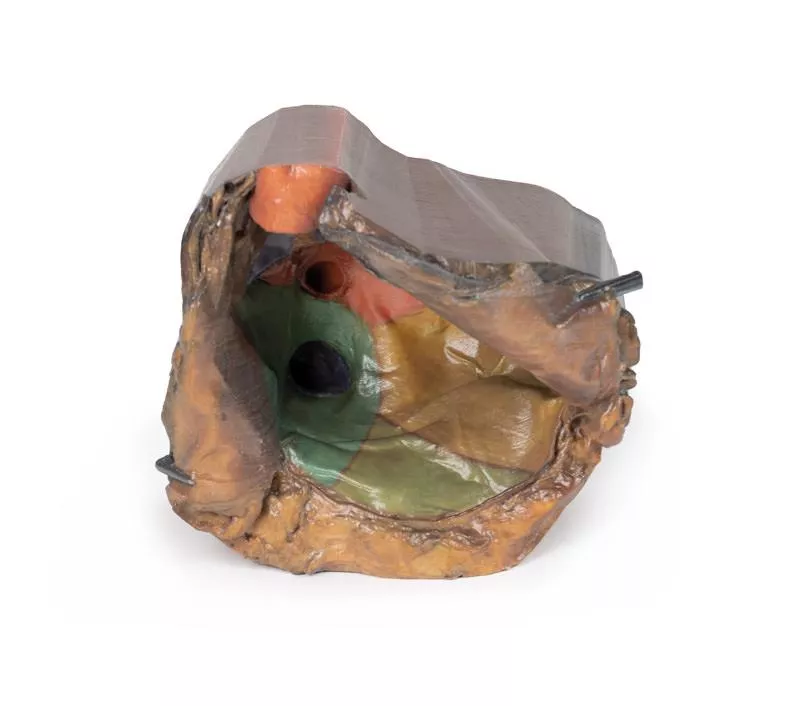
This high-resolution 3D model offers a detailed view of the left lung hilum and associated structures, sectioned sagittally through the cardiac notch. It clearly demonstrates the relationships between the bronchi, pulmonary vessels, pleura, and supporting structures, making it ideal for advanced anatomical education.

Key Features:
Hilum Anatomy:
- Entry/exit point for the pulmonary artery, superior and inferior pulmonary veins, main bronchus, lymphatics, and nerves.
- Shows the meeting point of visceral and parietal pleura, forming the pulmonary ligament—the lung’s sole anatomical connection to the body.

Pulmonary Circulation:
- Pulmonary artery (superior in position) carries deoxygenated blood from the heart.
- Pulmonary veins (anterior and inferior) return oxygenated blood to the heart.

Bronchial Structure:
Left main bronchus and its lobar branches are visible, located posteriorly within the hilum.

Additional Views:
- Oblique fissure along the lateral lung surface.
- Diaphragmatic surface at the base; costal visceral surface posteriorly.
- Pulmonary lymph nodes surrounding the hilum, both medially and laterally.
Documents
Datasheet MP1124 Download

0 of 0 reviews

Leave a review!

Share your experiences with other customers.


Erler-Zimmer

Erler-Zimmer GmbH & Co.KG
Hauptstrasse 27
77886 Lauf
Germany
info@erler-zimmer.de


Achtung! Medizinisches Ausbildungsmaterial, kein Spielzeug. Nicht geeignet für Personen unter 14 Jahren.

Attention! Medical training material, not a toy. Not suitable for persons under 14 years of age.


Documents
Datasheet MP1124 Download

Other customers also bought

Pericardial space
This detailed 3D anatomical model displays the pericardial cavity and reflections with the heart removed, allowing clear visualization of key structures within the middle mediastinum.Key Features:Pericardium:Shows the full extent of the parietal pericardium, continuous with the visceral layer (epicardium), and false-colored to indicate the positions of the atria, ventricles, and great vessels. Mediastinal Landmarks:Highlights the base, apex, diaphragmatic, and pulmonary surfaces of the heart by their impressions within the pericardial cavity.Great Vessels:- Aorta (ascending, arch, and descending)- Superior and Inferior Vena Cava- Pulmonary trunk and arteries- Pulmonary veins (4 total) - All shown in their natural positions relative to the pericardium.Pericardial Sinuses:- Transverse sinus: Located between arteries and veins; relevant for surgical access.- Oblique sinus: Posterior recess between pulmonary veins.Educational Use:- Ideal for teaching thoracic and cardiac anatomy, including pericardial reflections, mediastinal relationships, and surgical landmarks.- Useful in medical training, surgical planning, and radiological orientation.
Lung Slab, Hilum removed
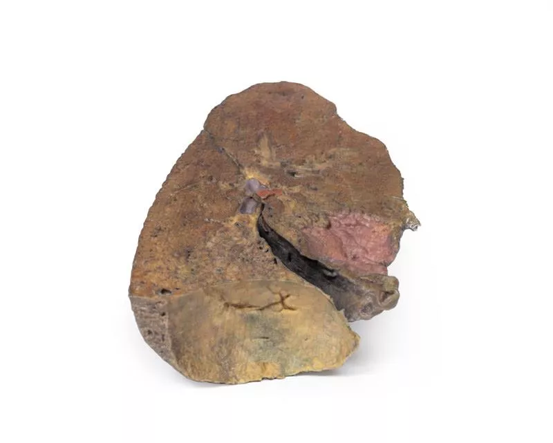
This 3D anatomical model presents a left lung dissected in a parasagittal plane, offering a unique internal view of pulmonary structures and anatomical landmarks. The mediastinal surface has been removed, allowing detailed observation of internal lung anatomy and its relationship to the heart and diaphragm.Key Features:Bronchial Tree (Internal View):- The primary bronchi are not visible due to prior branching.- Subdivided bronchi are preserved, though the dissection depth makes it unclear whether they represent secondary (lobar) or tertiary (segmental) bronchi. Vascular Structures:- Pulmonary arteries and veins are typically seen at the hilum, but precise levels of subdivision are undetermined in this section.Cardiac Impression:- A clear concave impression remains on the medial lung surface, formed by the left ventricle of the heart pressing against the lung.- Despite the dissection, this landmark remains distinctly visible.Diaphragmatic Surface:- The lung’s base is concave, resting atop the diaphragm.- While the pleura is not preserved, the model indicates where the diaphragmatic recess would form—between the costal and diaphragmatic pleura.
Stomach
This 3D model is an isolated stomach with two dissection windows to expose the rugae and pylorus. A small portion of the terminal oesophagus is preserved at the cardiac region, and a small portion of the proximal duodenum beyond the pyloric sphincter. The large window within the body of the stomach allows for a clear view into the fundus and the well-developed rugae on the posterior aspect of the wall of the organ. The smaller window, opened just at the pyloric region, allows for an appreciation of the thickening of the organ wall at the pyloric sphincter just proximal to the start of the duodenum.
Abdomen with bilateral Hernias
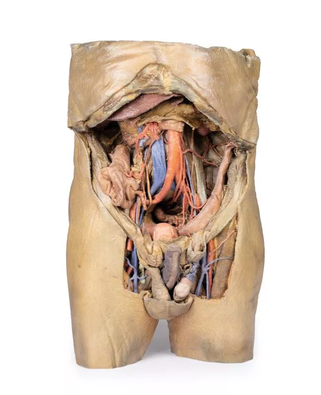
This 3D model represents one of the largest and most complex in the series, consisting of a partial torso from the diaphragm to the proximal thigh with a complete abdominal cavity preserving varying levels of dissection. This 3D model also records the rare, simultaneous occurrence of indirect and direct inguinal hernias allowing for a consideration of the anatomical underpinnings for both conditions. Given the scale of the dissection this 3D model description is divided into discrete parts based on views and regions.The diaphragmThe diaphragm is preserved on the model’s superior aspect, with both domes and costodiaphragmatic recesses visible despite some distortion from rib removal. The fibrous pericardium rests on the central tendon, with the terminal inferior vena cava seen in the caval foramen. Lateral to this lies the oesophagus in the oesophageal hiatus, and the descending thoracic aorta approaching the aortic hiatus near the vertebrae. The epigastric and hypochondriac regionsIn the abdomen, removal of the anterior wall, greater omentum, and much of the GI tract reveals retroperitoneal structures. The terminal oesophagus enters just left of the liver. With the stomach removed, the pancreas is fully exposed from head to tail, reaching the spleen in the left hypochondrium. Above it, the splenic and common hepatic arteries span the narrow space between pancreas, diaphragm, and liver. The tortuous splenic artery divides near the splenic vein; the common hepatic gives rise to the gastroduodenal and right gastric arteries, superficial to the portal vein. The superior mesenteric vessels pass near the pancreatic head, and the ileocolic artery leads to the caecum. The inferior mesenteric vein arises from the superior rectal vein and crosses the descending aorta. Below the liver, the gallbladder lies between the lobes. On the left, renal vessels pass deep to the pancreas, with ureters descending across the psoas muscles. The umbilical and lumbar regionsMost abdominal organs in the umbilical and lumbar regions have been removed to reveal the posterior abdominal wall. Centrally, the descending aorta and inferior vena cava are prominent, with testicular vessels traceable toward the inguinal region. Two right lumbar arteries branch from the aorta, and the inferior mesenteric artery gives rise to the left colic, sigmoid, and superior rectal arteries. On the right, subcostal, iliohypogastric, and ilioinguinal nerves are visible, along with the circumflex iliac artery.The hypogastrium and iliac regionsThe abdominal aorta bifurcates into the common, internal, and external iliac arteries, with matching iliac veins merging into the inferior vena cava. The obturator artery, ureters, and testicular vessels are visible. In the true pelvis, the peritoneum covers the bladder, while the rectum remains obscured. The right iliac fossa contains the terminal ileum, caecum, and appendix, with nearby vessels and nerves. On the left, the sigmoid colon crosses the iliac fossa, where an epiploic appendage extends into an indirect hernia near the inferior epigastric artery. The inguinal region and perineumThis model uniquely preserves both direct (right) and indirect (left) inguinal hernias, with the inferior epigastric vessels retained for anatomical orientation. The right hernia lies medial to these vessels; the left hernia sac extends laterally into the spermatic cord, containing an epiploic appendage. The perineum reveals the penis, testes, and spermatic cords. On the right, the cord remains intact; on the left, it’s opened, showing a varicose testicular vein linked to the indirect hernia. The thighThe femoral triangle has been dissected on both thighs. On the right, the femoral sheath was removed to reveal the femoral artery, vein, deep inguinal lymph nodes, and femoral nerve. On the left, a broader view exposes anterior and medial thigh muscles, with the femoral artery, profunda femoris, and circumflex iliac artery visible. The model ends mid-thigh, showing cross-sectional anatomy including the femoral shaft, vessels, and muscles in the subsartorial canal.
Head, Neck and Shoulder with angiosomes
This large, multipart 3D printed specimen showcases detailed anatomy of the head, neck, thorax, axillae, and upper limbs.Head and Neck:The calotte has been removed ~2?cm above the orbits to expose the brain and endocranial cavity. A transverse cerebral section shows grey and white matter, lateral ventricles, and choroid plexus. The right side retains skin and fascia, false-coloured to highlight facial and neck angiosomes. The left side reveals facial expression and mastication muscles, and infratemporal structures including the lingual nerve and terminal branches of the external carotid artery. The carotid sheaths are opened bilaterally, exposing the common, internal, and external carotid arteries, and vagus nerves. The sternocleidomastoid and internal jugular veins are mostly removed. On the right, the great auricular and hypoglossal nerves are visible, along with the stylohyoid ligament and supra-/infrahyoid muscles. The thyroid gland is prominent, with preserved superior and inferior thyroid vessels.Root of the Neck and Axilla:On the left, partial clavicle removal reveals the first rib, anterior scalene, and brachial plexus roots (C5–T1) forming trunks between scalene muscles. The subclavian artery passes posterior to scalenus anterior, transitioning to the axillary artery, closely related to the brachial plexus cords.The left axilla displays brachial plexus divisions and cords. The formation of the median nerve around the axillary artery is distinct. The ulnar, musculocutaneous, axillary, thoracodorsal, and long thoracic nerves are clearly identified with their courses and muscular targets.On the right, the clavicle and subclavius muscle are intact, showing the cervico-axillary canal. Pectoralis major and minor have been reflected, exposing deeper structures.Thorax:A window in the left thoracic wall reveals the mediastinum. The left lung has been removed. Intercostal spaces are visible beneath the parietal pleura; neurovascular bundles are identifiable posteriorly. The heart is exposed without pericardium, showing the left atrium and ventricle, pulmonary vessels, aorta, and both left vagus and recurrent laryngeal nerves. The right thoracic wall remains intact, displaying intercostal and upper limb muscles. From below, the right lung, pleural cavities, and diaphragmatic heart surface are visible. Posterior thoracic skin and fascia are intact, showing cutaneous nerve distribution.Size: 50 x 20 x 41 cm

Continuous innovation

Social responsibility

Active customer orientation

Understanding quality

Sustainable actions

ISO 9001 certification

Your last viewed products