Product information "Chondrosarcoma of scapula"
Clinical History
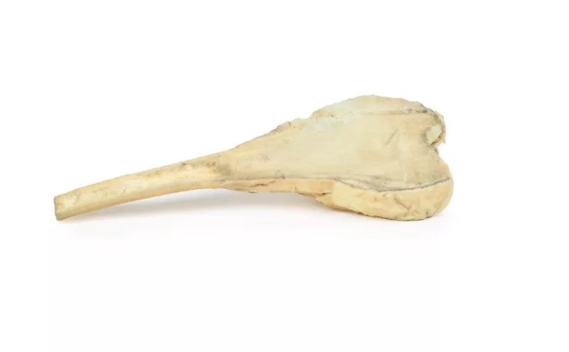
A 60-year-old female presented with a 12-month history of recurrent pain and swelling in her right shoulder. Examination revealed a palpable mass over the superior scapula, with limited abduction and external rotation. X-ray showed a mass involving the superior scapula, which was biopsied and led to complete scapular excision.
Pathology
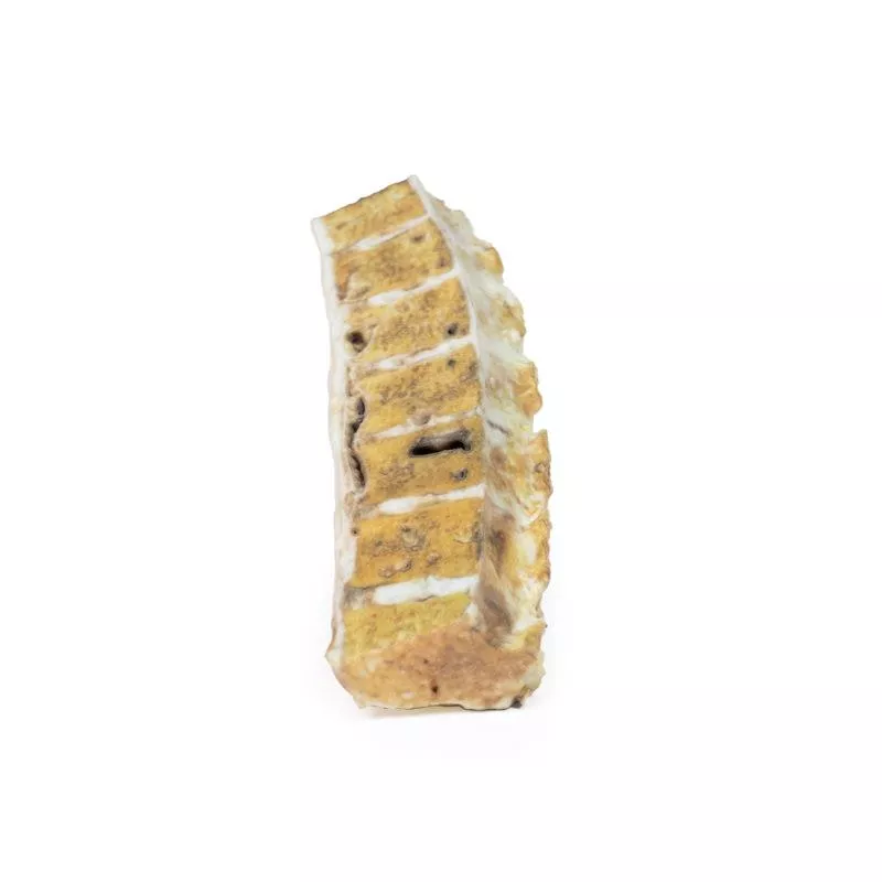
The excised scapula showed an irregular, lobulated tumour measuring 11?cm, extending to the acromion and coracoid process. The tumour had a mottled pale-yellow to brown appearance with haemorrhagic areas and had replaced normal bone. Histology confirmed chondrosarcoma, with pleomorphic spindle cells, mitotic figures and cartilage formation.
Further Information
Chondrosarcomas are malignant bone tumours producing cartilage and are the third most common primary bone malignancy. Most are conventional type, while rare subtypes include clear cell, dedifferentiated, and mesenchymal variants. Some originate from benign lesions such as enchondromas or osteochondromas. Genetic mutations commonly involve IDH1, IDH2 and CDKN2A.
Men are affected twice as often as women. The axial skeleton is commonly involved, with around 5% affecting the scapula. These tumours grow slowly, presenting as painful, enlarging masses. Most are low-grade at diagnosis and rarely metastasise, though the lungs are the most frequent site of spread. The 5-year survival rate is about 90% for grade 1 and drops to 43% for grade 3.
CT and MRI are preferred imaging modalities. Diagnosis is confirmed by biopsy. The treatment of choice is complete surgical resection, as these tumours generally do not respond to chemotherapy or radiotherapy.
Erler-Zimmer
Erler-Zimmer GmbH & Co.KG
Hauptstrasse 27
77886 Lauf
Germany
info@erler-zimmer.de
Achtung! Medizinisches Ausbildungsmaterial, kein Spielzeug. Nicht geeignet für Personen unter 14 Jahren.
Attention! Medical training material, not a toy. Not suitable for persons under 14 years of age.


