Product information "Osteochondroma"
Clinical History
A 61-year-old male with prostate cancer attended a pre-operative clinic ahead of his planned prostatectomy. He reported chronic right knee pain, previously attributed to osteoarthritis. To rule out bony metastases, a knee x-ray was performed, revealing a pedunculated lesion on the medial side of the right femoral shaft. The prostatectomy proceeded, but the patient unfortunately died from a postoperative pulmonary embolism.
Pathology
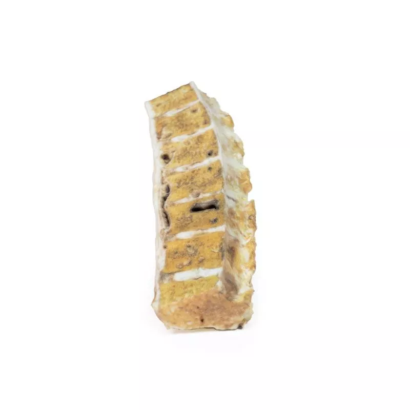
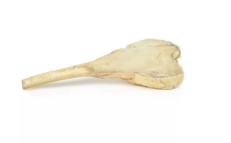
The specimen shows the lower right femur, cut in the coronal plane. A 2?cm pedunculated bony growth arises from the medial shaft, 7?cm above the medial condyle. It consists of normal bone with a hyaline cartilage cap. This lesion is a classic osteochondroma.
Further Information
An osteochondroma (also known as exostosis) is a benign cartilaginous tumour made of bone and capped with cartilage. It is the most common benign bone tumour. Most are sporadic, but some occur in hereditary multiple exostosis syndrome or after radiotherapy. They typically arise near the growth plate and are common in the knee or proximal humerus, especially in young males.
Symptoms depend on the location and size. Many osteochondromas are asymptomatic, but larger ones may cause pain, nerve compression, or fractures. Diagnosis is typically via x-ray, though MRI helps rule out malignancy. EXT1 and EXT2 gene mutations are linked to hereditary forms. Growth stops once the growth plate fuses. Treatment is only needed if symptoms are severe. Malignant transformation to chondrosarcoma is rare (<1%) but more frequent in hereditary cases (up to 20%).
Erler-Zimmer
Erler-Zimmer GmbH & Co.KG
Hauptstrasse 27
77886 Lauf
Germany
info@erler-zimmer.de
Achtung! Medizinisches Ausbildungsmaterial, kein Spielzeug. Nicht geeignet für Personen unter 14 Jahren.
Attention! Medical training material, not a toy. Not suitable for persons under 14 years of age.


