


Product information "Chronic hydrocoele"
Clinical History
An 80-year-old male with alcoholic liver cirrhosis and oesophageal varices presented with haematemesis. Examination showed spider naevi, large ascites, and a scrotal swelling that transmitted red light on transillumination. He had another severe haematemesis and died shortly after admission.
Pathology
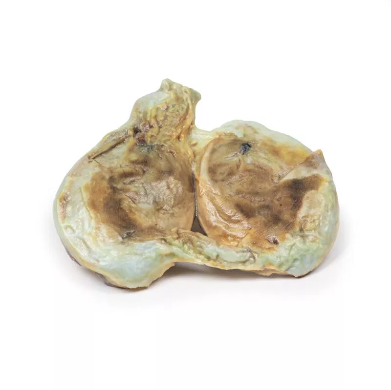
The specimen includes the testis, tunica vaginalis, and spermatic cord. The tunica vaginalis is thickened with a distended cavity, while the testis is normal. This represents a chronic secondary communicating hydrocele.
Further Information
A hydrocele is fluid between the parietal and visceral layers of the tunica vaginalis. Hydroceles can be communicating (connected to the peritoneal cavity) or non-communicating. Communicating hydroceles result from failure of processus vaginalis closure and may appear at birth or later due to increased intra-abdominal pressure, like heart or liver failure. Non-communicating hydroceles arise from fluid imbalance due to infections, tumors, trauma, or lymphatic obstruction.
Patients present with a scrotal mass that may be uni- or bilateral. Communicating hydroceles can be reducible and change size with pressure; non-communicating are usually fixed. Swellings are generally painless unless complicated. Larger hydroceles may cause skin irritation or infections.
Diagnosis is clinical, aided by transillumination and ultrasound to exclude other testicular conditions. Tumor markers (AFP and B-HCG) may rule out cancer. Many congenital hydroceles resolve by age 2; persistent or symptomatic hydroceles require surgical repair to avoid hernia risk. Treating the underlying cause can also resolve reactive hydroceles.
An 80-year-old male with alcoholic liver cirrhosis and oesophageal varices presented with haematemesis. Examination showed spider naevi, large ascites, and a scrotal swelling that transmitted red light on transillumination. He had another severe haematemesis and died shortly after admission.
Pathology
The specimen includes the testis, tunica vaginalis, and spermatic cord. The tunica vaginalis is thickened with a distended cavity, while the testis is normal. This represents a chronic secondary communicating hydrocele.
Further Information
A hydrocele is fluid between the parietal and visceral layers of the tunica vaginalis. Hydroceles can be communicating (connected to the peritoneal cavity) or non-communicating. Communicating hydroceles result from failure of processus vaginalis closure and may appear at birth or later due to increased intra-abdominal pressure, like heart or liver failure. Non-communicating hydroceles arise from fluid imbalance due to infections, tumors, trauma, or lymphatic obstruction.
Patients present with a scrotal mass that may be uni- or bilateral. Communicating hydroceles can be reducible and change size with pressure; non-communicating are usually fixed. Swellings are generally painless unless complicated. Larger hydroceles may cause skin irritation or infections.
Diagnosis is clinical, aided by transillumination and ultrasound to exclude other testicular conditions. Tumor markers (AFP and B-HCG) may rule out cancer. Many congenital hydroceles resolve by age 2; persistent or symptomatic hydroceles require surgical repair to avoid hernia risk. Treating the underlying cause can also resolve reactive hydroceles.
Login
Erler-Zimmer
Erler-Zimmer Medical GmbH
Hauptstrasse 27
77886 Lauf
Germany
info@erler-zimmer.de
Achtung! Medizinisches Ausbildungsmaterial, kein Spielzeug. Nicht geeignet für Personen unter 14 Jahren.
Attention! Medical training material, not a toy. Not suitable for persons under 14 years of age.
Other customers also bought

Osteochondroma
Clinical History A 61-year-old male with prostate cancer attended a pre-operative clinic ahead of his planned prostatectomy. He reported chronic right knee pain, previously attributed to osteoarthritis. To rule out bony metastases, a knee x-ray was performed, revealing a pedunculated lesion on the medial side of the right femoral shaft. The prostatectomy proceeded, but the patient unfortunately died from a postoperative pulmonary embolism.PathologyThe specimen shows the lower right femur, cut in the coronal plane. A 2?cm pedunculated bony growth arises from the medial shaft, 7?cm above the medial condyle. It consists of normal bone with a hyaline cartilage cap. This lesion is a classic osteochondroma.Further InformationAn osteochondroma (also known as exostosis) is a benign cartilaginous tumour made of bone and capped with cartilage. It is the most common benign bone tumour. Most are sporadic, but some occur in hereditary multiple exostosis syndrome or after radiotherapy. They typically arise near the growth plate and are common in the knee or proximal humerus, especially in young males.Symptoms depend on the location and size. Many osteochondromas are asymptomatic, but larger ones may cause pain, nerve compression, or fractures. Diagnosis is typically via x-ray, though MRI helps rule out malignancy. EXT1 and EXT2 gene mutations are linked to hereditary forms. Growth stops once the growth plate fuses. Treatment is only needed if symptoms are severe. Malignant transformation to chondrosarcoma is rare (<1%) but more frequent in hereditary cases (up to 20%).

Osteosarcoma of femur
Clinical HistoryA 57-year-old man presented with recurrent right thigh pain. Clinical examination showed no palpable abnormality. X-ray revealed bony resorption and periosteal reaction in the proximal femur. CT confirmed a mass in the proximal femur. Following a biopsy, the upper femur was excised and replaced with a prosthesis.PathologyThe specimen includes the head, neck, and upper shaft of the femur. Within the medullary cavity, a 6.5 cm ovoid tumour is seen. It is unencapsulated, with a haemorrhagic and cystic cut surface. Histology confirmed a low-grade chondrosarcoma.Further InformationChondrosarcomas are malignant bone tumours that produce cartilage and rank as the third most common primary bone cancer. The conventional type makes up 90% of cases, with less frequent subtypes including clear cell, mesenchymal, and dedifferentiated forms. Some cases develop from benign tumours like enchondromas.Mutations commonly involve IDH1/IDH2 or CDKN2A, with EXT gene mutations in multiple osteochondroma syndromes. Men are twice as often affected. The axial skeleton is most often involved, but around 20% affect the femur.These tumours are slow-growing and typically present with a painful, enlarging mass. Most are low grade and rarely metastasize. The lungs are the most common site for spread. 5-year survival for grade 1 is nearly 90%, but drops to 43% for grade 3.Diagnosis relies on CT and MRI, with biopsies for confirmation. Surgical resection is the main treatment, as chemotherapy and radiotherapy are generally ineffective due to the tumour’s slow growth.

Osteosarcoma of femur
Clinical HistoryA patient presented with a tender swelling above the right knee. Blood tests revealed elevated alkaline phosphatase. X-ray showed periosteal reactive changes in the distal femur, raising suspicion of malignancy. MRI and CT confirmed the findings. After neoadjuvant chemotherapy, the distal femur was resected. The patient made a full recovery.Pathology The excised distal femur showed a large pale tumour (10 cm), infiltrating through the periosteum near the joint surface. Diagnosis: osteosarcoma.Further InformationOsteosarcoma is the most common primary malignant bone tumour, producing osteoid matrix. It primarily affects the distal femur, followed by tibia and humerus. Most cases occur in children and adolescents, with a second peak in older adults (secondary osteosarcomas, often due to conditions like Paget’s disease or prior radiation).Typical symptoms include a painful, enlarging mass, sometimes pathological fractures. X-ray signs include sunburst appearance and Codman’s triangle. Diagnosis is supported by MRI (local staging), CT (metastases), and biopsy. The lungs are the most common site for metastases. Treatment includes chemotherapy and surgery. 5-year survival is 60–70% for localized disease, but <20% if metastasized.

Metastatic Malignant Melanoma
Clinical History A 65-year-old male with a history of melanoma on his left foot (treated surgically and with radiotherapy) presented with left groin pain. He was cachexic, had a hard, enlarged liver and a discharging sinus in the groin surrounded by black nodules. He passed away from a hospital-acquired pneumonia.Pathology The excised proximal right femur showed numerous tumour deposits in the medullary cavity, varying from pale brown to black, destroying cancellous bone up to 3?cm in size. Cortical bone remained intact except for some discoloration at the neck-shaft junction. These are metastatic deposits from a skin melanoma.Further InformationMelanoma is a malignant skin cancer linked to UV radiation exposure. Risk factors include fair complexion, numerous naevi, childhood sunburns, and immunosuppression. Although only about 5% of skin cancers, it has the highest mortality rate. It typically appears as a changing pigmented lesion with irregular borders, variegated colour and asymmetry.Common mutations include CDKN2A, BRAF, PI3K and TERT. Immunotherapy that targets melanoma antigens shows promising results.Melanoma often spreads to lungs, liver, brain, bone, and regional lymph nodes. Bone metastases occur in 25–50% of cases and often affect the axial skeleton, causing pain or pathological fractures. Diagnosis includes biopsy, blood tests (e.g., elevated ALP, calcium, LDH), and imaging (X-ray, CT, MRI, PET). Treatment is stage- and profile-dependent and includes surgery, chemotherapy, immunotherapy, radiotherapy or combinations.

Chondrosarcoma of scapula
Clinical History A 60-year-old female presented with a 12-month history of recurrent pain and swelling in her right shoulder. Examination revealed a palpable mass over the superior scapula, with limited abduction and external rotation. X-ray showed a mass involving the superior scapula, which was biopsied and led to complete scapular excision.Pathology The excised scapula showed an irregular, lobulated tumour measuring 11?cm, extending to the acromion and coracoid process. The tumour had a mottled pale-yellow to brown appearance with haemorrhagic areas and had replaced normal bone. Histology confirmed chondrosarcoma, with pleomorphic spindle cells, mitotic figures and cartilage formation.Further InformationChondrosarcomas are malignant bone tumours producing cartilage and are the third most common primary bone malignancy. Most are conventional type, while rare subtypes include clear cell, dedifferentiated, and mesenchymal variants. Some originate from benign lesions such as enchondromas or osteochondromas. Genetic mutations commonly involve IDH1, IDH2 and CDKN2A.Men are affected twice as often as women. The axial skeleton is commonly involved, with around 5% affecting the scapula. These tumours grow slowly, presenting as painful, enlarging masses. Most are low-grade at diagnosis and rarely metastasise, though the lungs are the most frequent site of spread. The 5-year survival rate is about 90% for grade 1 and drops to 43% for grade 3.CT and MRI are preferred imaging modalities. Diagnosis is confirmed by biopsy. The treatment of choice is complete surgical resection, as these tumours generally do not respond to chemotherapy or radiotherapy.

Tertiary Syphilis
Clinical History A 66-year-old male with deafness and non-verbal communication presented with epigastric pain after eating. Examination revealed a tender epigastrium and nodular lesions on the forehead and scalp. Blood tests showed low haemoglobin, impaired liver function, and were positive for anti-treponemal antibodies. He later developed a large gastrointestinal bleed and died despite medical intervention.Pathology The skull shows multiple necrotic lesions up to 3–4?cm on the external surface, eroding the outer table of the skull. The surrounding periosteum is thickened with inflammation. These are gummatous lesions typical of tertiary syphilis.Further InformationSyphilis is a chronic infection caused by Treponema pallidum, mainly transmitted sexually or congenitally. Risk factors include HIV infection, IV drug use, and male-to-male sexual contact. The disease progresses in three stages: - Primary syphilis: painless chancre at the infection site. - Secondary syphilis: general symptoms like skin rashes, condylomata lata, and lymphadenopathy. - Tertiary syphilis: may lead to cardiovascular syphilis, neurosyphilis or gummatous lesions in skin, bone, or organs. Gummas are chronic inflammatory lesions that may cause bone destruction and pathological fractures, more commonly seen in HIV-positive patients.

Tuberculosis
Clinical History A 37-year-old female with untreated HIV and pulmonary tuberculosis presents with worsening thoracic back pain, low-grade fevers, chills, and weight loss. Examination showed tenderness over multiple thoracic vertebrae. Blood tests revealed elevated serum calcium and ESR. Spine X-ray showed lytic lesions. She developed urosepsis and died during admission.Pathology The thoracic vertebrae specimen shows multiple osteolytic lesions with caseous necrosis and bone sclerosis. The tuberculous infection spread to intervertebral discs and paravertebral tissues. This is tuberculous osteomyelitis of the spine (Pott’s disease).Further InformationTuberculosis (TB) is a chronic infectious disease caused by Mycobacterium tuberculosis, transmitted by inhalation of droplets. Risk factors include immunosuppression (HIV, steroids), malnutrition, and living in endemic areas. Most infections remain latent but can reactivate, especially in immunocompromised patients. Osseous TB occurs in 1–3% of cases, with Pott’s disease representing 40% of bone TB infections. It causes bone destruction, vertebral collapse, and neurological symptoms from cord compression. Diagnosis involves history, chest X-ray, sputum cultures, and biopsies.

Suppurative arthritis of the knee
Clinical HistoryAn 82-year-old man with a history of tuberculosis of the left knee in childhood presented with a painful and swollen knee that developed ten days before hospital admission. Examination revealed an inflamed and painful joint that slightly improved with antibiotics and rest. X-ray showed a severely disorganized knee joint, leading to a mid-thigh amputation three days after admission. Post-operative recovery was uneventful.PathologyThe specimen includes the femur and tibia with exposed joint surfaces. These are severely eroded, brown, and irregular, with shaggy adhesions and yellow necrotic plaques. No normal articular cartilage remains. Bony projections up to 1?cm are present on the femoral condyles. Staphylococcus aureus was cultured from the joint. This represents a case of suppurative arthritis in a knee previously affected by tuberculosis.Further InformationSuppurative (septic) arthritis is a bacterial joint infection, diagnosed through synovial fluid analysis, microscopy, and culture. Staphylococcus aureus is a frequent cause. In patients with monoarthritis and a history of TB, tuberculous arthritis should be considered, especially if chest radiographs or eosinophil counts are abnormal.Musculoskeletal TB occurs in up to 19% of TB cases, more commonly in children due to higher bone marrow content. In adults, TB most often affects the spine (40%), followed by the hip (25%) and the knee (8%). While extrapulmonary TB accounts for 15–20% of cases in immunocompetent individuals, initial presentation as a joint infection is rare.

Chondrosarcoma of femur and ilium
Clinical History A teenage boy presented with groin pain after horse-riding. Examination revealed a large, deep lump. After biopsy and imaging, chondrosarcoma was diagnosed, leading to radical surgical resection of his right leg.Pathology The specimen includes the upper femur and pelvis joint. A lobulated pale grey tumour with cavitation, necrosis, and haemorrhage replaces the femoral neck, head, and much of the ilium. The tumour extends beyond bone into soft tissues and is encapsulated. Macroscopic signs such as infiltration, necrosis, and haemorrhage confirm malignancy.Further InformationChondrosarcoma is a rare primary malignant bone tumour with cartilaginous differentiation, representing about 20% of bone cancers. The only effective treatment is surgical excision; adjuvant therapies are ineffective. Pelvic tumours pose special challenges for surgery and reconstruction. Chondrosarcoma usually arises in the bones of limbs or pelvis but can occur wherever cartilage is present. It can develop de novo or from benign tumours like enchondroma or osteochondroma. Subtypes include conventional, clear cell, myxoid, mesenchymal, and dedifferentiated chondrosarcoma.

Continuous innovation

Social responsibility

Active customer orientation

Understanding quality

Sustainable actions

ISO 9001 certification