Product information "Osteosarcoma of femur"
Clinical History
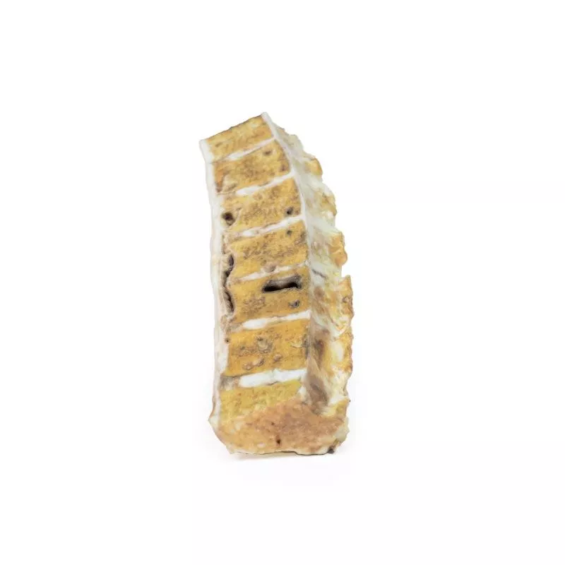
A 57-year-old man presented with recurrent right thigh pain. Clinical examination showed no palpable abnormality. X-ray revealed bony resorption and periosteal reaction in the proximal femur. CT confirmed a mass in the proximal femur. Following a biopsy, the upper femur was excised and replaced with a prosthesis.
Pathology
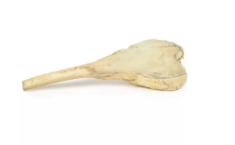
The specimen includes the head, neck, and upper shaft of the femur. Within the medullary cavity, a 6.5 cm ovoid tumour is seen. It is unencapsulated, with a haemorrhagic and cystic cut surface. Histology confirmed a low-grade chondrosarcoma.
Further Information
Chondrosarcomas are malignant bone tumours that produce cartilage and rank as the third most common primary bone cancer. The conventional type makes up 90% of cases, with less frequent subtypes including clear cell, mesenchymal, and dedifferentiated forms. Some cases develop from benign tumours like enchondromas.
Mutations commonly involve IDH1/IDH2 or CDKN2A, with EXT gene mutations in multiple osteochondroma syndromes. Men are twice as often affected. The axial skeleton is most often involved, but around 20% affect the femur.
These tumours are slow-growing and typically present with a painful, enlarging mass. Most are low grade and rarely metastasize. The lungs are the most common site for spread. 5-year survival for grade 1 is nearly 90%, but drops to 43% for grade 3.
Diagnosis relies on CT and MRI, with biopsies for confirmation. Surgical resection is the main treatment, as chemotherapy and radiotherapy are generally ineffective due to the tumour’s slow growth.
Erler-Zimmer
Erler-Zimmer GmbH & Co.KG
Hauptstrasse 27
77886 Lauf
Germany
info@erler-zimmer.de
Achtung! Medizinisches Ausbildungsmaterial, kein Spielzeug. Nicht geeignet für Personen unter 14 Jahren.
Attention! Medical training material, not a toy. Not suitable for persons under 14 years of age.


