Product information "Pituitary Adenoma"
Clinical History
A 29-year-old male presented with a 22-month history of headaches and blurred vision. Examination revealed a bitemporal hemianopia and a left sixth nerve palsy. Skull X-ray showed erosion of the sphenoid body, with some of the dorsum sellae and anterior clinoid process intact. Carotid angiography demonstrated upward and lateral displacement of the anterior and middle cerebral arteries. Pneumoencephalography showed upward displacement of the lateral and third ventricles. A craniotomy was performed, but the patient died immediately after surgery.
Pathology
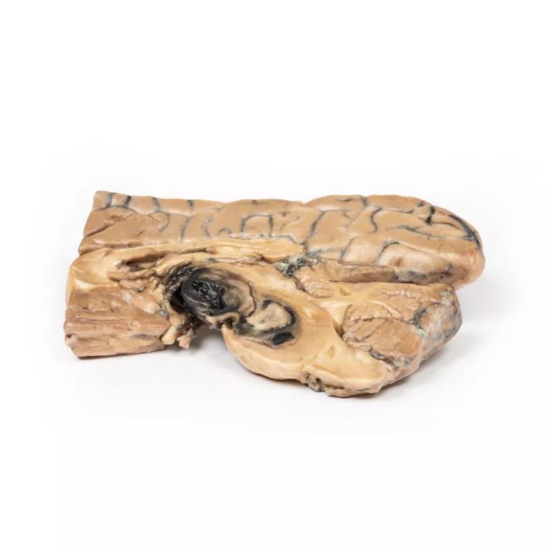
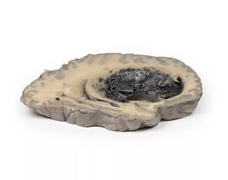
The pituitary gland has been completely replaced by a 4 cm round tumour, seen in a sagittal brain section to the right of the falx cerebri. The cut surface of the tumour is pale brown and homogeneous, with a small area of haemorrhage likely caused by surgical trauma. The tumour caused upward displacement of the midbrain and erosion of the sphenoid bone, enlarging the sella turcica. The optic chiasma is compressed. Histological diagnosis: chromophobe adenoma of the anterior pituitary.
Further Information
This case used now outdated methods such as pneumoencephalography. Today, a CT followed by a brain MRI would be standard. Pituitary adenomas are the most common pituitary tumours, typically affecting adults between 35 and 60 years. Clinical signs relate to mass effect—including increased intracranial pressure, bony erosion, and optic chiasma compression—as well as hormonal activity. About 75% are functioning adenomas (e.g., prolactin, growth hormone, or ACTH secretion), while non-functioning tumours may present later and cause hypopituitarism due to compression of the remaining gland.
Erler-Zimmer
Erler-Zimmer GmbH & Co.KG
Hauptstrasse 27
77886 Lauf
Germany
info@erler-zimmer.de
Achtung! Medizinisches Ausbildungsmaterial, kein Spielzeug. Nicht geeignet für Personen unter 14 Jahren.
Attention! Medical training material, not a toy. Not suitable for persons under 14 years of age.


