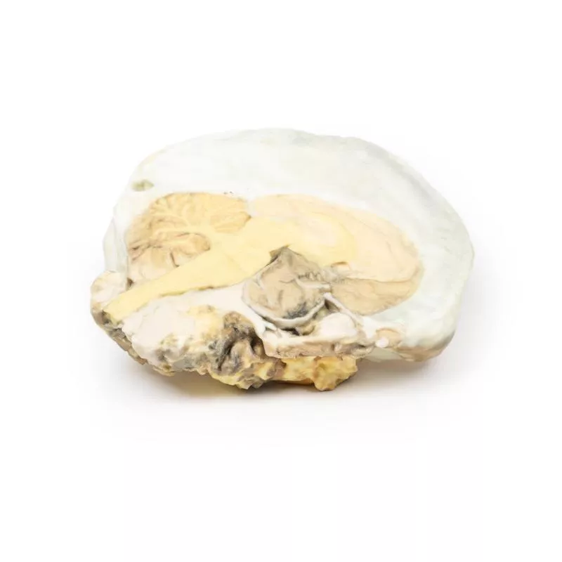
Product information "Ventriculitis, Secondary to Septicaemia"
Clinical History
A 50-year-old male with alcoholism was admitted with a 2-week history of weakness and shortness of breath. He initially presented with productive cough, chest pain, and blood-stained sputum. On examination, he was febrile, cyanosed, and drowsy, with grunting respiration and a friction rub over the right lower lobe. His condition worsened progressively. Shortly before death, a lumbar puncture yielded green opalescent fluid. Blood cultures confirmed Streptococcus pneumoniae.
Pathology
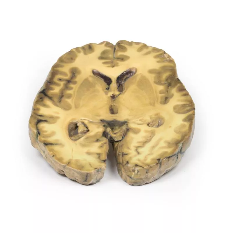
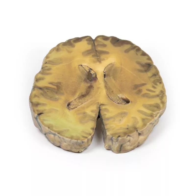
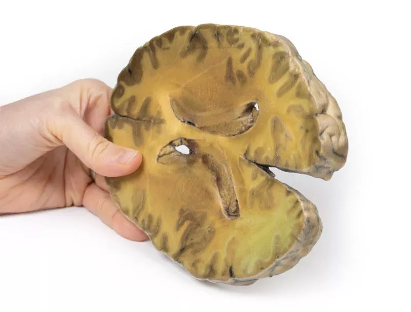
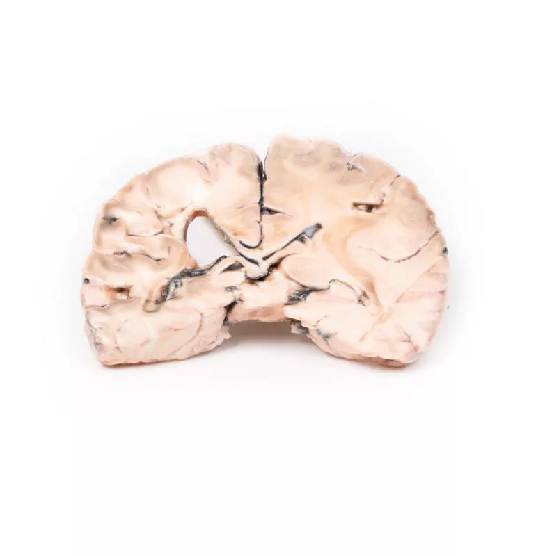
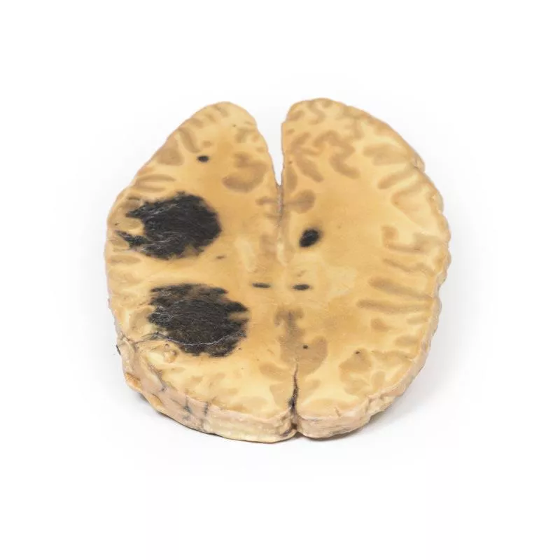
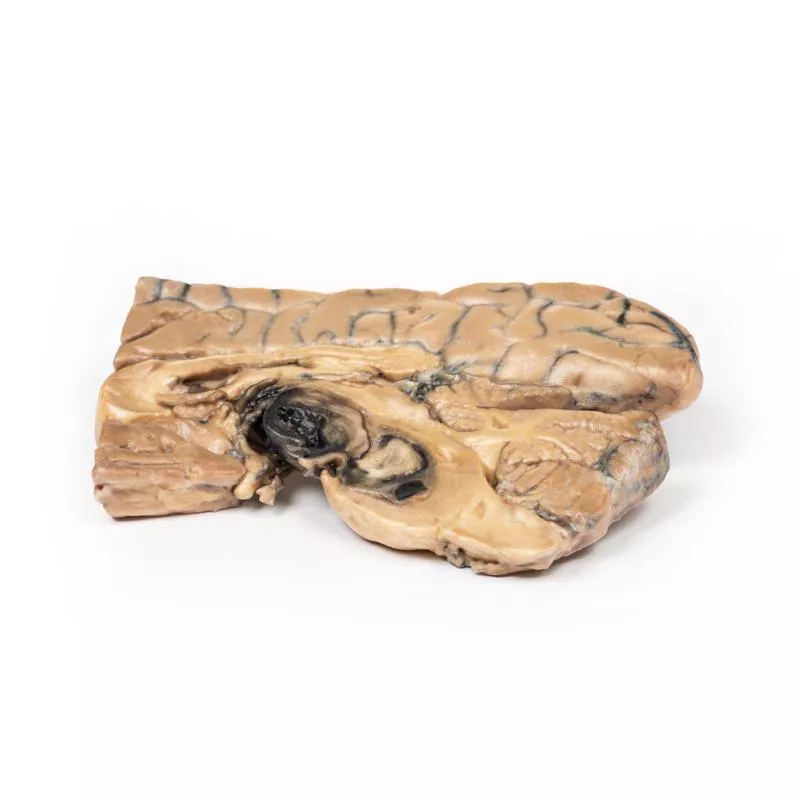
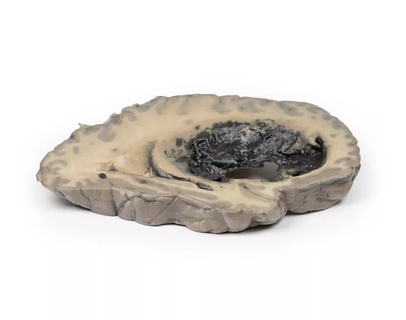
This is a case of ventriculitis, alongside pneumococcal meningitis and right basal pneumonia. A horizontal brain section shows both lateral ventricles with a thickened, rough ependymal lining and cellular debris near the choroid plexus and anterior horn. Structures like the caudate nucleus, lentiform nucleus and internal capsule are visible. Histology revealed extensive neutrophilic infiltration of the subarachnoid space, vascular wall involvement, and cerebral parenchymal inflammation causing haemorrhage and necrosis.
Further Information
Ventriculitis is a rare complication of intracranial infection. In adults, it usually occurs secondarily to surgery or trauma rather than primary meningitis. Common pathogens include staphylococci and resistant Gram-negative bacilli. In infants under 6 months, incidence is higher. Symptoms may mimic hydrocephalus due to aqueductal obstruction. Diagnosis relies on CSF analysis and imaging (CT, MRI). Treatment involves prolonged intravenous antibiotics that can penetrate CSF and brain tissue.
Erler-Zimmer
Erler-Zimmer GmbH & Co.KG
Hauptstrasse 27
77886 Lauf
Germany
info@erler-zimmer.de
Achtung! Medizinisches Ausbildungsmaterial, kein Spielzeug. Nicht geeignet für Personen unter 14 Jahren.
Attention! Medical training material, not a toy. Not suitable for persons under 14 years of age.


