Product information "Carcinoma of Breast"
Clinical History
A 76-year-old woman was admitted to the emergency department with a sudden loss of consciousness and signs of a left-sided stroke. She was intubated and treated in the ICU, where a fixed mass was noted in her left breast along with palpable axillary lymphadenopathy. She later died from pneumonia related to mechanical ventilation.
Pathology
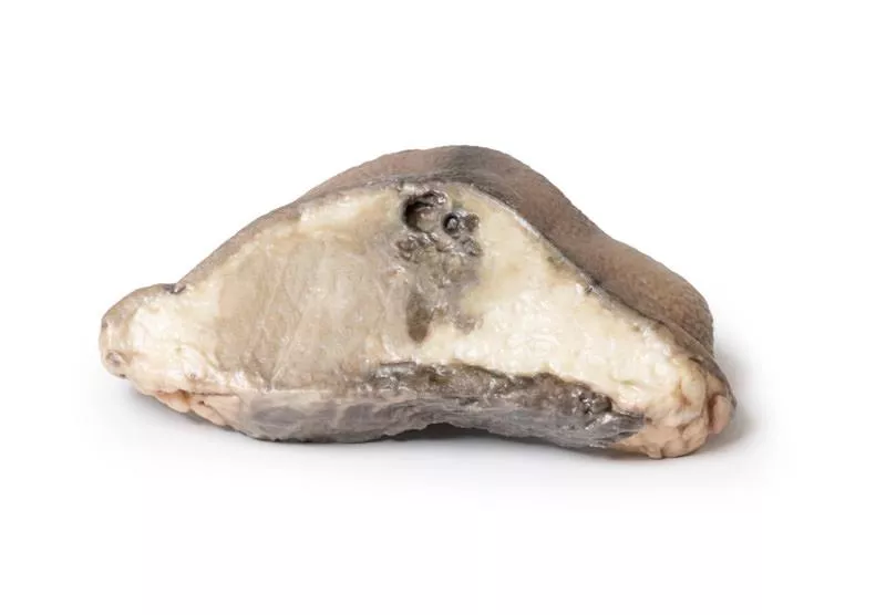
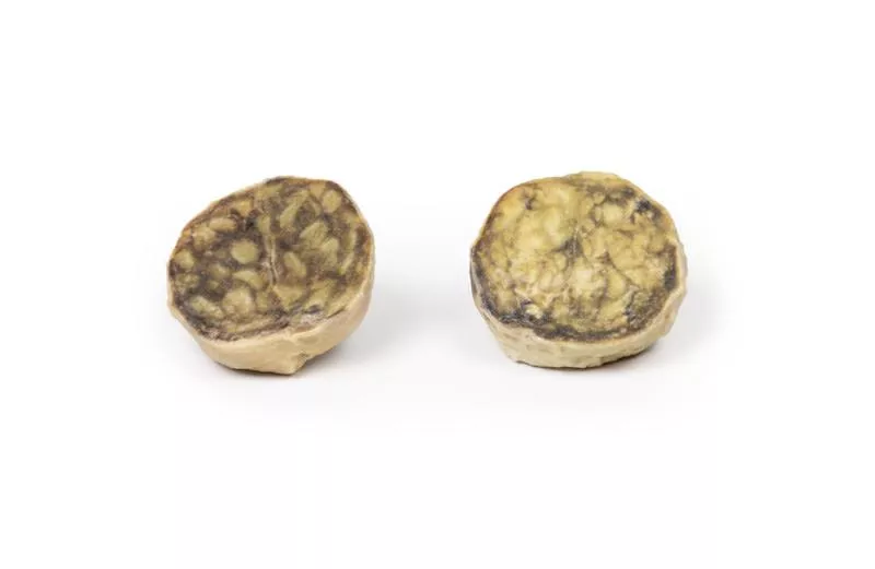
The specimen shows the left breast with a large, oval tumour mass (11?cm), located beneath and attached to the skin and adherent to the muscle. The cut surface is heterogeneous, showing areas of necrosis, haemorrhage, and cyst formation. Histology confirmed a breast adenocarcinoma with regional lymph node involvement.
Further Information
Breast cancer is the second most common cancer in women and mainly affects those over 30, peaking between 70 and 80 years. Risk factors include estrogen exposure, family history, nulliparity, obesity, and mutations in genes such as BRCA1, BRCA2, and others.
Most cases are adenocarcinomas originating in the duct or lobular system, often first appearing as DCIS. Subtypes are defined by hormone receptor status (ER) and HER2 expression, which guide treatment. Common metastatic sites are bone, liver, lung, and brain.
In countries with screening programs, most cases are found by mammography. Symptomatic signs include a hard, irregular, immobile mass, skin changes like dimpling (peau d’orange), and nipple retraction.
Treatment varies by stage and tumour profile and may include mastectomy, lumpectomy, radiotherapy, targeted therapies (e.g. trastuzumab for HER2+), anti-estrogen therapy (e.g. tamoxifen for ER+), and systemic chemotherapy.
Erler-Zimmer
Erler-Zimmer GmbH & Co.KG
Hauptstrasse 27
77886 Lauf
Germany
info@erler-zimmer.de
Achtung! Medizinisches Ausbildungsmaterial, kein Spielzeug. Nicht geeignet für Personen unter 14 Jahren.
Attention! Medical training material, not a toy. Not suitable for persons under 14 years of age.


