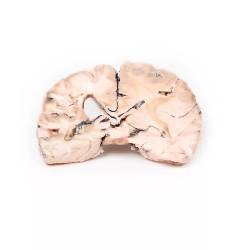
Product information "Berry Aneurism of Basilar Artery"
Clinical History
A 37-year-old patient presented with headache, vomiting, and disorientation after head trauma. CT revealed dilated lateral ventricles and a mass projecting into the third ventricle. A shunt was placed for hydrocephalus. Angiography identified a partially thrombosed 1 × 1 cm basilar artery aneurysm. The aneurysm enlarged over time, and multiple surgical attempts, including ligation and shunt revisions, were unsuccessful. The patient remained unconscious and later died.
Pathology
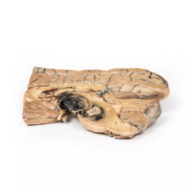
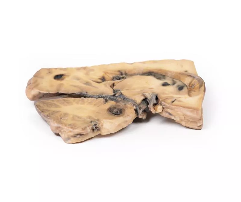
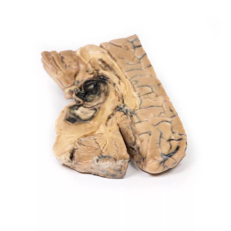
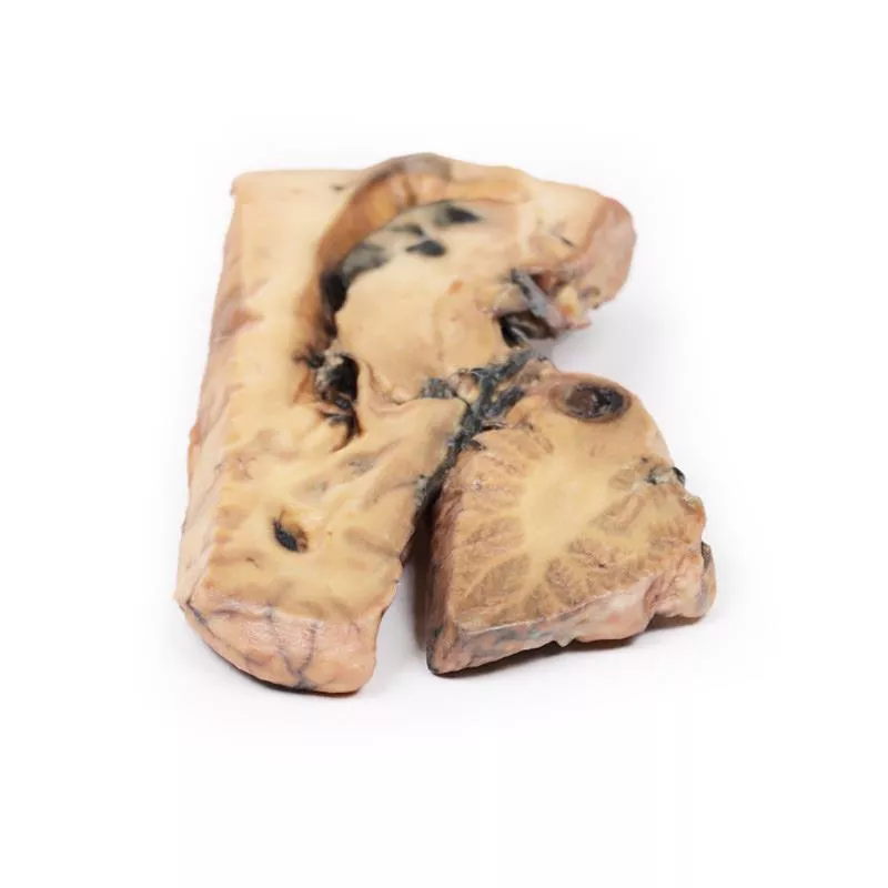
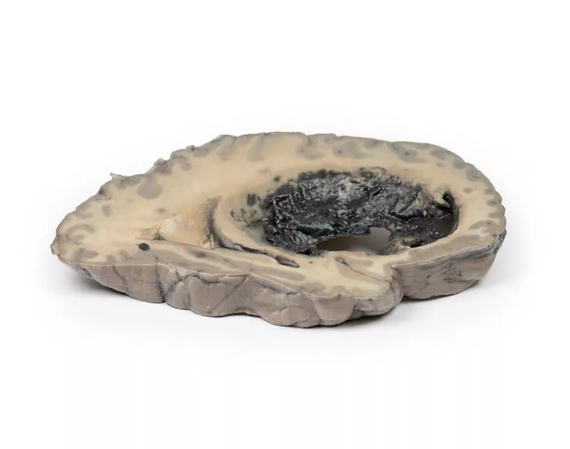
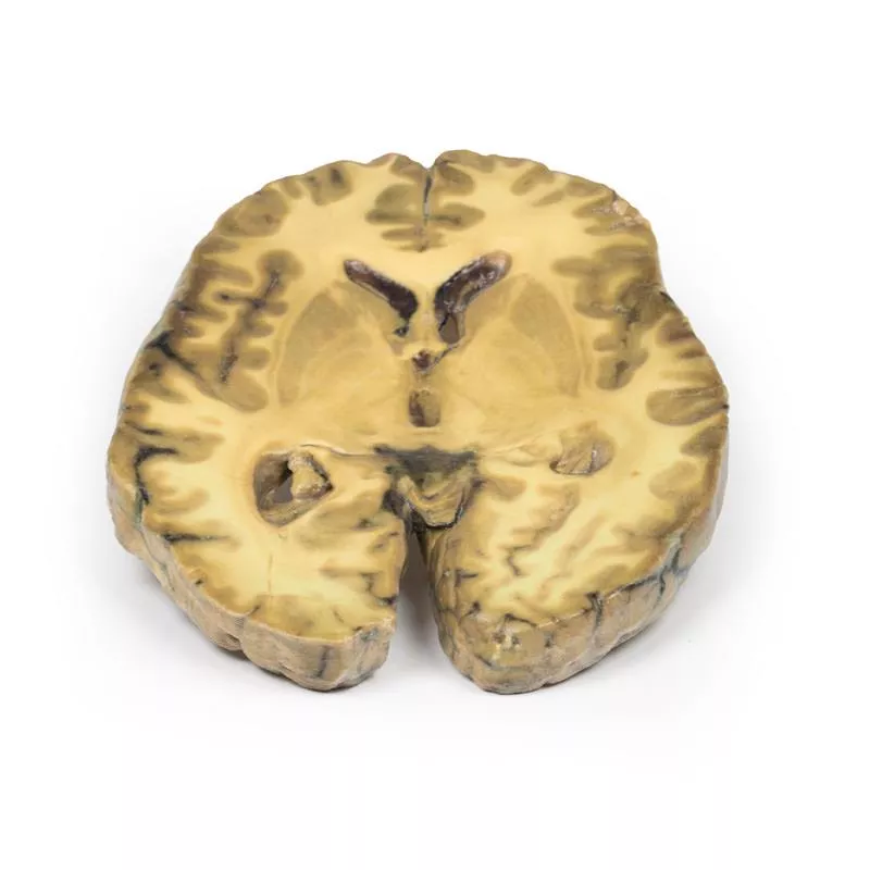
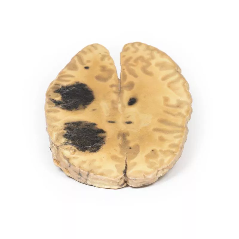
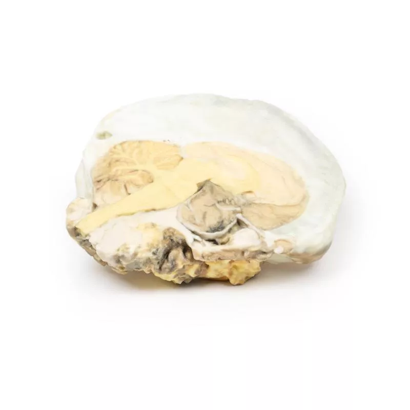
This mid-sagittal brain section (1 cm thick) shows a large, dark berry aneurysm (5 × 2 cm) from the basilar artery, eroding into the midbrain and pons, and compressing the third ventricle. The aneurysm is filled with a laminated thrombus, with blood visible in the third ventricle and signs of leakage. A mucoid degeneration (0.4 cm) is present in the pons. The lateral view shows ventricular dilatation, blood staining, and haemorrhagic infarction of the caudate nucleus, along with meningeal discolouration consistent with subarachnoid haemorrhage.
Further Information
Intracranial aneurysms occur in ~3.2% of the population, with rupture rates of 7.9 per 100,000 person-years. Posterior circulation aneurysms are less common and typically found at basilar, vertebral, or cerebellar artery junctions. Symptoms arise from subarachnoid haemorrhage or mass effect. Complications include raised intracranial pressure, hydrocephalus, re-bleeding, and vasospasm. Treatment options include surgical and endovascular interventions.
Erler-Zimmer
Erler-Zimmer GmbH & Co.KG
Hauptstrasse 27
77886 Lauf
Germany
info@erler-zimmer.de
Achtung! Medizinisches Ausbildungsmaterial, kein Spielzeug. Nicht geeignet für Personen unter 14 Jahren.
Attention! Medical training material, not a toy. Not suitable for persons under 14 years of age.


