


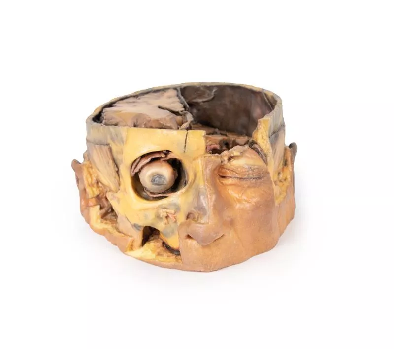
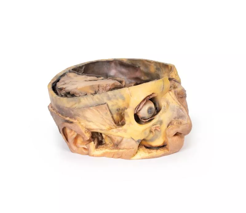

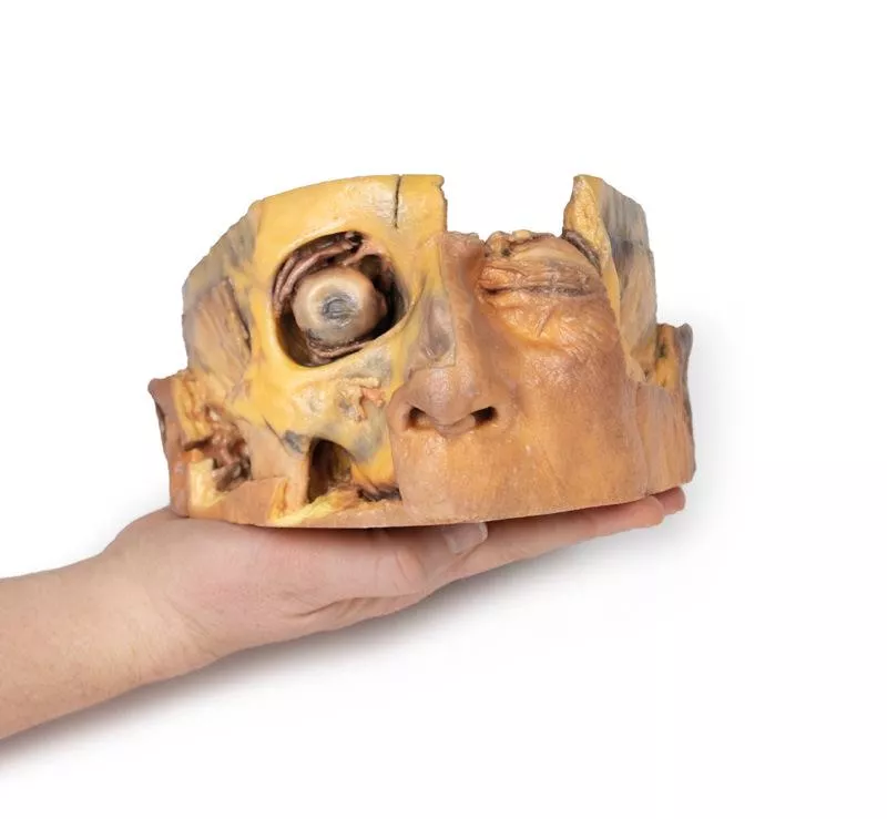


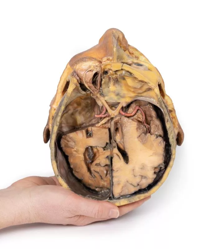

Product information "Transverse Section of the head"
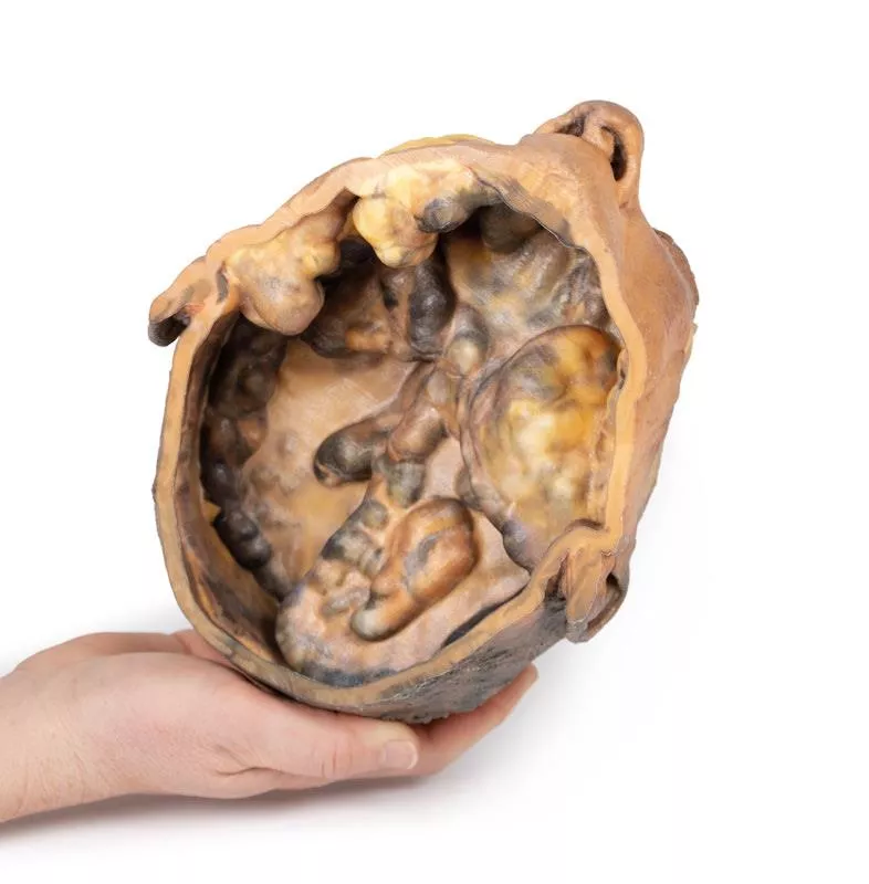
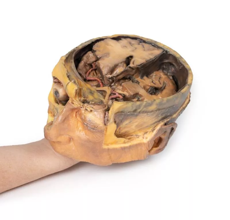
This 3D model features a transverse section through the cranial cavity with a deep dissection of the face, orbit, and temporomandibular joint (TMJ) region. It offers a comprehensive view of both intracranial and facial structures.
Key Features:
Cranial Cavity & Brain
- Partial dura mater removal; dissection reveals lateral and third ventricles, falx cerebri, choroid plexus, and optic pathways
- Middle cerebral artery visible in the lateral fissure
- Key vascular structures: internal carotid, anterior and middle cerebral arteries
Left Orbit
Roof removed; exposure of: Frontal nerve, lacrimal gland, superior oblique, medial rectus, nasociliary nerve
Right Orbit
Superficial tissues removed; shows: All extraocular muscles, including inferior oblique, and levator palpebrae superioris
Face & TMJ (Right Side)
- Exposed: infraorbital nerve/artery, masseter (both heads), and temporalis muscle near pterion
- Parotid gland dissected to show mandibular condyle in glenoid fossa and external ear alignment
Login
Erler-Zimmer
Erler-Zimmer Medical GmbH
Hauptstrasse 27
77886 Lauf
Germany
info@erler-zimmer.de
Achtung! Medizinisches Ausbildungsmaterial, kein Spielzeug. Nicht geeignet für Personen unter 14 Jahren.
Attention! Medical training material, not a toy. Not suitable for persons under 14 years of age.
Other customers also bought

Superficial Facial nerves & Parotid Gland
This 3D model provides a detailed view of the superficial anatomy of the face and head, expanding upon our HW 44 model with a broader dissection of the scalp, occipital region, and areas below the external ear.Key Features:Extended Facial AnatomyIncludes the terminal branches of the facial nerve (CN VII) traced from the parotid gland, with the platysma muscle preserved and extending from the mandible to the neck. Enhanced Posterior Dissection- Broader exposure across the posterior scalp and occipital region- Includes the retromandibular vein, great auricular nerve, and lesser occipital nerve- Shows the course of the occipital artery and vein near the trapeziusNeurovascular HighlightsImproved visualization of the supraorbital, supratrochlear, and superficial temporal arteries and nervesMusculaturePreserves fibers of the auricularis and occipitalis muscles, integrated into the epicranius (occipitofrontalis)

Lung Slab, Hilum removed
This 3D anatomical model presents a left lung dissected in a parasagittal plane, offering a unique internal view of pulmonary structures and anatomical landmarks. The mediastinal surface has been removed, allowing detailed observation of internal lung anatomy and its relationship to the heart and diaphragm.Key Features:Bronchial Tree (Internal View):- The primary bronchi are not visible due to prior branching.- Subdivided bronchi are preserved, though the dissection depth makes it unclear whether they represent secondary (lobar) or tertiary (segmental) bronchi. Vascular Structures:- Pulmonary arteries and veins are typically seen at the hilum, but precise levels of subdivision are undetermined in this section.Cardiac Impression:- A clear concave impression remains on the medial lung surface, formed by the left ventricle of the heart pressing against the lung.- Despite the dissection, this landmark remains distinctly visible.Diaphragmatic Surface:- The lung’s base is concave, resting atop the diaphragm.- While the pleura is not preserved, the model indicates where the diaphragmatic recess would form—between the costal and diaphragmatic pleura.

Stomach
This 3D model is an isolated stomach with two dissection windows to expose the rugae and pylorus. A small portion of the terminal oesophagus is preserved at the cardiac region, and a small portion of the proximal duodenum beyond the pyloric sphincter. The large window within the body of the stomach allows for a clear view into the fundus and the well-developed rugae on the posterior aspect of the wall of the organ. The smaller window, opened just at the pyloric region, allows for an appreciation of the thickening of the organ wall at the pyloric sphincter just proximal to the start of the duodenum.

Female pelvis deep dissection
This high-detail 3D model showcases a deep dissection of the female pelvis, isolated from surrounding regions, with emphasis on visceral, vascular, and ligamentous structures in relation to bony landmarks.Pelvic Organs & Peritoneal Structures- Sigmoid colon descends into the rectum over the pelvic brim, crossing the common and external iliac vessels.- Nearby: Sigmoid and superior rectal arteries, and the descending ureter.- Urinary bladder (collapsed) and uterus are positioned anteriorly in the true pelvis.- The broad ligament is retained, though ovaries, uterine tubes, ovarian and round ligaments are present but indistinct due to age-related atrophy.- Suspensory and round ligaments are detached from the peritoneum to expose surrounding vessels. Arteries & Veins- Internal iliac artery branches are visible bilaterally.- Median sacral artery is seen in the midline between the common iliac arteries.- Left side: Uterine artery only.- Right side: Uterine, superior vesical, and obturator arteries.- Inferior epigastric artery and vein arise from the external iliac vessels, visible near the inferior abdominal wall. Musculoskeletal Features- Right side: Entire femur and thigh muscles removed to expose:- Obturator membrane- Acetabular cartilage- Transverse acetabular ligament- Posterior dissection reveals:- Superior gluteal foramen and artery- Sacrospinous ligament (with sacrotuberous ligament removed)- Inferior rectal artery branches within the ischioanal fossa Nerves & Ligaments- Left sciatic nerve preserved within the greater sciatic foramen- Sacrotuberous ligament retained on the left- Ischioanal fossae on both sides show:- Inferior rectal artery branches- Pelvic diaphragm fibers- External anal sphincter integration with the rectal wall

Sagittal Section of Head and Neck with Infratemporal Fossa and Carotid Sheath Dissection
This 3D model complements the H11 and H12 head and neck specimens, offering a clear view of the endocranial cavity without the brain, alongside a lateral dissection of the face, infratemporal region, and neck.Key Features:Endocranial Cavity- Brain removed; dura mater, tentorium cerebelli, and superior sagittal sinus fully visible- Several cranial nerves (CN II, III, V, VI, VII, VIII) seen piercing the dura- Pituitary gland preserved in sella turcica, left vertebral artery visible in posterior fossa Lateral Facial & Infratemporal Region- Facial artery and vein retained, dissected free of superficial tissues- Partial removal of mandible and zygomatic arch reveals:- Inferior alveolar and lingual nerves, posterior deep temporal artery, and TMJ articulation- Visible branches of the external carotid artery, including maxillary and superficial temporal arteriesNeck Anatomy- Facial nerve (CN VII) near posterior belly of digastric- Dissected carotid sheath showing internal/external carotid arteries, internal jugular vein, and vagus nerve (CN X)- Hypoglossal nerve (CN XII) and facial artery near submandibular gland- Hyoid bone, thyroid gland, and larynx visible- Cervical plexus branches on scalene muscles; brachial plexus roots preserved near internal jugular vein

Abdomen with bilateral Hernias
This 3D model represents one of the largest and most complex in the series, consisting of a partial torso from the diaphragm to the proximal thigh with a complete abdominal cavity preserving varying levels of dissection. This 3D model also records the rare, simultaneous occurrence of indirect and direct inguinal hernias allowing for a consideration of the anatomical underpinnings for both conditions. Given the scale of the dissection this 3D model description is divided into discrete parts based on views and regions.The diaphragmThe diaphragm is preserved on the model’s superior aspect, with both domes and costodiaphragmatic recesses visible despite some distortion from rib removal. The fibrous pericardium rests on the central tendon, with the terminal inferior vena cava seen in the caval foramen. Lateral to this lies the oesophagus in the oesophageal hiatus, and the descending thoracic aorta approaching the aortic hiatus near the vertebrae. The epigastric and hypochondriac regionsIn the abdomen, removal of the anterior wall, greater omentum, and much of the GI tract reveals retroperitoneal structures. The terminal oesophagus enters just left of the liver. With the stomach removed, the pancreas is fully exposed from head to tail, reaching the spleen in the left hypochondrium. Above it, the splenic and common hepatic arteries span the narrow space between pancreas, diaphragm, and liver. The tortuous splenic artery divides near the splenic vein; the common hepatic gives rise to the gastroduodenal and right gastric arteries, superficial to the portal vein. The superior mesenteric vessels pass near the pancreatic head, and the ileocolic artery leads to the caecum. The inferior mesenteric vein arises from the superior rectal vein and crosses the descending aorta. Below the liver, the gallbladder lies between the lobes. On the left, renal vessels pass deep to the pancreas, with ureters descending across the psoas muscles. The umbilical and lumbar regionsMost abdominal organs in the umbilical and lumbar regions have been removed to reveal the posterior abdominal wall. Centrally, the descending aorta and inferior vena cava are prominent, with testicular vessels traceable toward the inguinal region. Two right lumbar arteries branch from the aorta, and the inferior mesenteric artery gives rise to the left colic, sigmoid, and superior rectal arteries. On the right, subcostal, iliohypogastric, and ilioinguinal nerves are visible, along with the circumflex iliac artery.The hypogastrium and iliac regionsThe abdominal aorta bifurcates into the common, internal, and external iliac arteries, with matching iliac veins merging into the inferior vena cava. The obturator artery, ureters, and testicular vessels are visible. In the true pelvis, the peritoneum covers the bladder, while the rectum remains obscured. The right iliac fossa contains the terminal ileum, caecum, and appendix, with nearby vessels and nerves. On the left, the sigmoid colon crosses the iliac fossa, where an epiploic appendage extends into an indirect hernia near the inferior epigastric artery. The inguinal region and perineumThis model uniquely preserves both direct (right) and indirect (left) inguinal hernias, with the inferior epigastric vessels retained for anatomical orientation. The right hernia lies medial to these vessels; the left hernia sac extends laterally into the spermatic cord, containing an epiploic appendage. The perineum reveals the penis, testes, and spermatic cords. On the right, the cord remains intact; on the left, it’s opened, showing a varicose testicular vein linked to the indirect hernia. The thighThe femoral triangle has been dissected on both thighs. On the right, the femoral sheath was removed to reveal the femoral artery, vein, deep inguinal lymph nodes, and femoral nerve. On the left, a broader view exposes anterior and medial thigh muscles, with the femoral artery, profunda femoris, and circumflex iliac artery visible. The model ends mid-thigh, showing cross-sectional anatomy including the femoral shaft, vessels, and muscles in the subsartorial canal.

Continuous innovation

Social responsibility

Active customer orientation

Understanding quality

Sustainable actions

ISO 9001 certification