Product information "Astrocytoma"
Clinical History
A 73-year-old woman was admitted with new left-sided hemiplegia. On further questioning, she reported a 3-month history of headaches, nausea, and worsening balance. CT identified an inoperable brain tumour. She passed away one week after admission.
Pathology
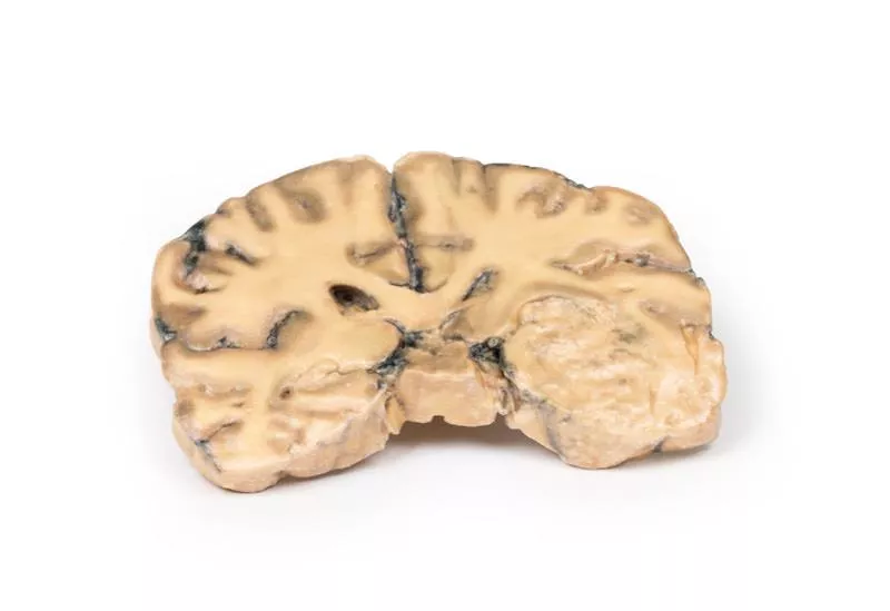
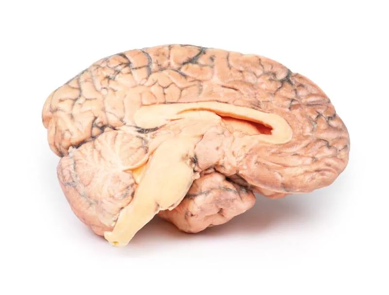
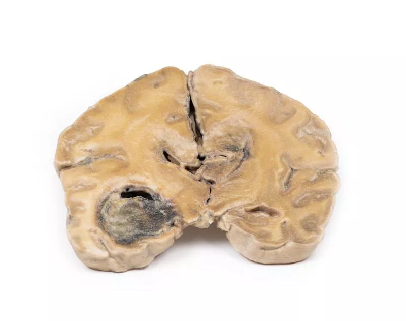
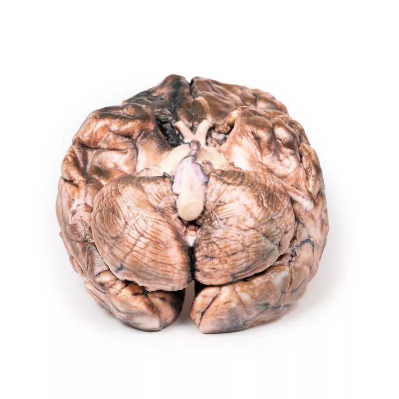
A coronal brain slice reveals a poorly demarcated tumour in the right temporal lobe, causing hemispheric enlargement and flattening of the gyri. The posterior view shows subfalcine herniation, and the mass displays areas of necrosis and haemorrhage. Histology confirmed an astrocytoma, Grade III/IV.
Further Information
Astrocytomas are a subtype of glioma, the second most common CNS cancer after meningioma. Originating from astrocytes, they are classified by grade—diffuse (II), anaplastic (III), to glioblastoma (IV). Typical histology shows gemistocytes with eosinophilic cytoplasm against a fibrillary background. Most occur in the hemispheres of patients aged 40–60 and present with seizures, headaches, nausea, or focal deficits. Without treatment, median survival for Grade III tumors is ~~18 months. Management involves surgery, radiotherapy, chemotherapy, or combinations tailored to the patient.
Erler-Zimmer
Erler-Zimmer GmbH & Co.KG
Hauptstrasse 27
77886 Lauf
Germany
info@erler-zimmer.de
Achtung! Medizinisches Ausbildungsmaterial, kein Spielzeug. Nicht geeignet für Personen unter 14 Jahren.
Attention! Medical training material, not a toy. Not suitable for persons under 14 years of age.


