Product information "Glioblastoma multiforme"
Clinical History
A 56-year-old male presented with a generalised seizure after which he remained unconscious and later died. A history revealed 6 months of progressive confusion, short-term memory loss, and personality changes.
Pathology
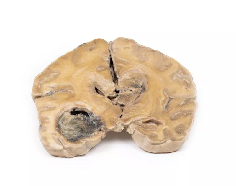
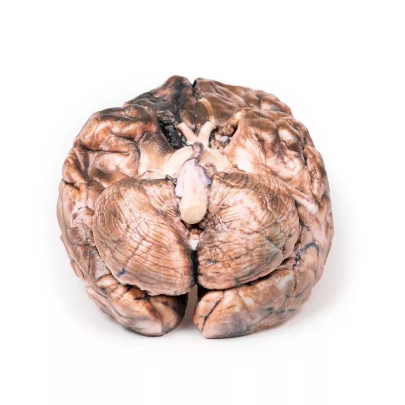
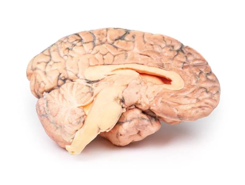
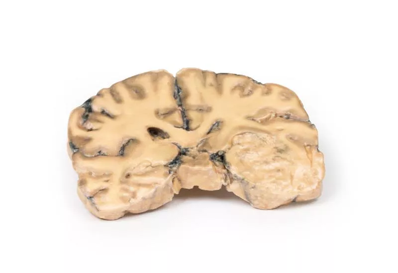
Post-mortem coronal brain sections show a 4 cm necrotic and haemorrhagic tumour invading from the inferior frontal lobe into the lateral ventricle. Meningeal spread is visible on the posterior aspect.
Further Information
Gliomas are the second most common central nervous system cancers after meningiomas. They originate from glial-like cells such as astrocytes, oligodendrocytes, or ependymal cells. Glioblastoma multiforme (GBM), a grade IV astrocytoma, arises from astrocytes and may develop de novo or from lower-grade tumours. GBMs typically show necrosis surrounded by anaplastic cells and hyperplastic blood vessels. More frequent in males, GBM commonly occurs in the 6th decade. Risk factors include neurofibromatosis type 1, Li-Fraumeni syndrome, and prior brain radiotherapy.
Symptoms depend on tumour location and include persistent headaches, visual disturbances, vomiting, appetite loss, mood and personality changes, cognitive decline, seizures, and speech difficulties. Diagnostic tools are CT and MRI. About 50% of GBMs involve more than one hemisphere, often invading ventricles or meninges, with rare spinal cord spread. Metastasis outside the CNS is uncommon.
Tumour growth leads to cerebral oedema and increased intracranial pressure. These aggressive tumours have a median survival of approximately 3 months if untreated. Treatment consists of surgery followed by radiation and chemotherapy.
Erler-Zimmer
Erler-Zimmer GmbH & Co.KG
Hauptstrasse 27
77886 Lauf
Germany
info@erler-zimmer.de
Achtung! Medizinisches Ausbildungsmaterial, kein Spielzeug. Nicht geeignet für Personen unter 14 Jahren.
Attention! Medical training material, not a toy. Not suitable for persons under 14 years of age.


