Product information "Cholecystitis and Cholelithiasis"
Clinical History
A 60-year-old man had experienced four episodes of severe gripping abdominal pain over the past year, each lasting around two hours and occurring after meals. He presented again with similar pain, now accompanied by vomiting and fever. This episode did not resolve spontaneously and led to a cholecystectomy.
Pathology
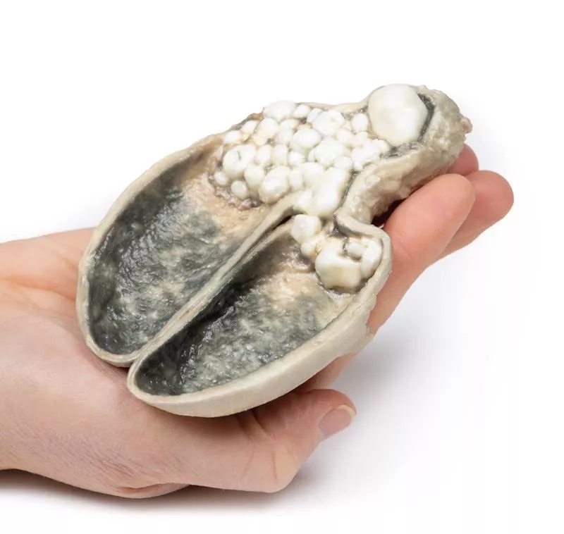
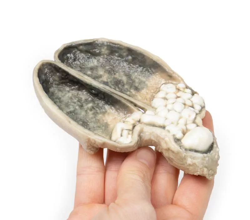
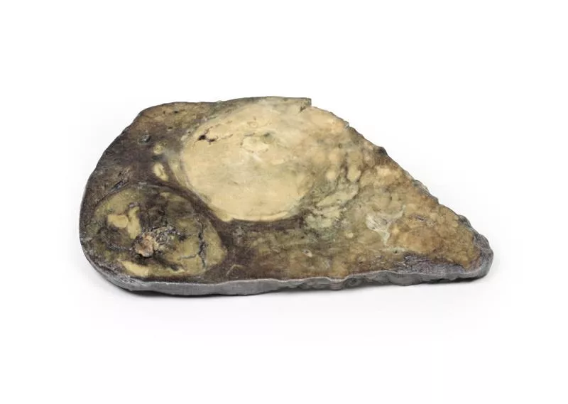
The gallbladder shows a thickened wall and haemorrhagic mucosa, with numerous irregular faceted gallstones inside. A large stone is impacted in the neck of the gallbladder. The outer surface is congested and dull. This is a classic example of cholecystitis caused by cholelithiasis.
Further Information
Gallstones are the leading cause of acute cholecystitis, responsible for about 90–95% of cases. Repeated episodes can result in chronic cholecystitis with fibrotic thickening of the gallbladder wall. About 6–11% of patients with symptomatic stones develop acute inflammation.
Lab findings often show leucocytosis, sometimes with cholestatic liver function changes. Imaging, particularly ultrasound, is key in diagnosis, typically showing stones, wall thickening, and a positive sonographic Murphy’s sign. Additional imaging may include MRCP, CT, or cholescintigraphy. ERCP can both diagnose and treat biliary obstruction.
If infection is present, common pathogens include E. coli, Enterococcus, Klebsiella, and Enterobacter. Complications may include gangrenous cholecystitis, perforation, cholecystoenteric fistula or gallstone ileus. The definitive treatment is surgical cholecystectomy.
Erler-Zimmer
Erler-Zimmer GmbH & Co.KG
Hauptstrasse 27
77886 Lauf
Germany
info@erler-zimmer.de
Achtung! Medizinisches Ausbildungsmaterial, kein Spielzeug. Nicht geeignet für Personen unter 14 Jahren.
Attention! Medical training material, not a toy. Not suitable for persons under 14 years of age.


