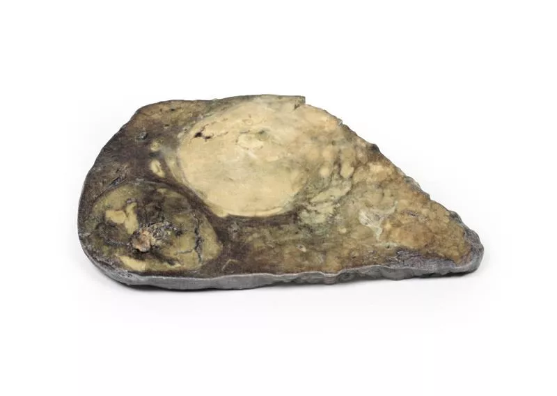
Product information "Adenocarcinoma of the stomach"
Clinical History
An 82-year-old woman presented with melena, along with a six-month history of dyspepsia, nausea, weight loss, and early satiety. Shortly after admission, she suffered a massive melena episode and passed away.
Pathology
The post-mortem specimen includes the esophagus, stomach, proximal duodenum, and pancreas. A 7×5?cm shallow gastric ulcer on the lesser curve features raised, rolled edges and necrotic debris. Dissection reveals pale tumor tissue elevating the edge. Two eroded arteries and recent hemorrhage are present, and the pancreas adheres to the ulcer’s serosal surface. Histology confirms a well-differentiated gastric adenocarcinoma invading directly into the pancreas.
Further Information
Gastric adenocarcinoma is the most common stomach malignancy, with incidence varying by region. Risk factors include smoking, high salt intake, H. pylori infection, GERD, atrophic gastritis, and intestinal metaplasia. There are two main types:
- Intestinal type: Gland-like, bulky, ulcerated or exophytic, common in endemic regions, often seen in men around 55 years old, may arise from dysplasia or adenomas.
- Diffuse type: Composed of signet-ring cells with mucin-vacuoles, infiltrative growth, rigid “leather bottle” stomach due to desmoplasia, equal across sexes and regions. Associated with CDH1 mutations, and higher risk in FAP patients with APC mutations.
Early symptoms include dyspepsia, dysphagia, and nausea. Advanced disease often presents with weight loss, early satiety, fatigue, anemia, or hemorrhage. Treatment depends on stage—surgical resection for early tumors, chemotherapy for advanced cases.
🔬 3D Anatomy Series - Human Body Replicas to Enhance Teaching!
August 26, 2025
Discover exclusive 3D-printed models of the human body – created directly from radiological data or real specimens.
Erler-Zimmer
Erler-Zimmer GmbH & Co.KG
Hauptstrasse 27
77886 Lauf
Germany
info@erler-zimmer.de
Achtung! Medizinisches Ausbildungsmaterial, kein Spielzeug. Nicht geeignet für Personen unter 14 Jahren.
Attention! Medical training material, not a toy. Not suitable for persons under 14 years of age.


