


Product information "Hepatocellular Carcinoma"
Clinical History
A 60-year-old male was admitted with jaundice, melena, and abdominal distension. He had a known history of untreated Hepatitis C due to previous intravenous drug use. On further questioning, he reported a 9-month history of fatigue, weight loss, nausea, and intermittent dull pain in the right upper abdomen. Liver ultrasound revealed two large lesions. The patient died shortly after admission due to suspected oesophageal variceal haemorrhage.
Pathology
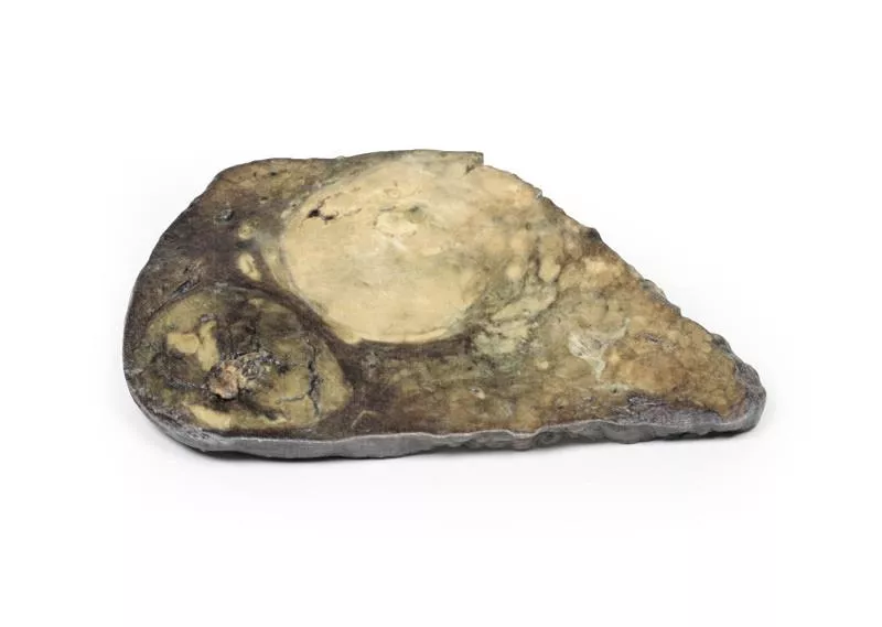
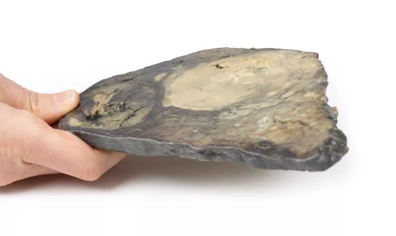
The liver specimen from the postmortem shows a macronodular cirrhosis with multiple nodules up to 2?cm in size, separated by narrow fibrous bands. Additionally, two large round tumours measuring 8?cm and 6?cm are present. Their cut surface shows areas of necrosis, haemorrhage, and bile staining. This is an example of hepatocellular carcinoma (HCC) arising in a cirrhotic liver.
Further Information
Hepatocellular carcinoma is the most common primary malignant liver tumour and originates from hepatocytes. Risk factors include Hepatitis B and C, liver cirrhosis, aflatoxin exposure, non-alcoholic fatty liver disease, haemochromatosis, and Wilson’s disease. The tumour often develops in the setting of genetic alterations, such as gain-of-function mutations in beta-catenin and loss-of-function mutations in p53. Clinical presentation may include abdominal pain, fatigue, weight loss, fullness, and in some cases jaundice or bleeding. HCC can metastasize via the bloodstream, especially to lungs, abdominal lymph nodes, and bones. Death typically occurs due to cachexia, haemorrhage, or liver failure. Treatment options depend on tumour stage and patient condition and may include surgical resection, tumour ablation, chemotherapy, or liver transplantation.
A 60-year-old male was admitted with jaundice, melena, and abdominal distension. He had a known history of untreated Hepatitis C due to previous intravenous drug use. On further questioning, he reported a 9-month history of fatigue, weight loss, nausea, and intermittent dull pain in the right upper abdomen. Liver ultrasound revealed two large lesions. The patient died shortly after admission due to suspected oesophageal variceal haemorrhage.
Pathology
The liver specimen from the postmortem shows a macronodular cirrhosis with multiple nodules up to 2?cm in size, separated by narrow fibrous bands. Additionally, two large round tumours measuring 8?cm and 6?cm are present. Their cut surface shows areas of necrosis, haemorrhage, and bile staining. This is an example of hepatocellular carcinoma (HCC) arising in a cirrhotic liver.
Further Information
Hepatocellular carcinoma is the most common primary malignant liver tumour and originates from hepatocytes. Risk factors include Hepatitis B and C, liver cirrhosis, aflatoxin exposure, non-alcoholic fatty liver disease, haemochromatosis, and Wilson’s disease. The tumour often develops in the setting of genetic alterations, such as gain-of-function mutations in beta-catenin and loss-of-function mutations in p53. Clinical presentation may include abdominal pain, fatigue, weight loss, fullness, and in some cases jaundice or bleeding. HCC can metastasize via the bloodstream, especially to lungs, abdominal lymph nodes, and bones. Death typically occurs due to cachexia, haemorrhage, or liver failure. Treatment options depend on tumour stage and patient condition and may include surgical resection, tumour ablation, chemotherapy, or liver transplantation.
Login
Erler-Zimmer
Erler-Zimmer Medical GmbH
Hauptstrasse 27
77886 Lauf
Germany
info@erler-zimmer.de
Achtung! Medizinisches Ausbildungsmaterial, kein Spielzeug. Nicht geeignet für Personen unter 14 Jahren.
Attention! Medical training material, not a toy. Not suitable for persons under 14 years of age.
Other customers also bought

Adenocarcinoma of the stomach
Clinical HistoryAn 82-year-old woman presented with melena, along with a six-month history of dyspepsia, nausea, weight loss, and early satiety. Shortly after admission, she suffered a massive melena episode and passed away.PathologyThe post-mortem specimen includes the esophagus, stomach, proximal duodenum, and pancreas. A 7×5?cm shallow gastric ulcer on the lesser curve features raised, rolled edges and necrotic debris. Dissection reveals pale tumor tissue elevating the edge. Two eroded arteries and recent hemorrhage are present, and the pancreas adheres to the ulcer’s serosal surface. Histology confirms a well-differentiated gastric adenocarcinoma invading directly into the pancreas.Further InformationGastric adenocarcinoma is the most common stomach malignancy, with incidence varying by region. Risk factors include smoking, high salt intake, H. pylori infection, GERD, atrophic gastritis, and intestinal metaplasia. There are two main types: - Intestinal type: Gland-like, bulky, ulcerated or exophytic, common in endemic regions, often seen in men around 55 years old, may arise from dysplasia or adenomas. - Diffuse type: Composed of signet-ring cells with mucin-vacuoles, infiltrative growth, rigid “leather bottle” stomach due to desmoplasia, equal across sexes and regions. Associated with CDH1 mutations, and higher risk in FAP patients with APC mutations.Early symptoms include dyspepsia, dysphagia, and nausea. Advanced disease often presents with weight loss, early satiety, fatigue, anemia, or hemorrhage. Treatment depends on stage—surgical resection for early tumors, chemotherapy for advanced cases.

Mesenteric Metastases from Cutaneous Malignant Melanoma
Clinical HistoryA 44-year-old man had a slow-growing skin lesion on his back. Years later, he presented at A&E with bone pain, hepatomegaly, and pleural effusion. He died shortly after admission.PathologyA loop of small intestine with attached mesentery reveals numerous dark brown nodules, ranging from pinhead size to 1 cm. Histology confirmed metastatic melanoma.Further InformationCutaneous melanoma arises from melanocytes, the pigment-producing cells in the skin. In men, it commonly appears on the back; in women, on the legs. Roughly 25% of melanomas develop from pre-existing moles. Warning signs include size increase, irregular borders, colour changes, itchiness, or ulceration.Key risk factors include UV radiation exposure, fair skin, multiple moles, childhood sunburn, and immunosuppression. Although melanoma represents just 5% of skin cancer cases, it has the highest mortality rate.Genetic mutations often involved include CDKN2A, BRAF, PI3K, and TERT. Recognition of melanoma by the immune system has led to progress in immunotherapy.Melanoma commonly metastasizes to lungs, liver, brain, bones, and lymph nodes. In the GI tract, it may cause bleeding, pain, obstruction or intussusception, particularly in the jejunum and ileum. Surgery is often used for symptom management in such cases.Diagnosis is based on excisional biopsy. Further evaluation may include blood tests (e.g. alkaline phosphatase, calcium, LDH) and imaging (X-ray, CT, MRI, PET). Treatment options include surgery, chemotherapy, radiotherapy, targeted therapy (like BRAF inhibitors), and immunotherapy, often in combination.

Hepatic duct calculi and Obstructive Biliary Cirrhosis
Clinical HistoryAn 85-year-old man presented with urinary retention due to benign prostatic hypertrophy. On admission, he was noted to be jaundiced with a cholestatic pattern in his liver function tests. He underwent a transurethral resection of the prostate but died five days later from pneumonia.PathologyA liver slice shows a slightly thickened capsule and a finely nodular surface. The intrahepatic bile ducts are dilated. On the posterior surface, a pigmented calculus (10 mm) is impacted in a distended hepatic duct, and a smaller one (3 mm) is dislodged. This represents secondary biliary cirrhosis due to obstruction of large bile ducts by hepatic stones.Further InformationHepatolithiasis involves the formation of intrahepatic gallstones, most commonly pigmented calcium bilirubinate stones. These obstruct bile flow, leading to bile duct dilation, portal inflammation, and progressive fibrosis. Histology would show feathery degeneration of periportal hepatocytes, cytoplasmic swelling, Mallory-Denk bodies, and bile infarcts. Chronic inflammation can result in biliary dysplasia and may progress to cholangiocarcinoma. Clinically, patients may experience recurrent cholangitis, intermittent pain, jaundice, or be asymptomatic. Treatment typically involves surgical removal of the calculi.

Pedunculated Adenoma of the Colon
Clinical HistoryA 50-year-old male underwent colonoscopy after a positive faecal occult blood screening. The examination revealed a pedunculated tumour in the descending colon, which was later resected.PathologyThe resected specimen from the descending colon shows a single dark, lobulated mass arising from the mucosal surface, attached to a 4 cm stalk. Histological analysis confirmed it as a tubular colonic adenoma, composed of connective tissue covered with hyperplastic colonic-type epithelium and areas of nuclear atypia.Further InformationColorectal adenomas are intraepithelial neoplasms that exhibit dysplasia and are considered precursors to adenocarcinoma, though not all progress to malignancy. These adenomas typically present as polyps—either pedunculated or sessile—and are more prevalent in men and in Western populations due to dietary and lifestyle factors.They are found in around 30% of individuals over 60 in the West, with increased risk linked to family history of colorectal cancer. Surveillance colonoscopy with polyp removal can significantly reduce cancer incidence.Colonic adenomas are classified into tubular (>75% tubular), tubulovillous (25–75% villous), and villous (>75% villous) types. Tubular adenomas are usually small, pedunculated, and composed of rounded or tubular glands.Adenomas less than 1 cm rarely progress, whereas those >4 cm have a progression rate of up to 40%. Most remain asymptomatic but may cause anaemia due to occult bleeding. In some cases, especially with villous types, excessive secretion of mucous and potassium-rich fluid can lead to hypokalemia.

Cholecystitis and Cholelithiasis
Clinical HistoryA 60-year-old man had experienced four episodes of severe gripping abdominal pain over the past year, each lasting around two hours and occurring after meals. He presented again with similar pain, now accompanied by vomiting and fever. This episode did not resolve spontaneously and led to a cholecystectomy.PathologyThe gallbladder shows a thickened wall and haemorrhagic mucosa, with numerous irregular faceted gallstones inside. A large stone is impacted in the neck of the gallbladder. The outer surface is congested and dull. This is a classic example of cholecystitis caused by cholelithiasis.Further InformationGallstones are the leading cause of acute cholecystitis, responsible for about 90–95% of cases. Repeated episodes can result in chronic cholecystitis with fibrotic thickening of the gallbladder wall. About 6–11% of patients with symptomatic stones develop acute inflammation.Lab findings often show leucocytosis, sometimes with cholestatic liver function changes. Imaging, particularly ultrasound, is key in diagnosis, typically showing stones, wall thickening, and a positive sonographic Murphy’s sign. Additional imaging may include MRCP, CT, or cholescintigraphy. ERCP can both diagnose and treat biliary obstruction.If infection is present, common pathogens include E. coli, Enterococcus, Klebsiella, and Enterobacter. Complications may include gangrenous cholecystitis, perforation, cholecystoenteric fistula or gallstone ileus. The definitive treatment is surgical cholecystectomy.

Continuous innovation

Social responsibility

Active customer orientation

Understanding quality

Sustainable actions

ISO 9001 certification