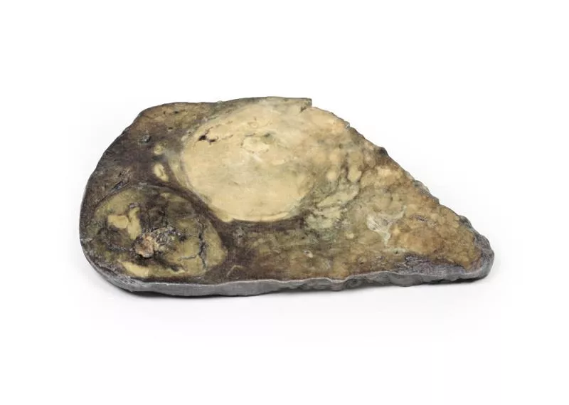
Product information "Hepatic duct calculi and Obstructive Biliary Cirrhosis"
Clinical History
An 85-year-old man presented with urinary retention due to benign prostatic hypertrophy. On admission, he was noted to be jaundiced with a cholestatic pattern in his liver function tests. He underwent a transurethral resection of the prostate but died five days later from pneumonia.
Pathology
A liver slice shows a slightly thickened capsule and a finely nodular surface. The intrahepatic bile ducts are dilated. On the posterior surface, a pigmented calculus (10 mm) is impacted in a distended hepatic duct, and a smaller one (3 mm) is dislodged. This represents secondary biliary cirrhosis due to obstruction of large bile ducts by hepatic stones.
Further Information
Hepatolithiasis involves the formation of intrahepatic gallstones, most commonly pigmented calcium bilirubinate stones. These obstruct bile flow, leading to bile duct dilation, portal inflammation, and progressive fibrosis. Histology would show feathery degeneration of periportal hepatocytes, cytoplasmic swelling, Mallory-Denk bodies, and bile infarcts. Chronic inflammation can result in biliary dysplasia and may progress to cholangiocarcinoma. Clinically, patients may experience recurrent cholangitis, intermittent pain, jaundice, or be asymptomatic. Treatment typically involves surgical removal of the calculi.
Erler-Zimmer
Erler-Zimmer GmbH & Co.KG
Hauptstrasse 27
77886 Lauf
Germany
info@erler-zimmer.de
Achtung! Medizinisches Ausbildungsmaterial, kein Spielzeug. Nicht geeignet für Personen unter 14 Jahren.
Attention! Medical training material, not a toy. Not suitable for persons under 14 years of age.


