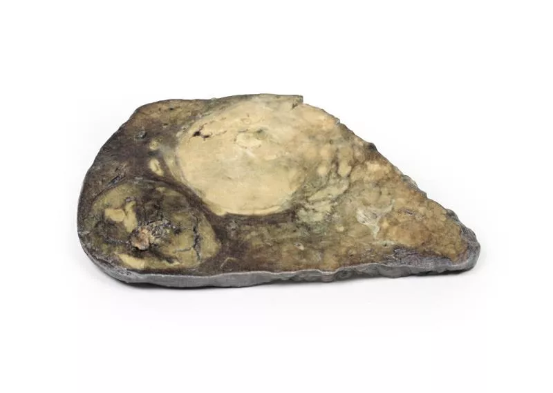
Product information "Mesenteric Metastases from Cutaneous Malignant Melanoma"
Clinical History
A 44-year-old man had a slow-growing skin lesion on his back. Years later, he presented at A&E with bone pain, hepatomegaly, and pleural effusion. He died shortly after admission.
Pathology
A loop of small intestine with attached mesentery reveals numerous dark brown nodules, ranging from pinhead size to 1 cm. Histology confirmed metastatic melanoma.
Further Information
Cutaneous melanoma arises from melanocytes, the pigment-producing cells in the skin. In men, it commonly appears on the back; in women, on the legs. Roughly 25% of melanomas develop from pre-existing moles. Warning signs include size increase, irregular borders, colour changes, itchiness, or ulceration.
Key risk factors include UV radiation exposure, fair skin, multiple moles, childhood sunburn, and immunosuppression. Although melanoma represents just 5% of skin cancer cases, it has the highest mortality rate.
Genetic mutations often involved include CDKN2A, BRAF, PI3K, and TERT. Recognition of melanoma by the immune system has led to progress in immunotherapy.
Melanoma commonly metastasizes to lungs, liver, brain, bones, and lymph nodes. In the GI tract, it may cause bleeding, pain, obstruction or intussusception, particularly in the jejunum and ileum. Surgery is often used for symptom management in such cases.
Diagnosis is based on excisional biopsy. Further evaluation may include blood tests (e.g. alkaline phosphatase, calcium, LDH) and imaging (X-ray, CT, MRI, PET). Treatment options include surgery, chemotherapy, radiotherapy, targeted therapy (like BRAF inhibitors), and immunotherapy, often in combination.
Erler-Zimmer
Erler-Zimmer GmbH & Co.KG
Hauptstrasse 27
77886 Lauf
Germany
info@erler-zimmer.de
Achtung! Medizinisches Ausbildungsmaterial, kein Spielzeug. Nicht geeignet für Personen unter 14 Jahren.
Attention! Medical training material, not a toy. Not suitable for persons under 14 years of age.


