Product information "Foot - Deep plantar structures"
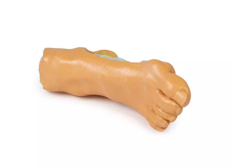
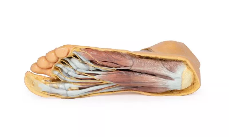
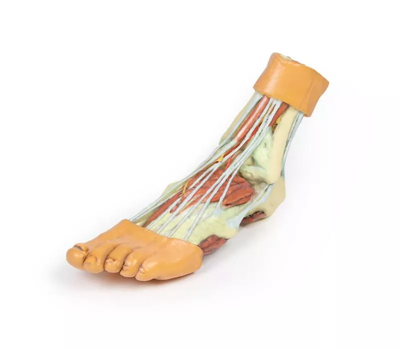
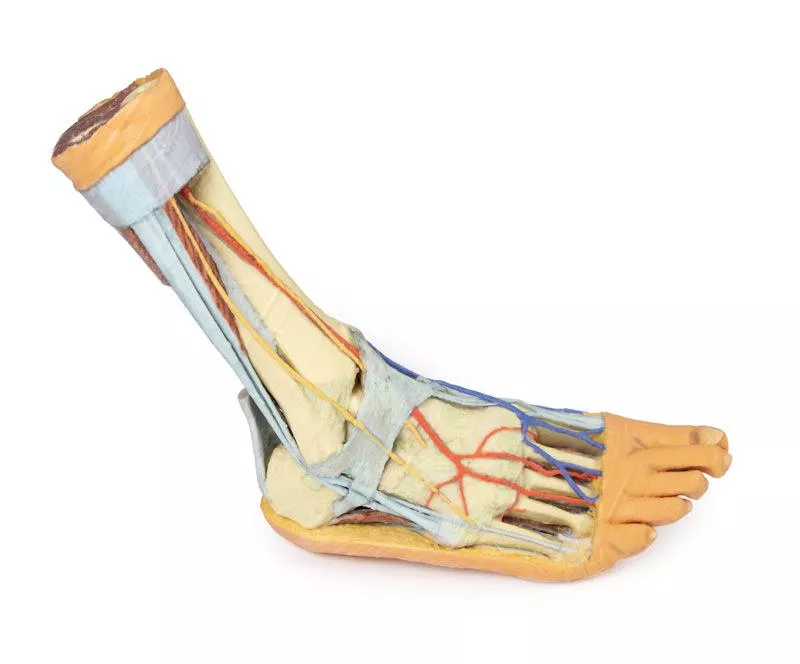
This 3D printed anatomical model offers a detailed view of the deep plantar structures of the foot.
Medial Structures and Vascular Pathways
On the medial side, the cut edge of the great saphenous vein is visible within the superficial fascia, positioned just anterior to the medial and lateral plantar arteries and nerves, which lie above the insertion of the tibialis posterior muscle.
Exposed Third Muscular Layer
The superficial fascia, plantar aponeurosis, and superficial muscles have been removed to reveal the third muscular layer. The cut edges of the first, second, and third layer muscles remain attached to the calcaneus for clear orientation. The cut tendon of the flexor digitorum longus and the distal tendons of both flexor digitorum longus and brevis are also exposed.
Key Muscles and Tendons
Visible beneath the flexor hallucis longus tendon are the transverse and oblique heads of the adductor hallucis, surrounded by a complete lateral and partial medial head of the flexor hallucis brevis. Plantar interosseous muscles can be seen deep to the adductor hallucis, adding depth to the muscular architecture.
Ligamentous Structures
Beneath the muscular layer, the model displays essential ligaments of the tarsal and metatarsal joint capsules, along with the long and short plantar ligaments and the plantar calcaneonavicular ligament, offering insight into structural support and joint integrity.
Lateral Aspect
Laterally, the abductor digiti minimi muscle has been sectioned to reveal the insertions of the peroneus longus and brevis tendons, completing a comprehensive view of the plantar foot anatomy.
Erler-Zimmer
Erler-Zimmer GmbH & Co.KG
Hauptstrasse 27
77886 Lauf
Germany
info@erler-zimmer.de
Achtung! Medizinisches Ausbildungsmaterial, kein Spielzeug. Nicht geeignet für Personen unter 14 Jahren.
Attention! Medical training material, not a toy. Not suitable for persons under 14 years of age.


