


Foot - Superficial and deep structures of the distal leg and foot
Question regarding article:
Product information "Foot - Superficial and deep structures of the distal leg and foot"
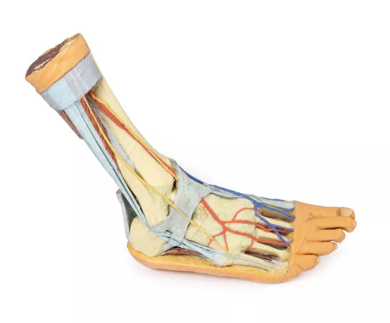
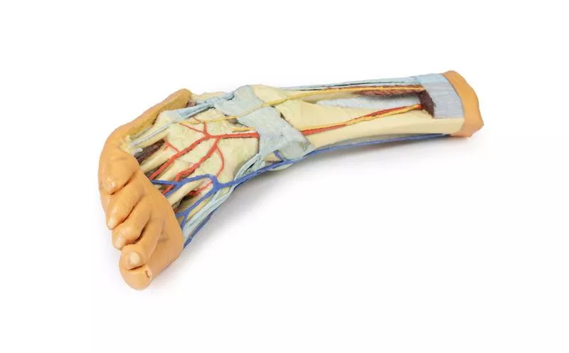
This 3D printed anatomical specimen showcases both superficial and deep structures of the distal leg and foot.
Posterior Compartment: Deep Musculature and Neurovascular Structures
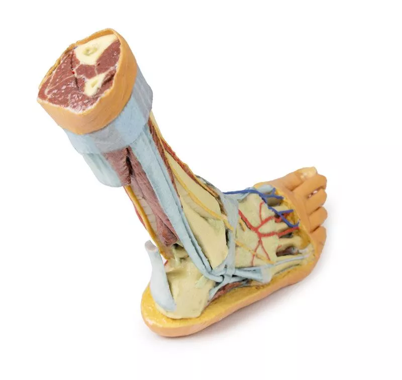
The posterior compartment of the leg has been dissected proximally to remove the triceps surae muscles and the tendocalcaneus, revealing the deep muscles: tibialis posterior, flexor digitorum longus, and flexor hallucis longus. The tibial nerve and posterior tibial artery can be traced down to their medial and lateral plantar branches at the level of the flexor retinaculum. To enhance visibility, the origin of the abductor hallucis brevis has been removed.
Venous Anatomy and Anterior Compartment Highlights
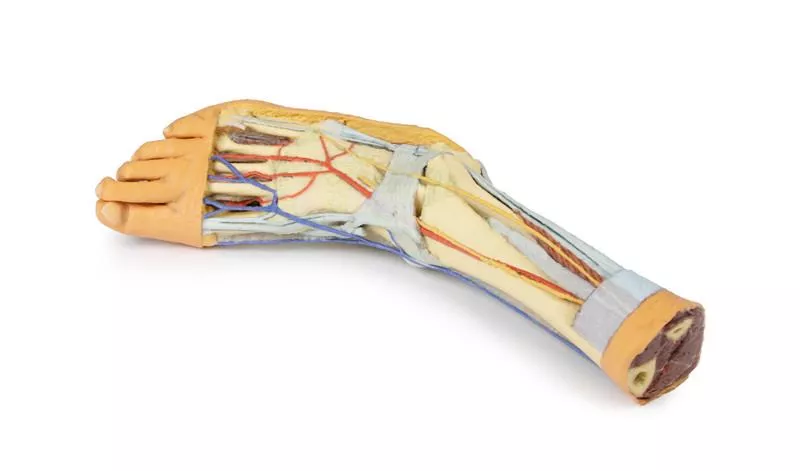
The great saphenous vein is preserved as it originates from the medial side of the dorsal venous arch, extending to the edge of the specimen. While the anterior compartment muscles have been removed to expose the interosseous membrane, the anterior tibial artery, and the deep fibular nerve, key tendons remain intact. These include the insertions of tibialis anterior, extensor hallucis longus, and the hallucal tendon of extensor digitorum longus, all passing deep to the inferior extensor retinaculum. The anterior tibial artery continues as dorsalis pedis, giving rise to the arcuate artery and dorsal metatarsal arteries. Removal of the dorsal interossei allows visualization of their terminal branches as they approach the plantar interossei.
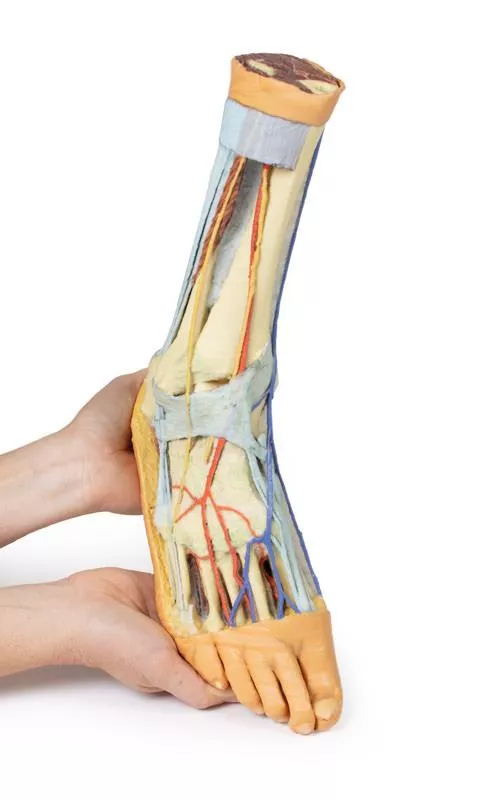
Lateral View: Muscles, Tendons, and Nerve Terminations
On the lateral aspect, the fibularis longus and fibularis brevis muscles and their tendons are clearly visible. The tendons pass beneath the superior fibular retinaculum (cut edge) and the intact inferior fibular retinaculum. Also visible are the extensor digitorum longus tendon to the fifth digit and the termination of the superficial fibular nerve. Near the entry of the fibularis longus tendon into the plantar foot, the abductor digiti minimi muscle origin is preserved.
Ligamentous Structures
Deep structures include key ligaments of the distal leg and foot, such as the anterior and posterior tibiofibular ligaments, calcaneofibular ligament, dorsal and posterior talonavicular ligaments, and the deltoid ligament, all providing essential support and insight into joint stability.
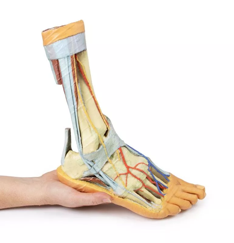
Posterior Compartment: Deep Musculature and Neurovascular Structures
The posterior compartment of the leg has been dissected proximally to remove the triceps surae muscles and the tendocalcaneus, revealing the deep muscles: tibialis posterior, flexor digitorum longus, and flexor hallucis longus. The tibial nerve and posterior tibial artery can be traced down to their medial and lateral plantar branches at the level of the flexor retinaculum. To enhance visibility, the origin of the abductor hallucis brevis has been removed.
Venous Anatomy and Anterior Compartment Highlights
The great saphenous vein is preserved as it originates from the medial side of the dorsal venous arch, extending to the edge of the specimen. While the anterior compartment muscles have been removed to expose the interosseous membrane, the anterior tibial artery, and the deep fibular nerve, key tendons remain intact. These include the insertions of tibialis anterior, extensor hallucis longus, and the hallucal tendon of extensor digitorum longus, all passing deep to the inferior extensor retinaculum. The anterior tibial artery continues as dorsalis pedis, giving rise to the arcuate artery and dorsal metatarsal arteries. Removal of the dorsal interossei allows visualization of their terminal branches as they approach the plantar interossei.
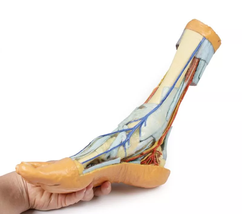
Lateral View: Muscles, Tendons, and Nerve Terminations
On the lateral aspect, the fibularis longus and fibularis brevis muscles and their tendons are clearly visible. The tendons pass beneath the superior fibular retinaculum (cut edge) and the intact inferior fibular retinaculum. Also visible are the extensor digitorum longus tendon to the fifth digit and the termination of the superficial fibular nerve. Near the entry of the fibularis longus tendon into the plantar foot, the abductor digiti minimi muscle origin is preserved.
Ligamentous Structures
Deep structures include key ligaments of the distal leg and foot, such as the anterior and posterior tibiofibular ligaments, calcaneofibular ligament, dorsal and posterior talonavicular ligaments, and the deltoid ligament, all providing essential support and insight into joint stability.
Login
Erler-Zimmer
Erler-Zimmer Medical GmbH
Hauptstrasse 27
77886 Lauf
Germany
info@erler-zimmer.de
Achtung! Medizinisches Ausbildungsmaterial, kein Spielzeug. Nicht geeignet für Personen unter 14 Jahren.
Attention! Medical training material, not a toy. Not suitable for persons under 14 years of age.
Other customers also bought

Foot - Deep plantar structures
This 3D printed anatomical model offers a detailed view of the deep plantar structures of the foot.Medial Structures and Vascular PathwaysOn the medial side, the cut edge of the great saphenous vein is visible within the superficial fascia, positioned just anterior to the medial and lateral plantar arteries and nerves, which lie above the insertion of the tibialis posterior muscle.Exposed Third Muscular LayerThe superficial fascia, plantar aponeurosis, and superficial muscles have been removed to reveal the third muscular layer. The cut edges of the first, second, and third layer muscles remain attached to the calcaneus for clear orientation. The cut tendon of the flexor digitorum longus and the distal tendons of both flexor digitorum longus and brevis are also exposed. Key Muscles and TendonsVisible beneath the flexor hallucis longus tendon are the transverse and oblique heads of the adductor hallucis, surrounded by a complete lateral and partial medial head of the flexor hallucis brevis. Plantar interosseous muscles can be seen deep to the adductor hallucis, adding depth to the muscular architecture.Ligamentous StructuresBeneath the muscular layer, the model displays essential ligaments of the tarsal and metatarsal joint capsules, along with the long and short plantar ligaments and the plantar calcaneonavicular ligament, offering insight into structural support and joint integrity. Lateral AspectLaterally, the abductor digiti minimi muscle has been sectioned to reveal the insertions of the peroneus longus and brevis tendons, completing a comprehensive view of the plantar foot anatomy.

Foot - Superficial and deep dissection of distal leg and foot
This 3D printed specimen presents a mixed superficial and deep dissection of the distal leg and foot, providing a detailed view of tendons, muscles, and neurovascular structures.Posterior and Medial AnatomyPosteriorly, the compartment muscles and neurovascular structures have been removed to highlight the tendocalcaneus and calcaneus body. Medially, the tibialis posterior, flexor digitorum longus, and flexor hallucis longus tendons are visible deep to the crural fascia, passing under the opened flexor retinaculum toward the medial foot. The adductor hallucis, medial head of the flexor hallucis brevis, and flexor digitorum brevis muscles are fully exposed on the medial aspect.Dorsal AnatomyOn the dorsum of the foot, both superior and inferior extensor retinacula are preserved, with anterior compartment muscles extending to their distal attachments, including the fibularis tertius. The anterior tibial artery is visible continuing as the dorsalis pedis artery. Deep to the long tendons, the extensor hallucis brevis, extensor digitorum brevis, and dorsal interosseous muscles are clearly visible. Lateral AnatomyLaterally, the fibularis longus and brevis muscles are visible beneath the crural fascia, with tendons passing under both superior and inferior fibular retinacula. The abductor digiti minimi muscle is exposed along the lateral margin of the foot.

Foot - Plantar surface & superficial dissection on the dorsum
This 3D printed specimen showcases the plantar surface of the foot with partial dorsal dissection, making it ideal for studying both superficial and deep structures.Plantar Surface AnatomyThe plantar aponeurosis has been largely removed to reveal the first layer of muscles, while a portion of the lateral band remains attached to the fourth metatarsal. The flexor digitorum brevis muscle and tendons overlie the flexor digitorum longus tendon, with divisions of the tendon and lumbricals visible approaching the flexor sheaths. Superficial branches of the medial and lateral plantar nerves radiate from the margins of the flexor digitorum brevis and divide into common and proper plantar digital branches. At the edges of the dissection, the abductors and flexors of the hallux and fifth digit are exposed, including the medial and lateral heads of the flexor hallucis brevis inserting on sesamoids beside the flexor hallucis longus tendon.Dorsal Surface AnatomyOn the dorsum, a window of skin has been removed to display the dorsal fascia and underlying tendons from the anterior compartment. The dorsal fascia over the lateral metatarsals reveals the extensor hallucis brevis, extensor digitorum longus and brevis tendons, and dorsal interosseous muscles. Proximal Leg StructuresThe distal tibia and fibula are visible, joined by the interosseous membrane. Leg compartment muscles and tendons, including the tendocalcaneus, are preserved. Both anterior and posterior tibial arteries with veins, the superficial fibular nerve, and the tibial nerve are visible in cross-section.

Foot - Structures of the plantar surface
This 3D printed specimen highlights the anatomy of the right distal leg and foot, including deep plantar structures.Proximal Leg StructuresIn cross-section, the tibia, fibula, interosseous membrane, and leg muscles are clearly visible. At the medial ankle, the long tendons of the dorsiflexors and plantarflexors pass superficial to capsular and extracapsular ligaments. The posterior tibial artery, veins, and tibial nerve are traced from the posterior leg to the plantar surface. Laterally, the fibularis muscles (longus, brevis, tertius) and their insertions are displayed.Dorsal Foot StructuresOn the dorsum of the foot, the anterior tibial artery and deep fibular nerve emerge deep to the extensor hallucis longus, lying superficial to the extensor hallucis brevis and extensor digitorum brevis. Plantar Surface AnatomyThe plantar aponeurosis and portions of superficial and deep muscles—flexor digitorum brevis, abductor hallucis, abductor digiti minimi, quadratus plantae—have been removed to reveal the tibialis posterior, flexor digitorum longus, flexor hallucis longus, and fibularis longus tendons. The origins of the flexor hallucis brevis and flexor digiti minimi brevis, as well as the lumbricals arising from the flexor digitorum longus tendons, are also visible.

Continuous innovation

Social responsibility

Active customer orientation

Understanding quality

Sustainable actions

ISO 9001 certification