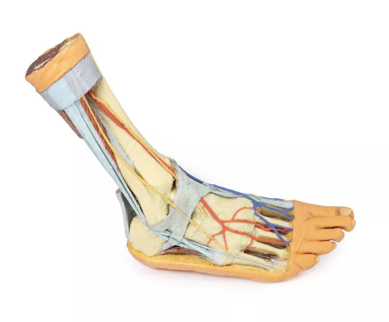
Product information "Foot - Plantar surface & superficial dissection on the dorsum"
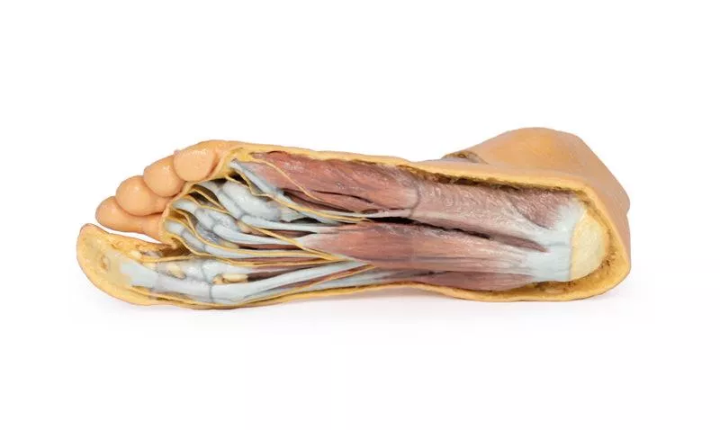
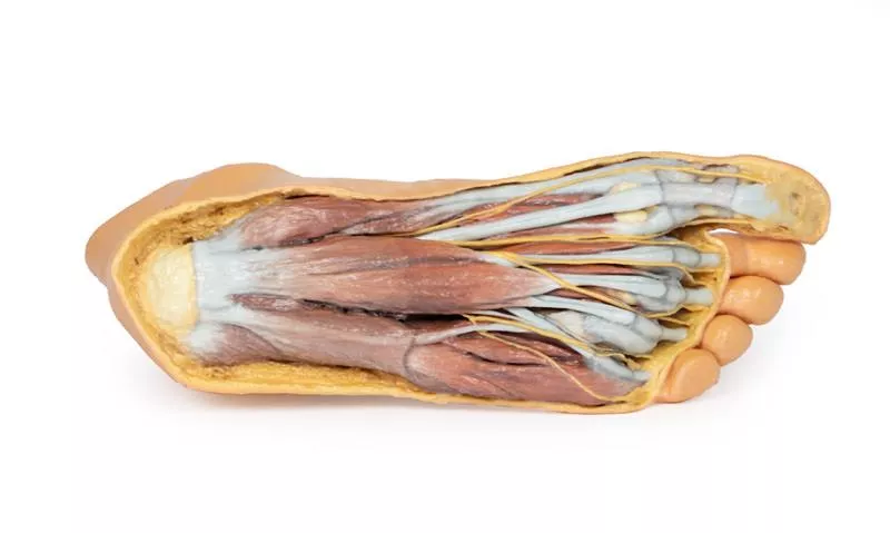
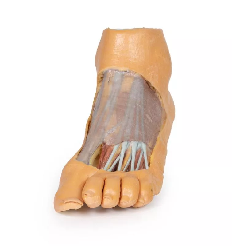
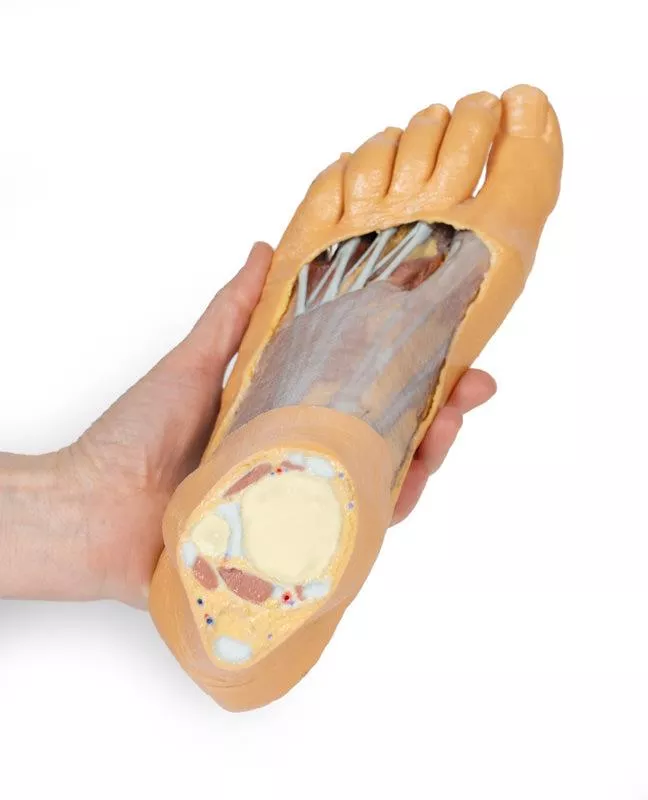
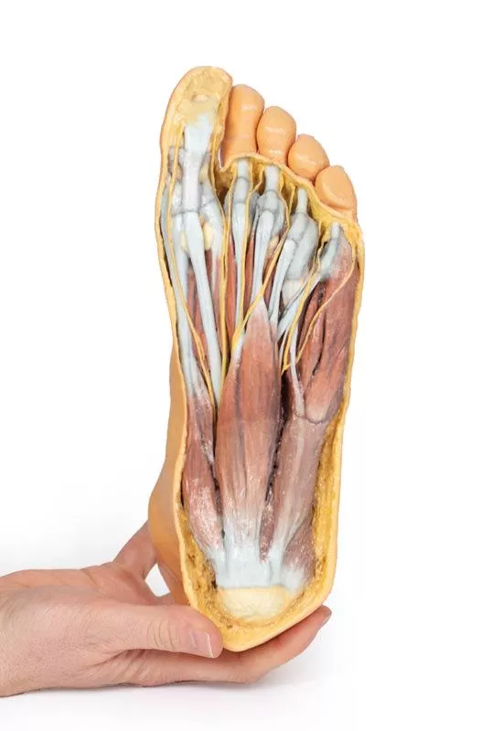
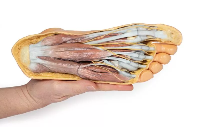
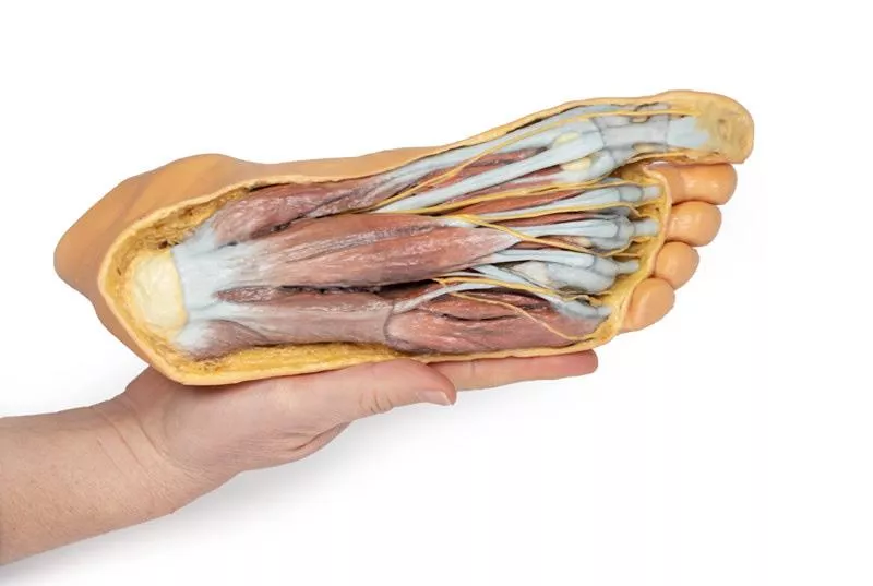
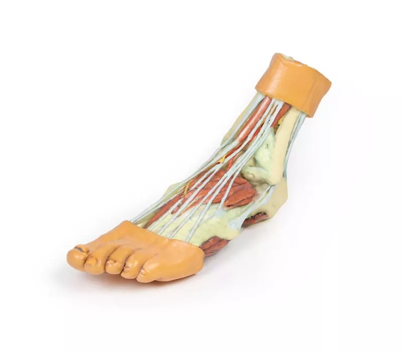
This 3D printed specimen showcases the plantar surface of the foot with partial dorsal dissection, making it ideal for studying both superficial and deep structures.
Plantar Surface Anatomy
The plantar aponeurosis has been largely removed to reveal the first layer of muscles, while a portion of the lateral band remains attached to the fourth metatarsal. The flexor digitorum brevis muscle and tendons overlie the flexor digitorum longus tendon, with divisions of the tendon and lumbricals visible approaching the flexor sheaths. Superficial branches of the medial and lateral plantar nerves radiate from the margins of the flexor digitorum brevis and divide into common and proper plantar digital branches. At the edges of the dissection, the abductors and flexors of the hallux and fifth digit are exposed, including the medial and lateral heads of the flexor hallucis brevis inserting on sesamoids beside the flexor hallucis longus tendon.
Dorsal Surface Anatomy
On the dorsum, a window of skin has been removed to display the dorsal fascia and underlying tendons from the anterior compartment. The dorsal fascia over the lateral metatarsals reveals the extensor hallucis brevis, extensor digitorum longus and brevis tendons, and dorsal interosseous muscles.
Proximal Leg Structures
The distal tibia and fibula are visible, joined by the interosseous membrane. Leg compartment muscles and tendons, including the tendocalcaneus, are preserved. Both anterior and posterior tibial arteries with veins, the superficial fibular nerve, and the tibial nerve are visible in cross-section.
Erler-Zimmer
Erler-Zimmer GmbH & Co.KG
Hauptstrasse 27
77886 Lauf
Germany
info@erler-zimmer.de
Achtung! Medizinisches Ausbildungsmaterial, kein Spielzeug. Nicht geeignet für Personen unter 14 Jahren.
Attention! Medical training material, not a toy. Not suitable for persons under 14 years of age.


